Inhibition of Contractile Vacuole Function by ... - Universität zu Köln
Inhibition of Contractile Vacuole Function by ... - Universität zu Köln
Inhibition of Contractile Vacuole Function by ... - Universität zu Köln

You also want an ePaper? Increase the reach of your titles
YUMPU automatically turns print PDFs into web optimized ePapers that Google loves.
information regarding the presence or absence <strong>of</strong> a SR or a<br />
similar structure.<br />
Mechanism <strong>of</strong> water uptake into CVs and LCVs<br />
Since LCVs are derived from CVs, the mechanism <strong>of</strong><br />
water uptake into both organelles is probably the same. Basically<br />
two models are generally considered. Water could enter<br />
the CVs and LCVs <strong>by</strong> osmosis, or ATP-energized water pumps<br />
could move water across membranes against an osmotic gradient<br />
(discussed in Allen and Naitoh 2002). The existence <strong>of</strong><br />
water pumps has been demonstrated (Zeuthen 1992). However,<br />
an involvement <strong>of</strong> water pumps with water transport into<br />
the CV has not been demonstrated yet. Water transport <strong>by</strong><br />
osmosis requires a higher osmolarity inside the CV than in the<br />
cytosol. Unfortunately, the composition <strong>of</strong> the CV liquid is<br />
not known. A long-held assumption is that the CV expels<br />
water. However, there is little direct evidence for the composition<br />
<strong>of</strong> the expelled liquid (reviewed in Allen 2000, Allen and<br />
Naitoh 2002). For the CV <strong>of</strong> the amoeba Chaos chaos, Riddick<br />
(1968) determined an osmolarity <strong>of</strong> 51 mOsmol (cytosol<br />
117 mOsmol), and similar values have been found for Amoeba<br />
proteus (Schmidt-Nielsen and Schrauger 1963). As pointed out<br />
already <strong>by</strong> Heuser et al. (1993), water should not accumulate<br />
under these conditions within the CV and therefore would<br />
require water pumps. Recent estimates <strong>of</strong> the osmolarity (based<br />
on Na + , K + Ca 2+ and Cl – activity measurements) <strong>of</strong> the CV and<br />
cytosol <strong>of</strong> Paramecium showed that the osmolarity <strong>of</strong> the CV<br />
was 1.5 times higher than that <strong>of</strong> the cytosol in Paramecium,<br />
supporting the hypothesis that water uptake into the CV is<br />
driven <strong>by</strong> osmosis in Paramecium (Stock et al. 2002a, Stock et<br />
al. 2002b). In this study, we have shown that under strong<br />
hyperosmotic conditions, LCVs shrunk, indicating that water<br />
within LCVs followed osmotic gradients and supporting the<br />
The contractile vacuole <strong>of</strong> S. dubia 209<br />
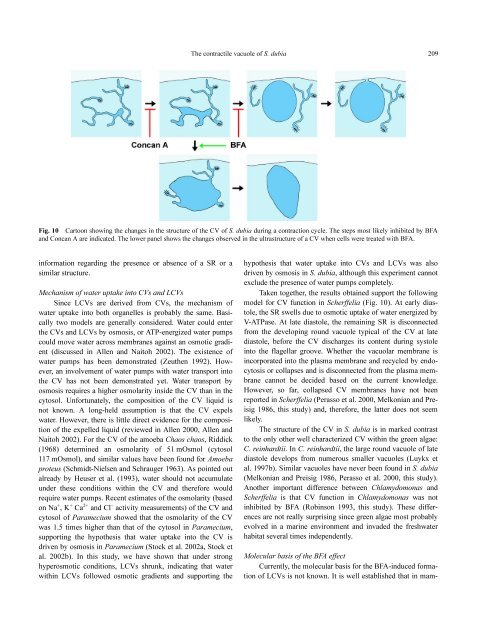
Fig. 10 Cartoon showing the changes in the structure <strong>of</strong> the CV <strong>of</strong> S. dubia during a contraction cycle. The steps most likely inhibited <strong>by</strong> BFA<br />
and Concan A are indicated. The lower panel shows the changes observed in the ultrastructure <strong>of</strong> a CV when cells were treated with BFA.<br />
hypothesis that water uptake into CVs and LCVs was also<br />
driven <strong>by</strong> osmosis in S. dubia, although this experiment cannot<br />
exclude the presence <strong>of</strong> water pumps completely.<br />
Taken together, the results obtained support the following<br />
model for CV function in Scherffelia (Fig. 10). At early diastole,<br />
the SR swells due to osmotic uptake <strong>of</strong> water energized <strong>by</strong><br />
V-ATPase. At late diastole, the remaining SR is disconnected<br />
from the developing round vacuole typical <strong>of</strong> the CV at late<br />
diastole, before the CV discharges its content during systole<br />
into the flagellar groove. Whether the vacuolar membrane is<br />
incorporated into the plasma membrane and recycled <strong>by</strong> endocytosis<br />
or collapses and is disconnected from the plasma membrane<br />
cannot be decided based on the current knowledge.<br />
However, so far, collapsed CV membranes have not been<br />
reported in Scherffelia (Perasso et al. 2000, Melkonian and Preisig<br />
1986, this study) and, therefore, the latter does not seem<br />
likely.<br />
The structure <strong>of</strong> the CV in S. dubia is in marked contrast<br />
to the only other well characterized CV within the green algae:<br />
C. reinhardtii. In C. reinhardtii, the large round vacuole <strong>of</strong> late<br />
diastole develops from numerous smaller vacuoles (Luykx et<br />
al. 1997b). Similar vacuoles have never been found in S. dubia<br />
(Melkonian and Preisig 1986, Perasso et al. 2000, this study).<br />
Another important difference between Chlamydomonas and<br />
Scherffelia is that CV function in Chlamydomonas was not<br />
inhibited <strong>by</strong> BFA (Robinson 1993, this study). These differences<br />
are not really surprising since green algae most probably<br />
evolved in a marine environment and invaded the freshwater<br />
habitat several times independently.<br />
Molecular basis <strong>of</strong> the BFA effect<br />
Currently, the molecular basis for the BFA-induced formation<br />
<strong>of</strong> LCVs is not known. It is well established that in mam-