Inhibition of Contractile Vacuole Function by ... - Universität zu Köln
Inhibition of Contractile Vacuole Function by ... - Universität zu Köln
Inhibition of Contractile Vacuole Function by ... - Universität zu Köln

Create successful ePaper yourself
Turn your PDF publications into a flip-book with our unique Google optimized e-Paper software.
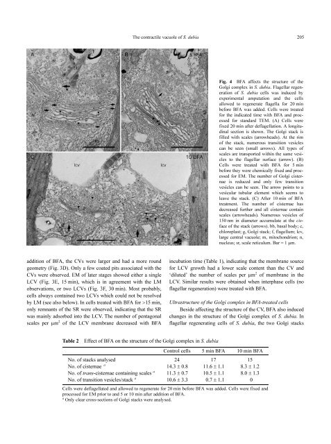
addition <strong>of</strong> BFA, the CVs were larger and had a more round<br />
geometry (Fig. 3D). Only a few coated pits associated with the<br />
CVs were observed. EM <strong>of</strong> later stages showed either a single<br />
LCV (Fig. 3E, 15 min), which is in agreement with the LM<br />
observations, or two LCVs (Fig. 3F, 30 min). Most probably,<br />
cells always contained two LCVs which could not be resolved<br />
<strong>by</strong> LM (see also below). In cells treated with BFA for >15 min,<br />
only remnants <strong>of</strong> the SR were observed, indicating that the SR<br />
was mainly adsorbed into the LCV. The number <strong>of</strong> pentagonal<br />
scales per µm 2 <strong>of</strong> the LCV membrane decreased with BFA<br />
The contractile vacuole <strong>of</strong> S. dubia 205<br />
Table 2 Effect <strong>of</strong> BFA on the structure <strong>of</strong> the Golgi complex in S. dubia<br />
Fig. 4 BFA affects the structure <strong>of</strong> the<br />
Golgi complex in S. dubia. Flagellar regeneration<br />
<strong>of</strong> S. dubia cells was induced <strong>by</strong><br />
experimental amputation and the cells<br />
allowed to regenerate flagella for 20 min<br />
before BFA was added. Cells were treated<br />
for the indicated time with BFA and processed<br />
for standard TEM. (A) Cells were<br />
fixed 20 min after deflagellation. A longitudinal<br />
section is shown. The Golgi stack is<br />
filled with scales (arrowheads). At the rim<br />
<strong>of</strong> the stack, numerous transition vesicles<br />
can be seen (small arrows). All types <strong>of</strong><br />
scales are transported within the same vesicles<br />
to the flagellar surface (arrow). (B)<br />
Cells were treated with BFA for 5 min<br />
before they were chemically fixed and processed<br />
for EM. The number <strong>of</strong> Golgi cisternae<br />
is reduced and only few transition<br />
vesicles can be seen. The arrow points to a<br />
vesicular tubular element which seems to<br />
leave the stack. (C) After 10 min <strong>of</strong> BFA<br />
treatment. The number <strong>of</strong> cisternae has<br />
decreased further and all cisternae contain<br />
scales (arrowheads). Numerous vesicles <strong>of</strong><br />
150 nm in diameter accumulate at the cisface<br />
<strong>of</strong> the stack (arrows). bb, basal body; c,<br />
chloroplast; g, Golgi stack; f, flagellum; lcv,<br />
large central vacuole; m, mitochondrion; n,<br />
nucleus; sr, scale reticulum. Bar = 1 µm.<br />
incubation time (Table 1), indicating that the membrane source<br />
for LCV growth had a lower scale content than the CV and<br />
‘diluted’ the number <strong>of</strong> scales per µm 2 <strong>of</strong> membrane in the<br />
LCV. Similar results were obtained when interphase cells (no<br />
flagellar regeneration) were treated with BFA.<br />
Ultrastructure <strong>of</strong> the Golgi complex in BFA-treated cells<br />
Beside affecting the structure <strong>of</strong> the CV, BFA also induced<br />
changes in the structure <strong>of</strong> the Golgi complex <strong>of</strong> S. dubia. In<br />
flagellar regenerating cells <strong>of</strong> S. dubia, the two Golgi stacks<br />
Control cells 5 min BFA 10 min BFA<br />
No. <strong>of</strong> stacks analysed 24 17 15<br />
No. <strong>of</strong> cisternae a<br />
14.3 ± 0.8 11.6 ± 1.1 8.3 ± 1.2<br />
No. <strong>of</strong> trans-cisternae containing scales a 11.3 ± 0.7 10.5 ± 1.1 8.0 ± 1.3<br />
No. <strong>of</strong> transition vesicles/stack a 10.6 ± 3.3 0.7 ± 1.1 0<br />
Cells were deflagellated and allowed to regenerate for 20 min before BFA was added. Cells were fixed and<br />
processed for EM prior to and 5 or 10 min after addition <strong>of</strong> BFA.<br />
a Only clear cross-sections <strong>of</strong> Golgi stacks were analysed.