Chapter 60 Traumatic Foot Injuries
Chapter 60 Traumatic Foot Injuries
Chapter 60 Traumatic Foot Injuries

You also want an ePaper? Increase the reach of your titles
YUMPU automatically turns print PDFs into web optimized ePapers that Google loves.
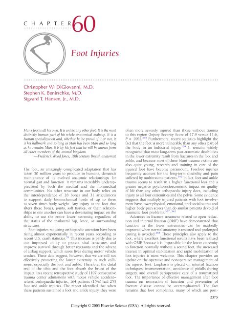
C H A P T E Rzzzzzzzzzzzzzzzzzzzzzzzzzzz<strong>60</strong><strong>Foot</strong> <strong>Injuries</strong><strong>Foot</strong> <strong>Injuries</strong>zzzzzzzzzzzzzzzzzzzzzzzzzzzzzzzzzzzzzzzzzzzzzzzzzzzzzzzzzzzzzzzzzzzzzzzzzzzzzzzzzzzzzzzzzzzzzzzzzzzzzzzzzzzzzzChristopher W. DiGiovanni, M.D.Stephen K. Benirschke, M.D.Sigvard T. Hansen, Jr., M.D.Man’s foot is all his own. It is unlike any other foot. It is the mostdistinctly human part of his whole anatomical makeup. It is ahuman specialization and, whether he be proud of it or not, itis his hallmark and so long as Man has been Man and so longas he remains Man, it is by his feet that he will be known fromall other members of the animal kingdom.—Frederick Wood Jones, 18th century British anatomistThe foot, an amazingly complicated adaptation that hastaken 30 million years to produce in humans, demandsmaintenance of its evolved anatomic relationships fornormal gait and function. It remains incredibly underappreciatedby both the medical and the nonmedicalcommunities. No other structure in our body relies onthe interdependence of 28 bones and 31 articulationsto support daily biomechanical loads of up to threeto seven times body weight. Any injury to the foot thatalters these bones, joints, soft tissues, or their relationshipsto one another can have a devastating impact on theability to use the entire lower extremity, regardless ofthe status of the ipsilateral hip, knee, or surroundingstructures.<strong>Foot</strong> injuries requiring orthopaedic attention have beenrising almost exponentially in recent years according torecent U.S. crash statistics. 35 This increase is partly due toour improved ability to protect vital structures andimprove survival through better restraints and the adventof airbag support, which saves lives during motor vehiclecrashes. These data suggest, however, that we are still noteffectively protecting the lower extremity in such collisions,especially the foot and ankle. Therefore, the distalend of the tibia and the foot absorb the brunt of theimpact. In a recent retrospective study of 1107 consecutivetrauma center admissions with motor vehicle accident–related orthopaedic injuries, 164 patients (15%) had 253foot and ankle injuries. The report identified that whenthese patients sustained a foot and ankle injury, they wereoften more severely injured than those without traumato this region (Injury Severity Score of 17.9 versus 11.6,P < .001). 355 Furthermore, recent statistics highlight thefact that the foot is more vulnerable than any other part ofthe body in an industrial injury. 109 It remains widelyrecognized that most long-term post-traumatic disabilitiesin the lower extremity result from fractures in the foot andankle, and because most of these blunt trauma victims arealso quite young, research and training in care of theinjured foot have become paramount. Forefoot injuriesfrequently account for the long-term disability and painsuffered by multitrauma patients. 342 In fact, foot and ankletrauma seems to result in a higher functional loss and agreater negative psychosocioeconomic impact on qualityof life than any other orthopaedic injury does, includinginjury to all four extremities and the pelvis. Some evidencesuggests that multiply injured patients with foot involvementhave lower physical, emotional, and social scores andhigher body pain scores than do similar patients devoid of337, 342traumatic foot problems.Advances in fracture treatment related to open reductionand internal fixation (ORIF) have demonstrated thatfunction in the lower extremities can be markedlyimproved when normal anatomy is restored and prolongedcasting is avoided. 283 These principles also apply to thefoot, where excellent functional results have been realizedwith ORIF. Because it is impossible for the lower extremityto function normally without a sound foot, the increasedinterest in optimal stabilization and rapid mobilization offoot injuries is most welcome. This chapter provides anupdate on the operative and nonoperative management ofthe injured foot. Emphasis is placed on internal fixationtechniques, instrumentation, avoidance of pitfalls duringsurgery, and overall perioperative care of a traumatizedfoot. The importance of effective management after foottrauma on restoration of function and prevention offracture disease cannot be overemphasized. The factremains that foot complaints, many of which are post-2375
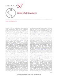
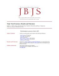
2376 SECTION V • Lower Extremitytraumatic in nature, are a leading cause of patient visits toan orthopaedic surgeon’s office today.Readers seeking information about relevant anatomy,biomechanics, or history or a review of treatment of footfractures are referred to excellent overviews by authorssuch as Heckman, 128 Sangeorzan, 285 and Myerson. 225Inman and colleagues, 145 Mann and Coughlin, 194 andothers have outlined the complex biomechanics of thefoot, which relies on normal motion, alignment, andstability. DeLee 64 has contributed a monumental historicalwork on the treatment of fractures, dislocations, and otherfoot injuries in Mann and Coughlin’s text. 194 From ahistorical standpoint, one of the most pertinent studies offoot function was published by the anatomist DudleyMorton in 1935. 218 Although much of his work has beenneglected or falsely discredited, his studies on theanthropology of the most advanced human musculoskeletalstructure, the foot, provide a comprehensive overviewof the specifics of foot structure and function that areimportant to consider when planning a reconstruction orORIF. The anatomy of the foot is actually complex andoften understudied by orthopaedic residents during training.It is advisable to have a skeletal foot model availablefor evaluation both before and during surgery to aid inunderstanding the proximity and three-dimensional relationshipsof these many structures when consideringinternal fixation or surgical exposure. We encourage ourresidents to use these simple models because they seem tocut down on the operative time required for safe exposure,proper hardware placement, and efficient C-arm use.Adaptation of internal fixation techniques to fracturesin the foot is consistent with the fundamental principles offracture treatment described by the AO/ASIF in the Manualof Internal Fixation. 219 Four goals of internal fixation areidentified by the AO group: (1) anatomic reduction of thefracture, (2) preservation of the blood supply duringsurgery, (3) application of stable internal fixation thataddresses the biomechanical demands of the affectedregion, and (4) mobilization of the injured limb as soonafter injury as possible. The last point is particularly salientto the foot because it spends its entire ‘‘life’’ supporting atremendous amount of weight in comparison to its sizeand thus seems to function even more poorly than otherparts of the musculoskeletal system if this environment isaltered for prolonged periods. By virtue of its location,function, distribution of stress per unit area, and articularinterdependence, the foot cannot tolerate some of thesmaller discrepancies in alignment or instability that caneasily be withstood in many of the more proximal lower orupper extremity regions. Consider the fact that, forexample, the metatarsal heads are normally in contact withthe ground up to 80% of the time during the stance phaseof normal gait. Thus, although 5° of extra-articularmalalignment might be well tolerated in many other partsof the body, this same amount of sagittal or even coronalincongruity in the metatarsal heads, calcaneus, or talusafter foot injury can translate to significant disability in43, 115active individuals on their feet much of the day.Not all fractures require internal fixation to heal withsatisfactory functional results. For example, treatment ofdiaphyseal tibia and humerus fractures by cast immobilizationfrequently results in satisfactory healing. Intraarticularfractures, on the other hand, require anatomicreduction of the joint surfaces and repair of surroundingtorn ligaments, tendons, and joint capsules if full functionalrecovery is to be expected. Precise anatomicrestoration to maintain the postural axis described byMorton and repair of surrounding tissues cannot beachieved without ORIF. The trend in fracture care of aninjured foot has recently evolved toward use of smallerand more low-profile devices; they not only have beenfound to provide adequate fixation but have also been lesssymptomatic than their larger predecessors. Their supportof periarticular surfaces has been acceptable and hasresulted in negligible subsidence over time.Full range of motion is required for normal function insome joints in the foot, but no correlation betweenmovement and normal function has been found in others.In general, joint motion may be sacrificed in the flat jointsof the midfoot without risk of functional impairment. Lossof motion in the intertarsal and tarsometatarsal (TMT)joints, which are predominantly flat joints, has little effecton the overall function of the foot. In marked contrast tothe fingers, loss of motion in the interphalangeal (IP) jointof the great toe and the proximal and distal IP joints of thelesser toes has very few consequences unless the toes aresignificantly deformed or cannot contact the ground.<strong>Injuries</strong> to these joints do not require precise openreduction.Two fundamental considerations should be kept inmind when choosing the appropriate treatment of a footfracture: motion is essential in some joints but not inothers, and early motion with at least some weight bearingis necessary for return of normal motion. These principlesare particularly important in navicular fractures and TMTand subtalar fracture-dislocations. Many of the bones andjoints in the foot are closely interrelated, and some jointsmust have nearly full range of motion for the rest tofunction normally. ORIF is indicated in articular injuriesand in areas that indirectly affect articular congruity. Thehindfoot joints, for example, must retain normal motion toevert and unlock and thereby provide a cushionedheel-strike, but they must later be able to act as a rigidplatform during inversion to shift weight smoothly to theforefoot during the late stance phase and push-off. Themidfoot, on the other hand, is required to remain stiff at alltimes to provide a fairly rigid and immobile arch throughwhich weight can be effectively transferred from posteriorto anterior and under which the neurovascular bundle canbe protected from impact during stance. The forefoot actsas the platform on which we generate formal locomotion,and it must contact the ground evenly to allow a normaldistribution of weight. During normal barefoot gait, theforefoot actually encounters three times as much loaddistribution as the hindfoot. 115 This load increases furtherin the event of a gastrocnemius contracture, which canoccur as a purely atavistic trait and not necessarily as aresult of any traumatic event or pathologic process in thelower extremity 71, 218 (Fig. <strong>60</strong>–1).The ‘‘essential’’ joints in the foot that require normal ornear-normal motion for proper foot function because theirmotion is obligatorily coupled to that of the othersurrounding joints are the ankle (fibulotalar and tibiotalar),subtalar, talonavicular, and lesser metatarsophalangeal
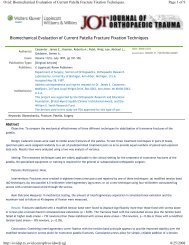
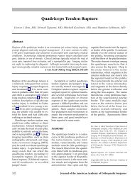
CHAPTER <strong>60</strong> • <strong>Foot</strong> <strong>Injuries</strong>2377Normalgastrocnemius(Normal)area of loading(Abnormal)area of loadingGastrocnemiuscontractureFIGURE <strong>60</strong>–1. Patients who exhibit evidence of gastrocnemius contractureon initial evaluation may have an increased risk for failure of fixation ornonunion in reconstructed midfoot and forefoot fractures or forinstability patterns because of undue stress on the foot during gait in theirrecovery period. A tight gastrocnemius muscle transfers greater stress tothe midfoot and forefoot during the stance phase of ambulation, asdepicted here in the normal versus abnormal (i.e., tight gastrocnemius)situation. Such tightness may have an impact on the outcome of injuryfixation in this region, and affected patients should be considered forconcomitant gastrocnemius release.(MTP) joints. The small translational ability of the flatcalcaneocuboid and the cuboid–fourth/fifth metatarsalarticulations is also important for optimal foot function. 242However, uninhibited motion of the calcaneocuboid jointis not needed for significant inversion and eversion of thehindfoot through the talocalcaneal and talonavicularjoints. In fact, incongruity or post-traumatic arthritis of thecalcaneocuboid joint is relatively well tolerated in mostindividuals, as opposed to other joints in the foot. Astionand associates 10 found by cadaveric study that simulatedfusion of the talonavicular joint eliminated all but 2% ofmotion in the hindfoot joint complex whereas subtalarfusion resulted in a 74% decrease in talonavicular and a44% decrease in calcaneocuboid motion. Fusion of thecalcaneocuboid joint, on the other hand, resulted in onlyminor impairment of motion (33%) in the talonavicularjoint and almost no change in subtalar joint motion. 10 Theremainder of the joints in the foot can be sacrificed orfused with little effect on overall function. For example,fusion of the first MTP joint in an optimal position iscompatible with near-normal function of the forefoot.When fixing fractures or joint incongruity of themidfoot or forefoot in particular, attention should also bedirected toward identification of concomitant gastrocnemiuscontracture. Gastrocnemius equinus is actuallycommon in non-neurologically impaired adults and, if leftuntreated in a trauma patient, might lead to a compromisedlong-term outcome by virtue of its potentiallydetrimental effect on midfoot and forefoot function, assuggested in a recent study by DiGiovanni and colleagues.71 When identified in a trauma patient who isconsidered at risk because of a midfoot or forefoot injurythat is either potentially unstable or anticipated to requireaprolonged period of healing, gastrocnemius contractureshould be released concomitantly during treatment of thefoot injuries. This procedure, called a gastrocnemius slide,is fast, safe, simple, and very effective in decreasing loadacross the front of the foot during gait (Fig. <strong>60</strong>–2), and itis especially important in trauma patients who have beenbedridden or off their feet (in or out of the hospital) for aprolonged period before discovery of a significant footinjury. Because foot injuries often initially take a back seatto more severe life-threatening injuries or are subtle innature and not noticed on admission of a multiply injuredpatient, this scenario is not uncommon. Such a period oftime in an unsplinted state predisposes patients to thedevelopment of gastrocnemius or Achilles contracture ifthey did not have preexistent gastrocnemius tightness, andit can be devastating for normal gait or foot recovery if notpromptly addressed.Because the foot is in a dependent position, it is proneto impaired circulation with inadequate venous andlymphatic return and, consequently, fracture disease.These conditions may lead to the gradual development ofsecondary complications such as joint stiffness, disuseosteoporosis, and muscle atrophy. Early motion andprotected weight bearing, even if allowed in only just aportion of the foot during the healing phase, may mitigatethese conditions. The foot is also prone to significantswelling immediately after a major operation and must beelevated slightly for 36 to 72 hours after surgery. ThisADFIGURE <strong>60</strong>–2. Gastrocnemius release (the ‘‘gastrocnemius slide’’ procedure)can be quickly and easily performed during the course of any footprocedure with the patient either supine or prone. This figure depicts thestandard location of the 1- to 2-inch posteromedial incision located at thegastrocnemius-soleus musculotendinous interval. This incision placementminimizes postoperative scarring around the sural nerve. It isdeepened through the superficial posterior compartment musculature,and the interval between the gastrocnemius (B) and soleus (C) isidentified anteriorly, along with the interval between the gastrocnemiusmuscle (B) and its tendinous attachment (E) distally. This interval is mosteasily identified with the surgeon’s finger in a sweeping motion fromproximal to distal (D). The sural nerve (A) is protected posteriorly whilethe release is made.BCE
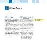
2378 SECTION V • Lower Extremityphase of recovery is called bed exercise rather than bedrestbecause patients are encouraged to perform gentle isometricfoot exercises of the plantar intrinsic musculature assoon as tolerable to prevent the development of edema.These muscles surround the large venous plexus located inthe plantar compartments of the foot, and their activity caneffectively improve venous outflow and decrease swelling.The amount of elevation must be monitored carefully;slight elevation is beneficial, but more is not better. Ideally,the foot is elevated 6 to 12 inches above the bed or justabove the level of the patient’s heart when lying in bed.Greater elevation does not further decrease venous pressurebut instead decreases arterial pressure. The cause of acompartment syndrome is commonly believed to be highintracompartmental pressure; however, it is in fact causedby a decreased differential between arterial and tissuepressure. Lack of arterial circulation in tissues is thepathogenic factor in necrosis. Elevation of the leg does notaffect tissue pressure, but it decreases arterial pressure andmay severely decrease arterial flow to the foot. Thiscondition is called elevation ischemia, and it frequentlyoccurs in patients who are in shock and in those withnormally low blood pressure. 353 Excessive elevation of thefoot, possibly combined with extrinsic pressure, can causeischemia. An early sign that this process has begun is theonset of discomfort when the leg is elevated and relief ofthe discomfort when it is lowered, the same as for a typicalcompartment syndrome.We do not generally use ice as a mainstay of therapy forpatients with foot injuries. Although ice is certainly acommon adjunct to decrease swelling in a traumatizedpatient, it is doubtful that it has much of an effect throughany padded splint or cast. Furthermore, ice bags can oftenbe heavy and uncomfortable for the patient and arefrequently not changed often enough to be of value. Ice isprobably much more useful in the absence of the denseexternal splinting or padding required for soft tissueimmobilization in most trauma cases. The point here isthat there is no better substitute for settling the soft tissueenvelope after foot injury than adequate external immobilization,elevation, pain control, and an appropriateobservation period. The presence of skin wrinkles is oftenthe best way to determine that a soft tissue envelope isamenable to surgical intervention. Early experience withfoot pumps that can be placed directly around the foot oreven within a posterior splint to decrease edema has alsobeen encouraging. 327 Patients seem to tolerate thesedevices fairly well, and they have been particularlyeffective in alleviating the swelling associated with hindfootand midfoot injuries to permit earlier surgicalintervention.Some general radiographic principles in treating footfractures should be mentioned. For any fracture of themidfoot or hindfoot, a set of three standard plainradiographs should always be obtained: an anteroposterior(AP) view, a lateral view, and a medial oblique view of theentire foot. Calcaneal fractures should also always have anaxial heel view (Harris) and a cone-down lateral view ofthe heel, and fractures or dislocations involving the talusor subtalar joint should always include specialized obliqueprojections of the foot as described in their respectivesections of this chapter, as well as AP and mortise viewsof the ankle. Any suspected Lisfranc injury should havestress views obtained in both the sagittal and transverseplanes. The standard set of three ‘‘trauma views’’ as justdescribed is usually adequate for all forefoot injuries unlessthe injury is clearly limited to a single toe, in which case atoe series is acceptable. When dealing with pathology ofthe first MTP joint, consideration should be given to bothan internal and an external oblique view, as well as an axialsesamoid view. The last-named is also useful in determiningdisplacement in a multiply injured forefoot. Computedtomography (CT) scans should be obtained for allcalcaneus, talus, and midfoot fractures or fracturedislocationsand for any complicated foot trauma that ispoorly understood on the radiographs available. Sections 1to 3 mm in thickness can be taken, depending on what thesurgeon is looking for. Axial views are the basic ‘‘workhorse’’plane for reconstruction and are most indicated forLisfranc’s joints, the calcaneocuboid joint, and the talonavicularaxis. Coronal cuts are most useful for imaging thedome of the talus, the ankle mortise, tibiotalar joint loosebodies, and the subtalar joint. Sagittal reconstruction viewsare best for visualizing talar neck fractures.The following sections outline the causes of injury,commonly seen complications, and the recommendedtreatment of various foot fractures, dislocations, or softtissue injuries. The sections on rehabilitation emphasizethe importance of early motion and partial weight bearingthroughout the healing phase in joints in which normalmotion is essential for foot function.INITIAL EVALUATION OF A PATIENTWITH A FOOT INJURYzzzzzzzzzzzzzzzzzzzzzzzzzzzzzzzzzzzzzzzzzzzzzzzzzzzzzzzzzzzThe foot is probably the most frequently overlooked aspectof a patient’s musculoskeletal system on arrival at theemergency department. One of the keys to identification ofsubtle foot injuries is understanding the mechanism ofinjury and taking a good history from the patient, ifpossible, or those responsible for transport. This informationserves to elevate the caregiver’s index of suspicion,when appropriate, regarding the likelihood and pathogenesisof foot injury. Knowledge of the magnitude, duration,mechanism, and location of trauma is imperative to permitefficient and accurate assessment of foot and ankle injury,as well as, of course, other associated injuries. A thoroughhistory of any previous injury or disease that could or hasalready affected the feet is also helpful, including queryingabout previous trauma, diabetes, venous stasis disease,deformity, use of assistive devices, and other conditions. Ifpossible, a patient’s ability to localize the pain with a singlefinger is extremely helpful in establishing a correctdiagnosis because patients will often be vague in theirdescriptions if allowed to be and many injuries arefrequently misdiagnosed as another, possibly more commonpathology in an adjacent area (some less commoninjuries often diagnosed late include lateral process talusfractures, anterior process calcaneal fractures, subtalarinstability, Lisfranc injury, navicular fracture, and compartmentsyndrome). Both feet and ankles should always be
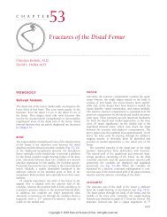
CHAPTER <strong>60</strong> • <strong>Foot</strong> <strong>Injuries</strong>2379FIGURE <strong>60</strong>–3. Fracture blisters around the foot as shownhere are a hallmark of severe injury to the soft tissueenvelope. They should be considered a major risk factorfor infection or wound complications if surgery isperformed too quickly in their vicinity before they havehad a chance to develop completely in terms of size andseverity, as well as to epithelialize. This process can oftentake up to 7 to 10 days. Note the clear versus the redblistering in this patient. Clear blistering representsmore superficial separation of the epidermal layers fromthe underlying skin with resultant serous fluid formationwithin. Hemorrhagic blisters, however, are an indicationof more severe injury because they occur when theentire epidermal layer has separated from the dermisbeneath. Thus, these latter blisters take a longer periodto epithelialize. The best way to manage them is stillcontroversial; some surgeons prefer to leave theseblisters intact, whereas others prefer unroofing them andcoating the underlying layer with silver sulfadiazine(Silvadene), xeroform, or other coating agents.completely exposed and evaluated in the course ofexamination.Physical examination of the foot must be meticulousand well documented in the chart. Pain control isimportant to allow the patient to assist the physician andcooperate with the clinical assessment. The skin is checkedfor puncture wounds, abrasions, blisters, skin tenting,lacerations, erythema, and swelling (Fig. <strong>60</strong>–3). Theexamination includes checking less obvious places such asthe plantar aspect of the feet, the back of the heels, andbetween the toes. Open wounds should generate areflexive response to administer first-generation cephalosporins(grade 1 and 2 injury) with the addition of anaminoglycoside (grade 3 injury) or clindamycin (barnyardinjury, severe contamination) as wound severity increases,after first verifying any drug allergies. Probing of wounds isnot necessary and is best done in a sterile operativeenvironment, except when determining the depth of aplantar puncture wound or deciding whether a woundviolates a nearby joint space (in which case, saline shouldbe sterilely instilled into the joint and observed forextravasation). Tetanus prophylaxis should also be administeredwhen necessary. Any open areas should be coveredwith a sterile povidone-iodine (Betadine)-soaked dressingand wrapped to minimize further contamination. <strong>Foot</strong>wound cultures in an emergency department setting areunreliable and amount to a useless, unnecessary expense.Any deformity of the foot in comparison to the oppositeside should be noted. The neurovascular status of the limbshould be documented, including assessment of (1) theintegrity and amplitude of the dorsalis pedis and posteriortibialis pulses with performance of an ankle-brachial indexand comparison to the opposite side if necessary, (2)capillary refill of the toes, (3) proprioceptive status, (4)sensation of all five nerves with a light touch examinationusing a paperclip or alternative instrument (and sometimestwo-point discrimination if indicated), and (5) motorfunction of all muscle groups in both the foot and leg. Anyirregularity in this portion of the examination should befollowed by an immediate similar examination of the moreproximal portion of the limb to determine causality andthe severity of involvement. Compartments should becarefully examined and compared with those on the otherside. Pain on passive extension (and flexion) of the toesshould be checked. Any suggestion of pathologic orworsening pain, swelling, numbness and tingling, orcoolness in the foot should be followed by promptreassessment of its neurovascular status and, if suspected,measurement of compartment pressure and comparisonwith the patient’s current pressure. Stability and alignmentof the foot and ankle should be checked, after which anappropriate routine radiographic trauma series (threeviews) of the foot or ankle (or both) should be obtained.This examination should not be impeded by dressings,pants, casting material, or other objects, if possible, ifhigh-quality films can be obtained. Poor films should beimmediately discarded and followed by a request forrepeat radiographic examination. In busy emergencydepartments, if one gets in the habit of accepting onlyquality radiographic views, they will eventually becomeroutine (and vice versa).Once this evaluation has been completed, the injuredextremity is splinted, braced, placed in a cast, or left aloneas the injury dictates, and the patient and family arecounseled about the severity of the problem, its prognosis,and the various treatment options available. Multiplyinjured patients and their families should also be told thatroughly 10% of occult traumatic injuries can go undetectedon initial evaluation (primary survey) and that inthe course of hospitalization and recovery (with secondaryand tertiary surveys), other injuries may be identified.Interestingly, studies have documented as high as a 30%incidence of delayed identification of foot and ankleinjuries in polytrauma patients.FRACTURES OF THE TALUSzzzzzzzzzzzzzzzzzzzzzzzzzzzzzzzzzzzzzzzzzzzzzzzzzzzzzzzzzzzOf all the bones in the foot, the most important one tostabilize anatomically with internal fixation and to mobilizesoon after injury is the talus. 134, 250 It alone providesthe link between the foot and the leg and is responsible fortransferring all weight from the body to the foot and
2380 SECTION V • Lower Extremitycoupling much of the motion and function from the foot tothe body. The surgical approach to a talar fracture, ifindicated, is determined by the fracture pattern and thestatus of the soft tissue envelope. In general, four workingapproaches to the talus are used: the medial utilityapproach with or without medial malleolar osteotomy, theanterolateral approach, the posterior approach, and acombined approach. Rarely, a fibular osteotomy or window,as described by Hansen, 120 can be used to access theposterolateral body of the talus. When faced with decisionsregarding treatment of a talus fracture, one must alwaysconsider the fracture pattern, the soft tissues, the tibiotalarjoint, and the talocalcaneal joint. For displaced talarfractures, a strong case can be made in favor of ORIF.When compared with closed treatment, this methodresults in a lower rate of nonunion, shorter time to union,earlier return to motion, earlier return to weight bearing,precise reduction of articular anatomy, enhanced revascularization,lower rate of detectable avascular necrosis, andlower rate of infection in the presence of open wounds.Talar fractures are open 15% to 20% of the time. Bonegrafting of major injuries is frequently required, about65% of the time in our experience. The goals of operativemanagement of these fractures are to restore joint congruity,prevent deformity, and avoid infection.ANATOMYThe articular surfaces of the head, the superior body, andthe inferior body of the talus make flexion and extensionof the ankle, inversion and eversion of the hindfoot,pronation during early stance, supination during latestance, and normal push-off possible. Most hindfootmotion is dependent on the integrity of the acetabulumpedis, a confluence of the talonavicular joint and anteriorfacet of the subtalar joint. More than <strong>60</strong>% of the surface ofthe talus is covered by cartilage and articulates in at leastseven different places with other bones; thus, normal gaitmechanics may be seriously disrupted by loss of motion inthese joints. 163 The unique structure, weight-bearingfunction, and articular anatomy of the talus demandindividualized treatment for the multiple potential fracturelocations and patterns that can occur in this singlebone. 93, 317 It is wider anteriorly than posteriorly andbroader inferiorly than superiorly as it fits within the anklemortise. The bone is extremely dense; accordingly, anyinjury to its neck or body should immediately suggest ahigh-energy mechanism. The neck is angled 15° to 20°medially in a proximal-to-distal direction and is one of thefew areas of the talus without cartilage; this area is alsodistinguished as being the major source of vascular inflowto the talus and, in addition, its most vulnerable site ofinjury. Because most of the bone must articulate with thesurrounding facets of the ankle, talonavicular, and subtalar200, 321joints, it has no muscular or tendinous attachments.Moreover, the relatively small perfusion zones of the talusrender it susceptible to disturbances in perfusion withmany injuries, especially dislocations (see Table <strong>60</strong>–1). Itstwo processes, posterior and lateral, help provide articularand ligamentous support to the surrounding structuresand will be discussed further in their fracture sections.BLOOD SUPPLYTalar blood supply is limited and easily compromised bytrauma. Thus, the talus is prone to vascular insufficiencyTABLE <strong>60</strong>–1zzzzzzzzzzzzzzzzzzzzzzzzzzzzzzzzzzzzzzzzzzzzzzzzzzzzzzzzzzzzzzzzzzzzzzzzzzzzzzzzzzzzzzzzzzzzzzzzzzzSusceptibility to Avascular Necrosis*Type IPeripheral fractures Circulation intact No necrosisProcessus fibularisProcessus posteriorDistal neckHeadType IICentral fractures without displacement Circulation mainly intact Seldom necrosisProximal neckBodyType IIICentral fractures with displacementProximal neckBodyType IVDislocation fracturesProximal neckBody dislocated in the ankle and/orsubtalar jointIntraosseous circulation interrupted, auxiliarycirculation intactInterosseous and auxiliary circulationinterruptedOften necrosisNearly always necrosiszzzzzzzzzzzzzzzzzzzzzzzzzzzzzzzzzzzzzzzzzzzzzzzzzzzzzzzzzzzzzzzzzzzzzzzzzzzzzzzzzzzzzzzzzzzzzzzzzzzzzzzzzzzzzz*As noted by Szyszkowitz and colleagues, the degree of avascular necrosis in the talus really depends on the ″personality″ of the fracture; thus when trying topredict the eventuality of osteonecrosis, one must consider not only the energy that went into the fracture but also its location.Source: Szyszkowitz, R.; et al: Clin Orthop 197:97–107, 1985.
CHAPTER <strong>60</strong> • <strong>Foot</strong> <strong>Injuries</strong>2381LATATSPTTCDPTCBand avascular necrosis, especially after subluxation ordislocation of the body. 255 Evidence suggests that immediatecompression fixation may reduce the ultimate extentof avascular necrosis, and ORIF should be considered fortreatment of all talar fractures. Blood flow to the talus issupported to some extent by all three major vessels thatcourse through the leg en route to the ankle. Becausebranches of these vessels traverse the talus to form ananastomotic sling around its neck in the region of the sinustarsi and tarsal canal, most blood flow must travel in adistal-to-proximal direction in the bone, and hence talarneck injuries result in a high level of avascular necrosiswithin the talar body. Although inflow support is variableand the talus enjoys only limited entry regions (neck andbody), multiple extraosseous sources are available, andintraosseous and extraosseous anastomoses are rich. Inorder of importance, the talus relies on the artery of thetarsal canal and deltoid branches (off the posterior tibialartery) to supply the body and on the dorsalis pedis artery(off the anterior tibial artery) and the artery of the tarsalsinus (off the perforating peroneal artery) to supply thehead, neck, and posterior process, respectively (Fig. <strong>60</strong>–4).Preservation of even one of these three major arteriescan at times provide enough collateral flow to sustainhealing and viability of the talus, depending on patientand injury factors, although in general, viability of thetalar body requires integrity of the posterior tibial102, 163, 220, 221, 295artery. The capsular and ligamentoussupports also contribute a rich vascular plexus of vesselssupplying collateral flow through the periarticular attachments,and tributaries between the three major arteriesprovide an intraosseous network. Medial malleolar osteotomyor fracture manipulation for fixation of talusfractures must be done with care to avoid injury to thedeltoid artery.DPPTTSLATFIGURE <strong>60</strong>–4. Knowledge of arterial perfusion to the injured talus isimperative when considering exposure for open reduction and internalfixation. These illustrations show the five major arterial supplies (variablyfrom each of the three major leg vessels) from dorsal (A) and plantar (B)views: the artery of the tarsal sinus (TS) from the dorsalis pedis orperoneal artery; the artery of the tarsal canal, usually from the posteriortibial artery; the deltoid artery from the artery of the tarsal canal (TC) orthe posterior tibial artery; the posterior direct branches (PT), usually fromthe peroneal artery; and the superomedial direct branches of the dorsalispedis (DP). Note that the talus is a largely cartilage-covered articularsurface and that important blood supply enters it wherever soft tissuesare attached to the bone.IMAGINGTalar injuries can usually be identified on a routine set ofankle plain films. Care should be taken to obtain a truelateral of the talus so that a nondisplaced talar neckfracture is not missed because of radiographic obliquity.With this latter view, the fracture line can often be seen atthe point where it exits the talus superiorly along the neckor inferiorly into the talocalcaneal joint. <strong>Foot</strong> views,however, should also be obtained because the talusarticulates with both the ankle and the foot and injury candisrupt the anatomic relationships of both. Canale andKelly have also described an additional view to visualizethe entire talar neck and shoulder as a true AP projection 37(Fig. <strong>60</strong>–5). The foot is maximally plantar flexed andeverted (pronated 15°) to swing the calcaneus underand away from the overlying talus, thereby allowing anunimpeded radiographic view of the talus in the AP plane.15 o 75 oAFIGURE <strong>60</strong>–5. The Canale view, as described by Canale and Kelly, is auseful en face image of the talus used to most accurately identify step-offor malalignment in talar neck fractures. When obtained correctly, itshould correct for the 15° declination of the talus and its overlap of thecalcaneus by everting and plantar flexing the foot in relation to the imagemachine as shown (A). This view brings the talus into a more orthogonalposition to the image plane and swings the calcaneus out fromunderneath to get an unimpeded view of the medial cortex and lateralaspect of the shoulder of the talar neck (B).
2382 SECTION V • Lower ExtremityA40 o10 o20 o30 o40 oresonance imaging (MRI) and bone scans are not ofparticular value in most acute foot injuries but becomemore useful in the chronic setting (6 weeks or more) whenpatients have unremitting ankle or hindfoot pain despitenegative studies. Often, these patients can have a previouslydifficult-to-recognize osteochondral talar injury,peroneal tear, or anterior process calcaneal fracture, all ofwhich can be manifested as swelling or discomfort aroundthe talus. Subtalar or ankle instability can also result fromthe same injury that affected the talus, although stiffness isfrequently a more common scenario in the subacute orchronic setting. AP and lateral stress views with comparisonwith the opposite foot are ideal in this situation(Fig. <strong>60</strong>–7).When evaluating fractures of the talar neck or body, itis important to remember that plain radiographs can bedeceptive. As a general rule of thumb, if one ‘‘sees’’ afracture line, it is generally ‘‘displaced.’’ Because closedtreatment of these injuries is reserved for truly nondisplacedvariants, this caveat plays a role in the workup ofany ‘‘borderline’’ fracture in this region.Fractures of the Neck of the TalusFIGURE <strong>60</strong>–6. The Broden view is taken best fluoroscopically because itoften requires multiple reposition attempts to get a ‘‘perfect view’’ of thesubtalar joint—varying both angulation with the C-arm from a verticalposition and rotation of the foot by the surgeon (A). This radiograph ismost useful to identify congruity of the posterior facet of the subtalarjoint, but with experience, it can also be used to evaluate the anterior andmiddle facets as well (B). Although the view provided shows a normalposterior facet, irregularity at the subchondral margin of the subtalar jointcan also easily be identified in this view, such as occurs afterintra-articular displacement from a calcaneal fracture.The x-ray tube is angled at 75° up from the horizontaltoward the center of the ankle joint (talus). Another viewthat can be used to examine articular congruity of theposterior facet is a Broden view (Fig. <strong>60</strong>–6). The foot isinternally or externally rotated 45° and the beam angledsequentially between 10° and 40° cephalad until anaccurate image of the posterior or middle facet (or both) isobtained. 28 Because of the need to vary beam positionand rotation, these views are most easily obtained intraoperativelywith a C-arm to minimize the need forrepetitive positioning and plain radiographs until anoptimal view is obtained. Despite the utility and reliabilityof routine plain films in the identification of talar injuries,more precise definition of talar injuries has been greatlyfacilitated by the use of CT scans. CT is particularly helpfulfor preoperative assessment of complex fractures, whenit can supplement the findings seen on plain radiographs,including oblique views of the ankle and foot. MagneticFractures in the body and neck of the talus usually resultfrom high-energy injuries. Fractures involving the head,midbody, and posterior body are less common but mayoccur after certain types of axial loading or high-energyinjuries. Osteochondral fractures are frequently associatedwith ankle sprains, subtalar sprains, and fracturedislocations.The type of talar fracture that is mostcommonly seen in trauma centers, however, is a fracture ofthe neck and the anterior body, 52, 321 which occurs in over50% of talar injuries. 165 Injury is usually caused bydorsiflexion impaction of the foot during a high-energycollision (the so-called aviator’s astragalus as described byColtart in 1952 after evaluation of over 200 talar injuries ofthe Royal Air Force during World War II). 51 Excessivedorsiflexion and axial loading are also applied to the footwhen it rests on the pedal of an automobile at the momentof a head-on collision or when it strikes the ground after afall from a height. These mechanisms lead to medialmalleolar fracture 20% to 25% of the time. 126 Talarfractures in this region can also occur as a result of forcedinversion, eversion, or rotation. Inokuchi and associatesdefined a talar neck fracture as one whose fracture lineexits laterally on the interior surface of the talus in front ofthe lateral process, regardless of its course anteromedially.148 Although the evidence suggesting that theseinjuries should be considered surgical emergencies whendisplaced is indeterminate, it is recommended that salvageabledisplaced talar neck fractures be treated within 4to 6 hours of identification to minimize complications. 281CLASSIFICATIONMany authors such as Coltart (1952), Mindell (1963), andMarti-Weber (1978) have provided classifications of talarfractures, but the Hawkins classification (1970) haswithstood the test of time as being the most useful to gradethe severity of injury and determine the best type of
CHAPTER <strong>60</strong> • <strong>Foot</strong> <strong>Injuries</strong>2383treatment of talar neck fractures. 126 This classification istraditionally used to grade talar neck fractures by theamount of dislocation that has occurred between the bodyfragments and the neck, the ankle, or the subtalar joint. Itis also used to predict the amount of avascular necrosisthat may be expected. 126 Depending on the severity of theinjury, the risk of avascular necrosis in the talar body runsfrom less than 5% to 90% or more. Although roughly 20%of talar injuries can be open with an anterolateral anklewound, almost 50% of Hawkins type III injuries occuras open wounds, and infection rates have been as highas 40%. 52, 198 The force necessary to create a talar neckfracture experimentally is extremely high, so any fractureof this remarkably hard bone needs to be recognized andaggressively treated. The extreme foot position and energydissipation resulting in Hawkins type IV injuries causeextrusion of the talar body fragment posteromedial to themedial malleolus. In these circumstances, the skin is oftentented or open with concomitant impingement of theneurovascular bundle, thus making this injury a trueorthopaedic emergency. Extreme care should be taken toavoid injury to the deltoid ligament in these situationsbecause it is usually both the only remaining soft tissueattachment to this fragment and the most importantprognostically regarding viability of the talar body. 251 TheHawkins classification is as follows:Type I: The talar neck fracture is undisplaced; the riskof avascular necrosis in the body is less than 10% (0%to 13%).FIGURE <strong>60</strong>–7. Subtalar instability is often subtle and can accompanyhindfoot injury or fracture. Although no absolute criteria can be used toreliably confirm its existence when not overt (i.e., dislocation), it is bestidentified by taking stress films when radiographs are otherwise normaland the clinical examination is equivocal. These views are similar to theanterior drawer and varus stress views of the ankle, although in this case,different parameters are being evaluated. Lateral gapping of the posteriorfacet of over 1 cm, or greater than 5 mm when compared with theopposite side, and 1 cm of anterior (forward) translation of the anteriorprocess of the calcaneus away from the lateral process of the talus suggestinstability of the subtalar joint. Such findings may require reconstructionof the interosseous ligament or subtalar fusion if symptoms warrant. Theimages are from a 20-year-old college student with a vague history oflow-energy trauma (sprains) to the foot but persistent complaints ofunsteadiness on uneven ground. He had no complaints about theankle—only the sinus tarsi. His unstressed (A) and anterior drawer (B)stress films suggest a stable ankle but excessive subtalar laxity, asindicated by lower markers. He eventually underwent subtalar arthroscopyto determine the nature of his problem and was found to haveabundant synovitis along the posterior facet with a small tear in theinterosseous ligament. Intraoperative arthroscopic images of the lateralaspect of the posterior facet during translational and varus stress showedabnormal motion when compared with the unstressed view shown here(C), where the edges of the talar (above) and calcaneal (below) facets lineup normally. The probe lies in the lateral gutter adjacent to the reflectionof the lateral talocalcaneal and calcaneofibular ligaments.
2384 SECTION V • Lower ExtremityPERCUTANEOUS FIXATION (HAWKINS TYPE I)Type II: The body is slightly displaced or dislocated fromgraphs. 294 allow direct visualization of the fracture, but it doesthe subtalar joint; the risk of avascular necrosis in thebody is less than 40% (20% to 50%).Type III: The body is displaced from both the ankle and thesubtalar joints; the risk of avascular necrosis in the bodyis greater than 90% (75% to 100%).Type IV: This category, added by Canale and Kelly 37 andnot part of Hawkins’ original classification, includessubluxation of the head at the talonavicular joint,dislocation of the body from the ankle and subtalarjoints, and extrusion of the body; the risk of avascularnecrosis in the body approaches 100%.Percutaneous fixation should be considered only if thefracture is nondisplaced or the resultant reduction isanatomic, and the use of a Canale view or CT shouldaccompany routine foot and ankle views to accuratelydetermine displacement and reduction because the accuracyof closed reduction is quite hard to verify. This plainfilm view permits assessment of angulation and shorteningnot noticeable on the other films, and it is also importantto know how to obtain this view intraoperatively to assessreduction, which often requires a team effort with theimaging technician (see Fig. <strong>60</strong>–5). With evidence of evenIt is important to note that many talar injuries are 1mmofstep-off or rotation in a talar neck fracture, it isunclassifiable, even with this scheme; therefore, results are recommended that it be openly reduced and fixed fordifficult to compare when looking at published series’ rates best results. It can be argued that even nondisplaced talarof avascular necrosis (multiple authors’ ranges are in neck fractures should be stabilized with screws to allowparentheses).earlier mobilization and range-of-motion exercises. Displacementof as little as 2 mm can significantly altercontact loads within the subtalar joint and affect motion ofCLOSED REDUCTIONClosed treatment is rarely advisable, even though it hasbeen recommended for undisplaced fractures. 250, 254 Anondisplaced Hawkins type I fracture, confirmed bythe hindfoot. A recent cadaveric biomechanical study bySangeorzan and colleagues found that displacement of 2mm resulted in significant weight-bearing shifts of thetalus on the calcaneal facets and subsequent changes incontact pressure that thereafter overloaded the posteriortomography or CT, may be managed through a posterior facet. 294 Malalignment in the dorsomedial or varusapproach with lag screw fixation. This fracture is the oneinstance when the posterior approach is ideal (described inmore detail in the section on percutaneous fixation). 200Rarely, nondisplaced fractures can be treated by closedposition resulted in the greatest displacement, the combinationof which happens to be the most commonmalposition after union. Another recent anatomic studyalso supports altered foot mechanics with varus hindfootmanipulation: the foot is distracted, plantar flexed, positioning, forefoot adduction, or both after talar neckinverted or everted to reduce the varus or valgus malunion. 59malposition, and subsequently compressed as it is returnedto a neutral position. If maintenance of reductionIf the talar neck fracture is truly nondisplaced eitherbefore or after reduction and surgery is anticipated, it canrequires a plantar flexed position, application of a be performed percutaneously either from an anteriornon–weight-bearing below-knee cast should be maintainedfor 3 weeks, after which it can usually be safelychanged to a neutral position. Even when this reduction isperfect, prolonged positioning in a plantar flexed postureis not optimal, and surgical intervention is usuallywarranted. Any residual skin tenting, at-risk soft tissue, ormalreduction demands rapid operative intervention. Inany case of closed management, non–weight-bearing castapproach with the patient supine on an image table orfrom a posterior approach with the patient prone. Somebiomechanical evidence suggests that fixation is improvedwith posteriorly directed screws, and this method ofinsertion avoids potential disruption of the major bloodsupply to the talar neck. Its disadvantages include limitedinspection of the fracture site and limited (no) access tothe subtalar joint. Anterior-to-posterior insertion allowsimmobilization should be pursued for at least 4 to 6 weeks visualization of the fracture edges (open anatomicand followed by 4 to 6 weeks of protected weight bearinguntil both radiographic and clinical evidence of bonyreduction) and subtalar débridement and is less prone topenetration of the subtalar joint, plantar flexion impingement,union. Usually, casting of nondisplaced or minimallyor flexor hallucis longus injury. 320 However, itdisplaced (less than 1 mm) talar neck fractures should beused only as a temporary measure to immobilize the footuntil internal fixation can be carried out or in cases inwhich surgery is contraindicated, such as in very elderly ornonambulatory patients. Regardless of the chosen form oftreatment, the vast majority of nondisplaced or minimallydisplaced fractures enjoy a favorable long-term outcome.does require dissection in the area of the blood supply,requires transchondral screw placement, and by laboratorystandards, provides weaker fixation in comparison.Posterior fixation can be performed through a verticalposterolateral or posteromedial approach. In either case,an incision is made just to one side of the Achilles tendonto avoid the adjacent lateral sural nerve or medialAlthough some authors suggest that up to 5° of malpositionposterior bundle, and regardless of the choice ofor 5 mm of displacement is an acceptable parameterfor closed reduction, we advocate consideration of closedreduction only when it can be anatomic (
CHAPTER <strong>60</strong> • <strong>Foot</strong> <strong>Injuries</strong>2385provide an avenue for relatively ‘‘percutaneous’’ screwapplication if indicated. Because the talus is alignedin a posterosuperolateral-to-anteroinferomedial directionabout 25° (10° to 40°) inthe transverse plane and about15° (5° to 50°) inthe sagittal plane, the posterolateralapproach is preferred.A countersunk, subchondral screw is introduced fromeither side of the posterior process of the talus so that itmisses the flexor hallucis longus. The tendency in recentyears has been toward smaller, lower profile fixation inthe foot to minimize the incidence of hardware irritation,and 2.7- or 3.5-mm screws are thus ideal for this purpose.Despite their seemingly shallow thread configuration,these small screws have strong shanks and a tremendousnumber of threads, which results in a high surface areaand allows excellent compressive strength in the typicallydense bone of the talus. Newer 2.4- and 4.0-mm screwdesigns may also be amenable to this application.Alternatively, the smaller 3.5- or 4.0-mm cannulatedscrews can be ideal for this purpose if limited exposure isrequired. Moreover, the head profile does not interferewith the ankle or subtalar joints, and in the case ofcannulated screws, definitive fixation can be placeddirectly over the K-wires if the reduction is anatomic.Although cannulated screws in this size range can makethe insertion process quicker, greater strength can beobtained with solid-core screws inserted after preliminaryreduction with two parallel K-wires, the holes of whichcan often be used for sequential screw insertion whenwires averaging 0.062 to 0.125 inch in diameter are used.They must be preliminarily drilled with a gliding hole andan appropriately sized bit, however, for compressive lagfixation. These newer solid screws are available asself-tapping screws, which can save operative time. Onewire should be left in place during insertion of the initialscrew to prevent rotation at the fracture site, notuncommon with the amount of torque generated duringscrew placement in this good bone stock. Placement ofonly one screw is not recommended because little time isrequired for a second one and the extra compression andresistance to rotation are certainly worthwhile. If ananterior approach is preferred, a screw can reliably beplaced percutaneously through stab incisions on both themedial and lateral aspects unless some displacementwarrants larger incisions. A small longitudinal medialutility incision between the posterior and anterior tibialtendons and a oblique one laterally in Langer’s lines(Ollier incision) can be used. A longitudinal lateralincision can similarly be used if extension is consideredlikely. Anteriorly placed hardware is less dangerous andmore accurate than posteriorly placed hardware withproper percutaneous insertion, although care must betaken during insertion to avoid interference with talonavicularfunction. If anterior, the screw heads must berecessed within the head of the talus or at the junction ofthe head and neck. One screw from each side is probablymore ideal fixation, although parallel screws from onlyone side can be considered (Fig. <strong>60</strong>–8). Regardless of thedirection of screw placement, the point of screw insertionis paramount in enabling anatomic and concentriccompression. Eccentrically inserted screws from eitherdirection, when tightened for compression, will allowFIGURE <strong>60</strong>–8. The ideal fixation of a talar neck fracture withoutsignificant comminution that has been reduced consists of one 3.5-mmlag screw for compression and another placed nearly parallel to it forrotatory control. Although K-wires can be used as alternative fixation,they do not provide acceptable enough compression to permit early rangeof motion and limited weight bearing. Standard AO technique is usedduring drilling to lag these cortical screws. They provide the best meansof fixation in this dense bone and, because of their thinner outerdiameter, produce less torque across the fracture site during introductionthan noted with other screws. A vertical posteromedial or preferably aposterolateral incision (because of talar angulation) provides the bestaccess. One or both screws can be placed through these incisions;occasionally, the second screw must be introduced through a nearby stabincision on the other side of the Achilles tendon.gapping of the fracture edges on the side opposite theirlocation if they are not placed in the midaxial plane of thetalus.If comminution is noted intraoperatively despite minimaldisplacement, use of a 2.0-mm blade plate as afixed-angle device should be considered to maintain lengthand rotation, which cannot be accomplished by screwsunder these circumstances. It is easily placed from thelateral side along the shoulder of the talus, and theentrance of the blade can be predrilled with a 1.5-mm drillbit or similar-diameter K-wire. Plate holders can positionthe device along the long axis of the talus, and a small bonetamp can be used to gently impact the fixed-angle bladeinto the predrilled hole. This hole should be made about 1cm proximal to the talonavicular joint, centered in the APplane. Imaging can be used to facilitate proper placementthrough the center of the anatomic neck of the talus. Afracture fixed firmly by this approach can adjust to stressescaused by early motion and rarely becomes displaced.Although stainless steel hardware is the strongest andeasiest to remove, one can consider titanium implants if
CHAPTER <strong>60</strong> • <strong>Foot</strong> <strong>Injuries</strong>2387FIGURE <strong>60</strong>–9. The medial and lateral approaches to the talar neck shouldbe used in all displaced talar neck fractures. The lateral exposure allowsevaluation of the subtalar joint for reduction of the inferior talar facetsand débridement; the medial approach allows excellent visualization ofthe ankle and talonavicular joints and can also be extended across themedial malleolus by osteotomy if necessary. Reduction of the talarfracture often requires looking through both incisions to identify wherethe best ‘‘read’’ is for assessing anatomic alignment and preventingmalreduction. This Hawkins type III talar neck posteromedial fracturedislocationin a young woman required posteromedial exposure inaddition to standard medial and lateral exposure to gain reduction. Thealmost extruded talus was causing an unusual neurovascular compromise.In this instance, good fixation was obtained with a screw placedthrough two of the three required incisions.under these circumstances helps maintain stable alignment.It should be kept in mind that these fractures oftenfail (malunite) in varus, thereby leading to a supinationdeformity, so the positioning of both bone graft and platesshould confer rigid structural integrity to support earlymotion. Often, a shoulder hook, much like the awls usedin shoulder surgery, and a headlight can facilitate reduction;the former has less of a tendency to break than theless stout dental picks traditionally used. Any screws usedoutside of a plate must be placed as perpendicular aspossible to the fracture line to provide firm anatomiccompression (Fig. <strong>60</strong>–12). A lateral screw can be insertedextra-articularly if the lateral flare of the neck is attached tothe distal fragment. This usually dense cortical boneprovides good fracture definition and accurate reduction.A medial screw obtains good purchase when it is placedthrough the tubercle in the neck or countersunk into themedial edge of the articular surface of the talonavicularjoint. These two incisions allow wide separation of thefixation and no intervening disruption in blood supply, aswell as optimal immobilization. It is ideal to maintainK-wire fixation of the fracture until the lag screws are wellacross the fracture site and compression is noted.Reduction of the body of the talus may be very difficultin displaced Hawkins type III or type IV (Canale and Kelly)fractures because visualization of the partially extrudedbody is obstructed by the flexor digitorum, the flexorhallucis, or the posterior tibial tendon. Nevertheless, anattempt should be made to replace the talus and to applycompression fixation. It is possible that the body may stillbe attached to the deltoid ligament even if it is completelyextruded and no soft tissue attachments are evident. In thissituation, the patient can be positioned prone if it alsofacilitates treatment of concomitant injuries and subsequentlyturned over after the talar body is repositioned, ora bump can be placed under the contralateral greater
2388 SECTION V • Lower Extremitytrochanter to permit easy exposure posteromedially toreduce the body fragment, followed by removal andreplacement of the bump under the ipsilateral buttock toallow equal anterior medial and lateral exposure for laterfixation. Such reduction should be performed on an imagetable with use of the C-arm. After positioning, thedisplaced body is directly exposed through a verticalposteromedial incision as previously described, with caretaken to avoid the neurovascular bundle, which is oftentented toward the incision from pressure of the fragment.The posterior tibial artery is a major source of blood to thebody of the talus, and it must not be disrupted afterdislocation of the talar body at the subtalar joint. The riskof avascular necrosis increases if this artery is severed.Vascularization of the body by vessels in the deltoidligament may eliminate the need for major arthrodesis inthe future. A temporary large K-wire or Schanz pin may beused as a joystick to manipulate the fracture fragmentsback into anatomic position. Often, a femoral distractor orexternal fixator frame can be a useful adjunct; if applied,these devices can be attached to pins in the tibial crest andcalcaneus from the medial side and subsequently used towiden the volume of the ankle joint for ease of talarrelocation.Instability in the ankle or subtalar joint that persistsafter screw fixation can be corrected by placement of a1⁄8-inch Steinmann pin buried under the skin, through thecalcaneus, across the subtalar and tibiotalar joints, and intothe distal end of the tibia from below. The pin should beleft in place for approximately 4 weeks to providesupplementary fixation; it may be removed when motionis initiated. However, if direct observation reveals severedamage to the articular surface of the subtalar joint,primary arthrodesis can be carried out with screwfixation. 112 When these fragments are reconstructible,however, as in the case of a large lateral process fracture,this fixation can often prevent further dislocation andimpart stability (Fig. <strong>60</strong>–13).POSTOPERATIVE CAREPostoperative wound closure can be performed in layeredfashion if soft tissue swelling permits. As is typical forhigh-energy injuries involving the talus, calcaneus, andankle (pilon fractures), once the tourniquet is deflated,approximately one-half hour is available for closure beforereperfusion edema prevents safe reapproximation of skinedges. In any of these cases, skin should not be handledwith forceps and, after closure, should be protected withindividually laid, uncut (full length) Steri-Strips withoutapplication agents such as benzoin. It makes no sense touse Steri-Strips for an expanded distribution of pressureacross the skin and wound and concomitantly cut them inhalf, which decreases their effectiveness in half. Similarly,using substances such as benzoin across the skin beforeSteri-Strip application prevents their ability to allow skinmovement underneath them should the swelling becometoo great. The worse alternative (in this case) instead forcesFIGURE <strong>60</strong>–10. Comminuted or severely displaced talar neck fracturesrequire formal open reduction and internal fixation through a twoincisiontechnique for best visualization and reduction. Often, acombination of plate-and-screw fixation provides rigid fixation to allowsafe early range of motion and maximize the chance for revascularization,which is probably best with early, anatomic, rigid internal fixation. A2.0-mm blade plate, as used in this case, can be nicely contoured alongthe lateral aspect of the shoulder or medial part of the neck of the talusas a strong construct, and 2.0-mm screws can be fanned out along thetalar neck and body for maximal, balanced fixation. Small 1.5- or,preferably, 2.0-mm minifragment plates can alternatively be used if thefracture anatomy is not amenable to simple screw placement. These platesallow maintenance of length, rotation, and alignment that is frequentlynot possible with screw fixation alone. This case involved an open talarneck fracture-dislocation, as well as a small body fragment (A, B), and theopen wound was extended intraoperatively to both débride and facilitatereduction of the fragments (C).
CHAPTER <strong>60</strong> • <strong>Foot</strong> <strong>Injuries</strong>2389FIGURE <strong>60</strong>–10 Continued. External fixation across the ankle to the midfoot also facilitated reduction of the dislocated fragments (D, E) and can be animportant adjunct in these cases to avoid the temptation of larger incisions and more devascularization (dissection) to alternatively obtain reduction.Postfixation images in the operating room should include an anteroposterior and lateral views of the foot, a Canale view, and a mortise view of the ankleto verify appropriate position of the implants (F–H).motion between the dermal and epidermal layers of theskin, which frequently results in massive blisters that cancompromise the integrity of the skin envelope and increasethe likelihood of infection. The patient should eventuallybe placed in a bulky Jones-type dressing and a wellpaddedposterior splint in neutral position. Sutures shouldbe removed 2 to 3 weeks after surgery and non–weightbearing maintained until evidence of early healing isidentified both radiographically and clinically at around 8to 10 weeks. This period is within the time frame at whichthe first sign of revascularization in subchondral bone isseen, approximately 6 weeks after surgery. At this time, amortise view of the talus should be examined for theHawkins sign—evidence of disuse osteoporosis of thesubchondral bone of the superior talar dome. Thisosteolysis is an excellent indication of revascularization,and weight bearing may be increased after it occurs.Sometimes, osteolysis appears only in the medial half ofthe talar body, but even this finding is an optimistic sign.As noted by Canale and Kelly in follow-up of over 70 talarneck fractures, avascular necrosis developed in only 1 of23 with a Hawkins sign versus 20 of 26 without a Hawkinssign. 37 Once union of a talar fracture is demonstratedradiographically at between 8 and 12 weeks after surgery,the patient is allowed to proceed gradually to full weightbearing. If weight bearing is increased in the absence of aHawkins sign, partial collapse of the talus may result.However, usually no amount of discouragement keepspatients from gradually increasing weight bearing after 8weeks if the foot is not symptomatic, and data correlatingearly weight bearing with the risk of avascular bonecollapse are lacking for the talus, hip, or knee. Fortunately,good function is usually regained despite partial collapseand minor arthrosis if it occurs. 52Like all injuries involving periarticular structures, theimportance of early range of motion should be consideredwhen deciding on how the fix a talus. Early motion isespecially important for this bone because every part of the
2390 SECTION V • Lower Extremityfoot rotates around it and motion of the ankle, subtalar,and transverse tarsal joints is coupled to its function.Outcome can be significantly affected by improper alignment,stiffness, or residual pain. Thus, as soon as the softtissues permit, fixation should allow active range-ofmotionexercises, edema control, and physiotherapy. Thisprogram can usually be started between 2 and 4 weeksafter surgery, and to facilitate mobilization, the patient canbe placed in a short leg bivalved removable cast. Ifavascular necrosis is suspected, as heralded by the absenceof a Hawkins sign or the presence of subchondral collapse,the patient can alternatively be fitted with a patellartendon–bearing (PTB) cast or double-upright ankle-footorthosis (AFO) to provide, at least in theory, some addedprotection against premature weight bearing until revascularizationis complete. A limited amount of avascularnecrosis is expected in all severely displaced talar neckfractures. If part of the deltoid ligament and its branch ofthe posterior tibial artery remain attached to the medialbody and the blood supply has not been disrupted, medialFIGURE <strong>60</strong>–11. Typical stable fixation that can be obtained during plate fixation of comminuted talar neck or body fractures (A, B). Note the proximalextension of the hardware along both the medial portion of the neck and the lateral aspect of the shoulder of the talus, without ankle impingement (C–E).This patient fell 25 ft because of a drug addiction and came in with a moderately comminuted Hawkins type III talar neck fracture that required aminifragment 2.0-mm blade plate and 2.7-mm L plate along the neck of the talus for rigid fixation. Motion was begun within a week of surgery becauseof the stability of the construct.
CHAPTER <strong>60</strong> • <strong>Foot</strong> <strong>Injuries</strong>2391AFIGURE <strong>60</strong>–12. Two3.5-mm cortical screws are placed across a fracture located at the base of the neck of the talus. The first screw, which is placed medially,is countersunk into the articular surface. The second screw is placed through a 3.5-mm gliding hole in the sinus tarsi and a 2.5-mm thread hole in thebody. A, In this case, both screws are inserted approximately perpendicular to the fracture line and along the midaxial line of the talar neck to avoid creatingdorsal or plantar gapping along the fracture line during compression. B, Lateral radiograph showing the talar neck fracture. C, D, Intraoperative lateraland dorsoplantar anteroposterior views showing screw location.vascularity may gradually spread and maintain the normalanatomy of the talus (i.e., prevent collapse) even afterpartial weight bearing has been started.COMPLICATIONSComplications are common after talar neck fractures, evenwhen reduction and fixation are performed in a timely andstable anatomic fashion. Their frequency is related to theinitial severity of the soft tissue injury, fracture andarticular displacement, vascular embarrassment, and timeelapsed before treatment. 58 Most patients with theseinjuries, however, enjoy an acceptable outcome withappropriate treatment. No prospective studies have investigatedoutcome after ORIF of talar neck injuries, nor has15, 95any compared open and closed treatment. Theliterature is replete with studies basing conclusions onsmall numbers, short follow-up, and comparisons ofapples and oranges. Data do suggest, however, that theHawkin’s classification is prognostic with regard to overalloutcome.Avascular Necrosis. Too much emphasis is placed onosteonecrosis of the talus after fracture fixation. Fortunately,avascular necrosis is usually an incomplete phenomenon,and the presence of partial necrosis guaranteesneither collapse nor a poor result. In fact, rigid lag screwfixation of the talar fragments may permit early revascularizationof the fracture site and limit the ultimate amountof avascular necrosis that occurs in the body. Traditionally,K-wires were used to immobilize talar neck fractures, but
2392 SECTION V • Lower Extremitythey are no longer recommended because they do notprovide sufficiently stable fixation for revascularization tooccur. In addition, the fixation accomplished by K-wiresalone is not sufficient to preclude cast immobilization. 320Optimal conditions for revascularization are obtainedwhen the fracture interfaces are completely immobilizedby compression screws or by plate fixation with afixed-angle device through two approaches to the talus assoon as possible after injury. Fixation with this combinationof hardware is significantly stronger than fixation withK-wires alone. Moreover, screw fixation is more beneficialto ultimate joint function because it allows early motion inFIGURE <strong>60</strong>–13. This patient sustained an open lateral subtalar fracture-dislocation with a medial wound and a large lateral process fracture after ahigh-energy motorcycle crash; closed reduction in the emergency department was unsuccessful (A). Initial operative open reduction was accompanied byplaster immobilization, but neither fixation of the lateral process nor pin stabilization. Within a few days of surgery, the joint was found to have redislocatedlaterally directly into the defect made by the persistently displaced lateral process because of incompetent medial soft tissue restraints (B). This case is anexample of the degree to which fixation of body fragments of the talus such as the lateral process can impart stability to a peritalar injury and preventrecurrent instability. Stabilized large lateral process fractures can effectively functionalize their many lateral ligamentous attachments and intrinsic bonyrestraint in lieu of alternative Steinmann pin fixation across the subtalar joint from the heel, as was eventually used here on return to the operating room(C). It should be noted that the lateral process can still be seen to be malunited, along with some subtalar joint space narrowing, on the latest follow-upfilm only 6 months after injury (D).
CHAPTER <strong>60</strong> • <strong>Foot</strong> <strong>Injuries</strong>2393the peritalar joints. It is not uncommon for a healed butavascular talus to remain asymptomatic for years andrequire no bracing or other interventional treatment,probably because the avascular process in the posttraumaticas opposed to the systemic (for example, steroidinduced) setting is spotty and leaves enough healthy bonebehind to support the structure and function of the talus.MRI is probably the most accurate means of assessing thepresence and severity of avascular necrosis in a talus afterfracture. Its usefulness is facilitated by using titaniumhardware at the time of initial treatment. 329If symptomatic avascular necrosis develops, the avascularportion of the talus can be replaced with a largetricortical block of bone from the posterior iliac crest orother cancellous bone. Either limited arthrodesis orpan-arthrodesis can then be performed with screw fixation.An anterior Blair fusion or in situ arthrodesis with theremaining vascularized portion of talus can also beconsidered, depending on how much viable bone remains.Recent encouraging results suggest that total-ankle arthroplastymay be beneficial when the volume of necrosis issmall and significant ankle arthrosis and pain exist. Thesedevices are still undergoing a process of design evolution,even for patients without a dysvascular talus, and shouldtherefore be considered experimental under these particularcircumstances. Tibiotalocalcaneal fusion results in afairly rigid hindfoot and produces disability in heavypatients and in those in whom accurate alignment was notoriginally achieved; a pan-talar arthrodesis maintains thenormal size and shape of the hindfoot and preserves themidtarsal joints. Much of the symptomatic discomfort thatremains may be alleviated by a well-cushioned rockerbottomshoe or a solid-ankle, cushioned-heel (SACH)appliance. However, significant arthrosis will occur inadjacent joints over the long run.Infection. The risk of infection increases with increasedfracture displacement, open wounds, and attemptsto close swollen tissues too early. Prompt reduction andtreatment can minimize many of these factors, andaggressive débridement of all open wounds should alwaysbe performed. Any open wound or questionable woundflaps after ORIF of an initially closed injury are better leftopen with antibiotic bead pouches and splinting, elevation,and return to the operating room in 3 to 5 days forrepeat débridement and delayed closure. Infection of atalar fracture almost always results in a poor outcome andsalvage surgery in the form of partial or total talectomycombined with fusion. 37Nonunion and Malunion. Although talar neck fracturescan often take 3 to 6 months or even longer to heal,the incidence of actual nonunion is quite low, between 0%126, 186and 4%, and delayed union occurs in under 10%.Protected weight bearing in this situation is advised andshould be followed by clinical examination and eventuallyevaluated by CT scanning if no progress is identified onplain films within 6 months. In our experience, it typicallytakes on average about 11 weeks for these injuries to heal.Symptomatic nonunion or nonunion persisting for over 12to 18 months can be effectively treated with either localcancellous bone grafting or the use of a corticocancellousdowel graft obtained from the iliac crest and insertedacross the nonunion site by transtalar drilling above thesinus tarsi. This grafting can be accompanied by appropriatehardware for stability of the construct to allow earlyrange of motion.Malunion is usually avoidable with stable and anatomicfixation through direct open medial and lateral approachesfor any displaced talus fracture. Typically, malunion resultsfrom impaction and shortening in a dorsal (especially) andmedial direction, with subsequent varus deformity, dorsiflexionimpingement at the ankle, shortening of the medialcolumn (adduction of the foot), and impaired peritalarmotion (Fig. <strong>60</strong>–14). Malunion can occur from settlingafter fixation of a comminuted fracture, inadequate initialfixation, malreduction as a result of incomplete visualizationof the fracture (for example, from only one side), orplacement of fixation on only one side of the talus, whichcan cause compression and shortening through thefracture on that side. The resultant foot deformity usuallycreates an abnormal lateral weight-bearing pattern and apainful gait. If the initial anatomic reduction is lost onserial films, early intervention with revision reduction andfixation can circumvent these problems before requiring arelatively higher risk osteotomy of the talus or a limitedfusion procedure. 59 Sometimes, simple exostectomy caneliminate the symptoms if they are localized. 37Arthrofibrosis. Stiffness after talar neck injury is alsocommon, particularly with higher energy injuries or thosethat required prolonged periods of immobilization. Earlyrange-of-motion exercises should begin as soon as possible,with a goal of maintaining at least 50% of preinjuryperitalar motion. Some degree of stiffness is inevitable withany displaced talar fracture requiring ORIF. Most patientsshould be able to obtain at least 50% of subtalar and 75%to 100% of ankle motion after injury. Post-traumaticarthritis is also common in this setting, although it ismore likely to be the result of initial osteoarticular damageat the subtalar, ankle, and talonavicular joints. Adjacentjoint stiffness can make the other, less affected jointsaround the talus more subject to wear and accelerateddegeneration. Hindfoot fusion is thus not an uncommonsalvage procedure after talar injury.SALVAGETalar neck injuries with poor results can be salvaged witha number of treatment options, depending on the reasonfor failure and the joints involved. Options includesubtalar arthrodesis, tibiotalar arthrodesis, pan-talar arthrodesis,and in rare cases, talectomy.Fractures of the Body of the TalusFractures of the talar body come in many shapes and sizesand are usually the result of axial compression. Theyaccount for over 25% of talar injuries and are probablybest described as being located within or behind the lateralprocess of the talus, which distinguishes them from neckfractures, where the inferior fracture line runs anterior tothis anatomic landmark. 1, 126 Fractures extending behindthe lateral talar process obligatorily involve the posteriorfacet of the subtalar joint and are hence most aptlyconsidered body fractures. Although these injuries have
2394 SECTION V • Lower ExtremityFIGURE <strong>60</strong>–14. Malunion of a talar neck fracture usually results in medial column shortening, varus malposition of the foot, and abnormal joint mechanics(A, B). Because a reduced talonavicular joint defines a ‘‘neutral foot,’’ it seems obvious that attention needs to be paid to reduction of talar injuries andthe surrounding articulations. This case is an example of such a problem. It required a high-risk takedown osteotomy of the talar neck with bone graftingof the defect to restore proper length and alignment (C, D). Follow-up on this patient is short, however, and her ultimate response to this surgery remainsunknown (E, F). This case is a good example of how an ounce of prevention is worth a pound of cure.been classified in the past, it seems most utilitarian to justperform a CT scan on them and base treatment on theirlocation and the extent of comminution instead ofdescribing the fracture configuration. They can occur in acoronal, sagittal, transverse, or comminuted (crushed)pattern. As with all talar injuries, prolonged casting iscounterproductive to outcome, and thus the vast majorityof these body fractures require ORIF through a standardset of exposures and instrumentation. Occasionally, a talarbody fracture is rendered non-reconstructible by virtue ofcomminution, in which case it is best left closed to allowconsolidation with the understanding that future reconstructionin the form of hindfoot or ankle fusion (or fusionof both) may be necessary. 331TREATMENTThe lateral and medial approaches described previouslymay not provide sufficient exposure of fractures in themidbody or posterior body of the talus. In this event, otherapproaches should be used to avoid excessive dissectionand keep the blood supply safe from injury. A fracture in
CHAPTER <strong>60</strong> • <strong>Foot</strong> <strong>Injuries</strong>2395the midbody can be adequately visualized through amedial transmalleolar approach (Fig. <strong>60</strong>–15). The surgicalapproach is made through a posterior extension of themedial utility incision that curves proximal and cephaladover the malleolus itself, followed by an osteotomy into theaxilla of the medial malleolus. The malleolus should bepredrilled and fixed with two 4.0-mm cancellous screwsbefore performing the osteotomy to facilitate later reduction.Once these screws are removed, the cut can be madewith a small microsagittal saw and completed with anosteotome so that the intra-articular area is protected andthe mobile segments key in well during fixation. Obviously,if the malleolus is concomitantly fractured, thisplane can be alternatively extrapolated for exposure of thetalus.A fracture of the posterior body may be visualizedthrough a vertical posterolateral or posteromedial approachas described earlier. The lateral body is approachedthrough a vertical incision just lateral to the heel cord andcontinued behind the sural nerve. Screws inserted fromthis posterolateral location are directed anteriorly andslightly medially and follow the normal anatomy of thehead and neck of the talus. A posteromedial talar bodyfracture is visualized through a posteromedial approach,with the neurovascular bundle and flexor hallucis longuslifted to access the subtalar joint. Small screws (2.0 and 2.4mm) are necessary to avoid the ankle and subtalararticulations. Care must be taken to protect the calcanealbranch of the tibial nerve during this procedure. A 4-mmhalf pin can be placed in the talar head from a medialapproach to assist in maintaining both length andalignment.Ahigh-energy Hawkins type III or IV fracture can resultin posteromedial or posterolateral extrusion of the bodyand is frequently associated with an open injury (40% to50%) necessitating aggressive irrigation, débridement, andantibiotic bead pouch placement. A long, vertical posteromedialincision provides the best approach for reduction ofthese injuries. As mentioned previously, the deltoidligament’s attachment to the medial body must not bedamaged because it carries the blood supply to this portionof the talus. Often, calcaneal pin traction on a femoraldistractor can facilitate reduction of the extruded fragment.The surgeon should also ensure that the patient isparalyzed during surgery, if possible, to help relieve anytension in the surrounding soft tissue.The size of the fracture fragments and the density of thebone determine the diameter of the screws to be insertedFIGURE <strong>60</strong>–15. Talar body fractures often require a medial malleolarosteotomy to adequately visualize the area of injury and reduce andstabilize it, often with countersunk subchondral screws. As opposed towhen the medial malleolar osteotomy is performed at or above the levelof the malleolar shoulder for vertical access and transplantation of theautogenous osteoarticular graft harvest during mosaicplasty for talardome osteochondritis dissecans, in this case the osteotomy should beperformed just below the level of the shoulder because it not only allowssufficient access for open reduction and internal fixation but alsobecomes a more stable construct after fixation. There remains a smallintact shoulder against which the talus can safely sit without stress to therepair, and weight bearing is thus also away from the area of osteotomy,theoretically decreasing the risk of symptomatic osteoarthritis. This caseof a talar body fracture (A) required medial malleolar osteotomy andosteoarticular screw fixation for adequate reduction (B, C). The typicallocation of the osteotomy is well visualized.
2396 SECTION V • Lower ExtremityFIGURE <strong>60</strong>–16. Talar head fractures are uncommon and easily missed. In this case, the patient sustained a significant abduction and impaction loadinginjury to the foot that caused a shear fracture through the talar head and an associated impaction fracture of the cuboid with loss of length of the lateralcolumn and abduction and peritalar subluxation through the medial column (A). An axial computed tomographic cut shows this shear injury well (B).The fracture of the talar head was disimpacted, reduced, and fixed, and the cuboid fracture was disimpacted, bone grafted, and plated (C).and the size of the holes to be drilled. A large fragmentmay accept 2.7- or 3.5-mm screws for compression andimmobilization of the fracture, as well as rotatory control.Asmall fragment may be adequately secured with a 2.4- or2.0-mm screw inserted through a gliding hole in theproximal fragment. All the screws are allowed to self-tapinto the cancellous bone. Any large defects should beconcomitantly filled with bone graft harvested from theiliac crest or Gerdy’s tubercle, depending on size requirements.This procedure should be done primarily onlywhen the soft tissue bed is considered healthy.OUTCOMEThe high incidence of complications with major talar bodyfractures has been well documented. 51, 315 These complicationsare similar to those listed for talar neck fracturesand follow the same treatment guidelines. 149 Openfractures are associated with a markedly inferior overallprognosis. Avascular necrosis can be anticipated in at least50% of patients, and in many, post-traumatic arthrosis willdevelop in the ankle, subtalar, or both joints and lead tochronic pain of varying severity.Fractures of the Head of the TalusFractures of the head of the talus are uncommon but mayoccur from compressive axial loads, extreme dorsiflexionmoments, or high-energy injuries such as motor vehicleaccidents. 51 These fractures typically occur in conjunctionwith more complex injuries, and partial dislocationor subluxation of the talus is common. Capsular andligamentous injuries may occur with subluxation ordislocation and are frequently associated with fractures ofthe talar head. The talonavicular joint is often damagedand can be rendered unstable if this fracture involves halfof the talar head. 332 A Chopart joint injury can also extendinto the lateral column and involve the calcaneocuboidarticulation, thus suggesting an even more severe injury.These injuries are frequently missed on routine radiographs.TREATMENTSmall, impacted articular fragments or nondisplacedfractures are best treated conservatively with a short periodof casting for 4 to 6 weeks, followed by early mobilizationand progressive weight bearing. Consideration can begiven to orthotic arch support after full weight-bearingstatus and cast removal to decompress stress across thetalonavicular joint until the patient is asymptomatic.Surgery is indicated when fragments are displaced, inassociation with talonavicular incongruity, or are large insize. The surgical approach for the treatment of fracturesand dislocations of the head of the talus is similar to thatused for other talar fractures and is typically anteromedial.Depending on size, fracture fragments are secured to thehead with 1.5-, 2.0-, 2.4-, or 2.7-mm cortical screws oreven headless (Herbert or Accutrax) or bioresorbablescrews inserted perpendicular to the fracture line andcountersunk into the cartilage (Fig. <strong>60</strong>–16). Bone graftingmay be necessary for severely impacted fragments toprevent settling and articular incongruity after fixation.Fragments that are too small to be replaced or heldsecurely by fixation because of comminution should beexcised. Impacted articular fragments, usually caused bynavicular impression, may be buttressed by miniplates
CHAPTER <strong>60</strong> • <strong>Foot</strong> <strong>Injuries</strong>2397(2.0, 1.5 mm) after support by cancellous bone grafts. Byitself, a fracture in the head of the talus does not threatenvascularity in the body, although a dislocation associatedwith a fracture may present this risk. Occasionally afterreconstruction of the talar head, the talonavicular jointmay still be unstable and can be secured in anatomicposition with a large axial K-wire at least 0.062 inch indiameter, and the capsule is sutured in place for additionalstability. As a last resort, talonavicular fusion may berequired in the event of persistent instability, nonunion, orpost-traumatic arthrosis.A fracture or dislocation of the talar head requiringoperative intervention usually heals after 6 to 8 weeks ofimmobilization in a partial weight-bearing cast. If mobilizationcan be permitted with this or other talar injuriesafter fixation, a dorsiflexion night splint or similar devicethat is easily removable can help prevent a resting equinuscontracture, protect the limb during healing, and providefree access during therapy.Chip and Avulsion FracturesThese small fractures involve various segments of the talusand are usually the result of a twisting-type sprain of thefoot or ankle. They are typically low energy and accountfor 25% of talar fractures. Usually, these fractures areclinically insignificant in size and result from the pull ofa ligamentous attachment. They are more important toidentify as a radiographic sign of a potentially significantsoft tissue or ligamentous injury that might require furtherevaluation, as opposed to worrying about a bony abnormalitythat might require ORIF.Osteochondral Fractures of the TalusSmall osteochondral fractures of the talus are not alwaysvisible on standard radiographs. They may also be missedon physical examination because their symptoms sometimesmimic those of a sprain syndrome. However, thepresence of osteochondral fracture fragments should besuspected in all type III ankle sprains or syndesmoticinjuries and all partial or complete subtalar fracturedislocations.5 Furthermore, a recent retrospective study of50 supination–external rotation type IV ankle fracturesidentified osteochondral fractures in 38% of cases. 317 Nodifference was noted between bony and ligamentousinjury patterns, and inspection of the talar dome wasrecommended in all ankle fractures. The presence ofosteochondral fragments may be confirmed by Broden’sradiographic views and by careful palpation of the ankle 28(see Fig. <strong>60</strong>–6). Additionally, mortise views of the ankle inmaximal dorsiflexion and plantar flexion should also beobtained because of their increased sensitivity andbecause these injuries are often posterolateral or anteromedialon the talar dome. If left untreated, even small,undisplaced fragments may precipitate symptoms ofinternal derangement of the ankle or secondary subtalararthritis that can eventually necessitate subtalar fusion.CT, scintigraphy, and MRI are helpful in making a difficultdiagnosis.TREATMENTSmall osteochondral fractures 5 mm or smaller in diametermay occur along the margins of the talar dome or aroundthe head. These fragments are composed primarily ofcartilage and should be removed by open surgery orarthroscopy. If they are anterior enough, they can easily beremoved by a medial or lateral arthrotomy. Most of them,however, are best managed by arthroscopic interventionthrough standard anterior, lateral, and sometimes posterolateralankle portals with a 30°,2.7-mm scope camera12, 87(Fig. <strong>60</strong>–17). Occasionally, a 1.9-mm, 30° or a 2.7-mm,70° angled camera is required for osteochondral fragmentsin difficult positions. Once the area is débrided, its baseshould be either curetted or drilled transmalleolarly with0.054- or 0.062-inch K-wires because smaller wires runthe risk of intra-articular breakage. Various vector guidesare available to facilitate this technique through a singleportal that violates the cartilage only once. Larger fragmentsmay be anatomically repositioned and secured with2.0-, 2.4-, or 2.7-mm cortical screws or Herbert screws assoon as possible after injury. The screws are countersunkunder the articular surface. Every effort should be made todébride free non-reconstructible osteochondral or chondralfragments if surgically accessible.Postoperatively, patients are kept in a removable controlledankle motion (CAM) walker and kept non–weightbearing for 4 to 6 weeks, during which time they undergoaggressive mobilization and therapy. Such managementallows sufficient time for a fibrocartilage healing response,after which patients can begin weight-of-leg weight bearing,which can rapidly progress to unrestricted ambulationguided by patient comfort. Mosaicplasty, a recentlydescribed technique to replace injured or displacedhyaline cartilage in a defined anatomic region withsimilar cartilage from the knee, remains experimental butpromising. It requires a fair amount of surgery, usuallythrough a windowing osteotomy of either the medialmalleolus or distal end of the fibula, and should probablybe reserved as salvage if the arthroscopic intervention asoutlined earlier fails. Thus, it is best considered in themore chronic situation. 119 Newer techniques relying oncartilage restoration by osteoarticular allograft transplantation113 or cartilage regeneration through host chondroblastinjection beneath periosteal flaps sewn over thearticular surface defects 45, 213 are promising but as of yetunproven methods of managing this difficult problemaround the talus (Fig. <strong>60</strong>–18). Patients frequently remainsymptomatic, not so much because of the size of thesedefects (most of them are actually quite small and aresurrounded by otherwise normal hyaline tissue), butrather because of the location of these defects in a majorweight-bearing area.Fractures of the Posterior Process of theTalusAcute osteochondral fractures involving the subtalar jointare usually more difficult to diagnose and treat, partlybecause of the confusing anatomy and more limitedimaging. CT scanning and a high index of clinical
2398 SECTION V • Lower Extremitysuspicion are most helpful in this regard. The presence offragments here, if left untreated, usually leads to latearthrosis in the subtalar joint and eventually necessitatessubtalar arthrodesis. A fracture in this location is generallycaused by a direct plantar flexion compressive injuryresulting in impaction or by an indirect tension injury(sprain) resulting in avulsion by pull of the posteriortalofibular ligament. 249 They are frequently missed oninitial examination.ANATOMYThe posterior process is probably the most commonlyinjured region of the talus, although it is rare for it to befractured in its entirety and injury almost always involvesone of its adjacent tubercles, primarily the lateralone. 44, 234 This prominence is composed of a larger, lateraltubercle (so-called Shepherd’s fracture) and a smallermedial one; the sulcus between these tubercles providesFIGURE <strong>60</strong>–17. A talar dome osteochondral fracture can often beaddressed by arthroscopic methods if it is displaced, if it is causingmechanical symptoms, or if conservative treatment fails. Ideally, if thefragments have intact cartilage and bone at the level of or beyond thesubchondral plate, they can be repaired with 1.5-mm minifragmentor headless screws, as seen here (A, B), or with resorbable pins thatresemble K-wires. If the fragments are mostly cartilaginous or arefragmented/unstable to an extent precluding fixation, they should bedébrided arthroscopically. The resultant defect can then be treated byeither drilling, curettage, or a microfracture technique. The lattersituation was found in this semiprofessional football player who hadchronic locking and pain in his ankle after a bad ankle sprain a fewmonths before the initial evaluation. His displaced osteochondralfracture can be identified on the initial plain radiograph at the lateralaspect of the shoulder of the talus (C) and is better seen as a step-offon the initial arthroscopic evaluation (D). After excision of a large,unstable lateral defect (E), this patient was treated by drilling (F). Todate, he still has some intermittent pain and swelling in his ankle, butthe procedure has relieved 80% of his pain and all of his mechanicalsymptoms, and he is back playing football.
CHAPTER <strong>60</strong> • <strong>Foot</strong> <strong>Injuries</strong>2399FIGURE <strong>60</strong>–18. Osteochondral transplantation is a relatively new and promising technique for treating chronic and possibly acute full-thickness cartilagedefects or osteochondral fractures. In the foot, it has been most widely used for contained talar dome injuries. Experiments are being performed withosteocartilaginous allograft transplantation, chondral regeneration, and most commonly, autograft mosaicplasty as seen here. With the last-namedtechnique, dowel-type autogenous donor grafts composed of cartilage, subchondral plate, and cancellous bone approximately 1 mm longer and thickerthan the cored-out receiver site defects are harvested from the ipsilateral knee and inserted into the talar dome recipient sites. Malleolar osteotomiesare usually necessary to provide access during these procedures. The grafts are left proud approximately 1 mm, and non–weight-bearing, earlyrange-of-motion exercises are begun to nourish the cartilage and avoid stiffness. Although computed tomography best identifies the exact anatomy of thedefect, it is less sensitive than magnetic resonance imaging in predicting an intact cartilaginous surface preoperatively or identifying the edematous changesthat correlate with subclinical impaction injuries not seen on plain films. The case provided shows the typical lateral fibular window and postoperativefixation used to treat a posterolateral osteochondral defect in the talus (A, B). The arthroscopic picture (C) was taken 6 months after transplantation duringhardware removal and demonstrates a nicely filled-in defect at the talar level.passage for the flexor hallucis longus before it traversesunderneath the sustentaculum. The process can bedescribed by various terms, depending on its appearance.Acute fracture must be distinguished from a congenital ostrigonum, essentially a well-rounded synchondrosis ofthis process to the posterior talar body that is found justbehind the lateral tubercle. 99, 235, 296 An os trigonum ispresent bilaterally in 50% to <strong>60</strong>% of patients, andcomparison views are therefore often helpful. Both can besymptomatic after an extreme plantar flexion momenton the foot that drives the posterior process into theinferior tibial plafond (posterior malleolus), an injurybest described as ‘‘posterior impingement syndrome ofthe ankle.’’ Pain in this case results from a combinationof soft tissue impingement and inflammation, as wellas from bone bruises or fracture as a consequence of theimpaction. The patient can have a fracture, symptomaticos trigonum, or simply soft tissue swelling and tendernessto palpation located posterolaterally or posteromediallyin the soft spot behind the ankle. It may be morecommon with two alternative variations of this anatomy,for example, when the posterior process is large, theso-called Stieda process or, if the os trigonum is fused, theso-called trigonal process. Both these processes are joinedto the posterior talar body by synostosis. The best testis probably a positive provocative plantar flexion maneuverof the foot in the face of a negative dorsiflexionmaneuver. Patients are often locally tender to palpationin either the medial or lateral posterior fossa of theankle joint. Acute fractures can be differentiated fromthese other congenital abnormalities by an irregularcontour on radiographs or by use of a bone scan, CT,or MRI.TREATMENTAll causes of posterior impingement syndrome exceptdisplaced, large articular fragments are best treated acutelywith a RICE (rest, ice, compression, elevation) protocol for2 to 3 weeks with immobilization, followed by rapidprogression of weight bearing and range-of-motion exercisesover the ensuing few weeks. Full recovery is usuallythe rule, but sometimes a diagnostic (and occasionallytherapeutic) injection, as advocated by Hamilton, 118 oreven open or arthroscopic excision of a symptomatic ostrigonum or ununited tubercle fracture is necessary formaximal relief. 10, 346 Arthroscopically, either tubercle canbe accessed through a two-portal posterior approach(trans-Achilles and posterolateral or superoposterolateraland inferoposterolateral), although if a limited openapproach is preferred, direct exposure on the side of theinjury through either a small posterolateral or posteromedialexposure is recommended. Excision is typically verysuccessful in relieving pain and restoring motion and a201, 346more normal gait pattern.Large displaced fragments have strong ligamentousattachments superolaterally with the posterior talofibularligament and the fibuloastragalocalcaneal ligament ofRouvière and Canela Lazaro (on the lateral tubercle)and inferomedially with the flexor retinaculum, posteriortalocalcaneal ligament, and deltoid ligament (on themedial tubercle). Thus, fractures of the posterior processnot only represent significant intra-articular involvementof the ankle and subtalar joints (25% of thefacet) but may affect joint stability as well. These fracturesare therefore probably best fixed openly through aposteromedial or, preferably, a posterolateral vertical
2400 SECTION V • Lower Extremityincision. Small 1.5- or 2.0-mm screws are best, andoccasionally a minifragment T plate is required. 234The posteromedial talar body is often involved in asubtalar dislocation or high-energy axial load on avarus-positioned foot. The medial subtalar dislocation thatresults is often open anterolaterally because the skin isliterally torn by the force of the dislocation. Access to theposteromedial body is difficult and requires elevation ofthe neurovascular bundle and flexor hallucis longus, withlimited visualization of the subtalar joint. Large fragmentsshould be repaired to facilitate subtalar stability andpotentially limit the often rapid onset of subtalar arthritis.Fixation is usually with small screws (2.0 or 2.4 mm) toavoid transgressing the ankle and subtalar joints because ofthe limited room for implants in this area (Fig. <strong>60</strong>–19).Fractures of the Lateral Process of theTalusLateral process fractures (the so-called snowboarder’sfracture) are frequently mistaken for sprains and diagnosedlate in the course of treatment. 129, 238 They are missedabout 33% of the time at initial examination. The tip-off tothese injuries is often a history of an inversion sprain thatnever seems to become pain free. Late symptoms arecommon after these injuries, regardless of the initialmethod of treatment. The ability to successfully treat thesefractures with ORIF, if necessary, to maximize the outcomedecreases after approximately 4 weeks from injury. Afterthis period, excision becomes the only feasible treatmentalternative in symptomatic patients and is usually successfulin alleviating some, but not all symptoms of pain andstiffness in the hindfoot. Symptomatic nonunion isunfortunately common and usually requires operativeintervention.The lateral process or ‘‘shoulder’’ of the talus forms theposterior wall of the sinus tarsi and the anterolateral cornerof the posterior facet of the talus. This structure isunderappreciated functionally, but it incorporates somemajor ligamentous attachments of the lateral talocalcanealligament, the anterior talofibular ligament, and the posteriortalofibular ligament. 296 It also provides some bonystructural support and resistance to valgus stress throughits superior articulation with the lateral malleolus andinferior articulation with the posterior facet of thecalcaneus. It is frequently larger than appreciated on plainfilms and represents some of the biggest osteochondralfractures surrounding the talus. These fractures aretypically caused by axial loading with elements ofdorsiflexion and eversion and have been identified to havea high prevalence in snowboarders as a result of theparticular stresses put on the foot and ankle in theboot-binding complex. 170 Physical examination usuallyreveals point tenderness over the anteroinferior aspect ofthe distal end of the fibula, in the immediate vicinity of thelateral ligament complex of the ankle. Although thesefractures can usually be seen on plain films of the ankle,FIGURE <strong>60</strong>–19. Fractures of the posterior processof the talus frequently denote a more significantinjury to the ankle or hindfoot and are oftenmissed. When they involve a considerable portionof the posterior process or are displaced (A, B),they should be reduced anatomically and fixed,as in this case, usually with a 1.5- or 2.0-mmminifragment plate and screws from a posteriorapproach (C–E). As with lateral process talusfractures, these small-body fragments lend significantstability to the talus after fixation because oftheir stout ligamentous attachments.
CHAPTER <strong>60</strong> • <strong>Foot</strong> <strong>Injuries</strong>2401FIGURE <strong>60</strong>–20. Isolated lateral process fractures are difficult to diagnose. Because of their vastligamentous attachments, they should be fixed if larger than 1 cm or displaced more than 2 mmto lend stability and allow early range of motion. Fixation can proceed with minifragment 1.5-or 2.0-mm lag screws or, as in this case, with a small, straight minifragment 2.0-mm platethrough an Ollier approach and early motion.especially on a mortise view with a plantar flexed foot, aCT scan may be warranted in those with a high index ofclinical suspicion or to detect the presence of an undisplacedbody fracture. Hawkins has classified these injuriesinto three groups: (1) an extra-articular avulsion fracture,(2) a large fragment with a single fracture line traversingboth the superior and inferior articular surfaces, and (3) alarge fragment that is comminuted. 125TREATMENTSmall or minimally displaced (less than 2 mm) fractures ofthe lateral process can be treated with short leg castimmobilization and non–weight bearing for 4 to 6weeks. 129 Because this fracture fragment transfers much ofthe 16% to 17% of weight borne in the leg through thefibula, early weight bearing risks further displacement.Generally good results can be expected with nondisplacedfragments treated according to this regimen. Closedreduction is usually difficult when displacement is present.Alarge osteochondral fracture that is not comminutedor one that may be smaller but significantly displaced(greater than 2 mm) requires anatomic open reduction andcompression with one or two 1.5-, 2.0-, or 2.4-mm screwsor a small miniplate (Fig. <strong>60</strong>–20). Consideration should begiven to primary excision of comminuted fracture fragments,especially those associated with an undisplacedbody fracture, because of the ill effects that closedtreatment of such injuries can have on subtalar motion. 217Excision should also be considered in the event of a delayin diagnosis (chronic). If a body fracture is present, itshould be repaired at the same operation. Frequently,patients are seen with chronic ankle or sinus tarsi painafter a previous sprain, with unremarkable plain films,but in actual fact these patients have ununited, oftenundisplaced lateral process fractures. Unless the fragmentsare very large and the joint space is in good shape, theyshould probably be excised for symptomatic relief. 340Instability is not usually a problem at this juncture(although it certainly can be in the acute setting) becausemost of these patients have some degree of stiffness thatoverrides the instability. As with all talar fractures, any gapafter fixation should be treated by autogenous bonegrafting.
2402 SECTION V • Lower ExtremityDespite appropriate management in many of thesecases, a large number of patients treated by both closedand open means remain persistently painful and stiffthrough their subtalar joint and require either subtalararthroscopy or even subtalar fusion for minimization oftheir symptoms. Subtalar arthrosis is a common eventualityin this scenario.Postoperative Care and RehabilitationIn general, the postoperative regimen for talar fractures isthe same as for other major injuries in the foot. The foot iseither wrapped in a bulky compression splint or immobilizedin a cast. The patient is restricted to bed exercise withthe leg slightly elevated for 2 to 3 days.Patients should actively wiggle their toes (intrinsicmuscle exercises) and perform isometric exercises of theankle and other parts of the foot to stimulate circulationand control swelling. On the third postoperative day, aremovable splint may be applied and patients may beginactive range-of-motion exercises of the ankle and subtalarjoints, provided that these joints are stable. Weight-of-legweight bearing is also begun if the fixation is strongenough to tolerate the weight. The foot must be elevatedfrequently during the next 2.5 to 3 weeks. The original castand sutures are removed at the scheduled time, and awell-molded short leg bivalved cast or a commerciallymade removable splint is applied. A bivalved cast orremovable splint should be used only on a reliable patientwho is expected to follow the exercise regimen and replaceit afterward. The splint or cast must be applied firmly in aneutral position at night to prevent the foot from droppinginto an equinus posture during sleep. Weight-of-leg weightbearing and active motion of the toes, ankle, and subtalarjoint should be continued, if fixation and bone stock arereliable, until healing is complete, usually at about 10 to12 weeks after surgery. Passive range of motion of the MTPjoints, especially in extension, can help prevent long toeflexor clawing. Early motion is extremely important for asatisfactory result. Active, but nonresistive inversioneversionexercises are performed at first, followed byinversion-eversion exercises with the use of rubber tubingfor resistance. If a length of rubber tubing is looped overthe toes of both feet, one foot can work against the otherin eversion. If the loop of tubing is wrapped around a solidobject such as a table leg, the foot may be inverted againstresistance. Return of normal motion in the foot is verygradual and may take up to a year.Fractures that are susceptible to avascular necrosisshould be evaluated by radiograph at 6 weeks to look forHawkins’ sign. 126 If disuse osteoporosis, which is associatedwith intact vascularity, occurs only in the medial partof the talar body, the patient should continue partialweight bearing until evidence is seen of either healing andincreased vascularity progressing laterally or slight collapse.Some surgeons rely on a PTB brace to preventexcessive weight bearing. A brace does not guaranteeprevention of excessive weight bearing because patientcompliance is difficult to ensure and these orthoses are noteasy to fit. The brace creates an obligatory leg lengthdiscrepancy that makes normal gait essentially impossible.It is hard to keep a patient on crutch-protected weightbearing for longer than a few months anyway, althoughenough vascularity usually remains in the body fragmentsto heal the fracture even when weight-bearing restrictionsare disregarded. In fact, Szyszkowitz and associates 321 andothers have demonstrated that complete avascular necrosisis rare when immediate reduction and compressionfixation are followed by an active, but low-impactpostoperative regimen. A removable bivalved cast or splintshould be used at night for at least 3 months to prevent thedevelopment of an equinus contracture under thesecircumstances. Thereafter, the act of weight bearingpassively continues to stretch the gastrocnemius-soleuscomplex, and splinting can be more safely terminated.Anatomic reduction, stable fixation, and early motion canachieve near-normal function in many cases.TARSAL DISLOCATIONSzzzzzzzzzzzzzzzzzzzzzzzzzzzzzzzzzzzzzzzzzzzzzzzzzzzzzzzzzzzSubtalar DislocationANATOMY AND DIAGNOSISAbout 1% to 2% of all dislocations involve the subtalarjoint. 176 Isolated subtalar dislocations classically involveincongruity of the talocalcaneal and talonavicular joints,whereas the calcaneocuboid and ankle joints remainunaffected. 47, 66 Occasionally, surrounding hindfoot injuriessuch as a talar neck fracture can accompany thedislocation and should be carefully looked for on plainfilms, although more commonly, simple avulsion orimpaction fractures of the talar processes or head, hindfoottarsals, fifth metatarsal base, or malleoli are identified in atleast half of cases. 66 Osteochondral fractures can be foundin most of these injuries, particularly lateral dislocations,thus making a ‘‘pure’’ dislocation fairly unusual.Subtalar dislocations are the result of a high-energymechanism 75% of the time, and their description is basedon the relationship of the resultant foot to the remainingleg and ankle. When compared with low-energy mechanisms,the high-energy variant is associated with a poorerprognosis and a higher incidence of lateral dislocation,intra-articular fracture, and open injury. 142 The subtalarjoint can dislocate in any direction, most commonly in amedial (80%) and, to a lesser extent, a lateral (15%)direction. These injuries can also have posterior or anteriorcomponents to them, although pure sagittal plane dislocationsare rare. The main ligamentous support involvesthe interosseous, superficial deltoid, and fibulocalcanealligaments, all of which need to tear for a typical coronalplane dislocation. The calcaneonavicular ligament isoften spared during this injury because of its increasedtensile strength and thus may also indirectly contribute tothe reason why the talocalcaneal and not other surroundingtarsal joints dislocate with such frequency in thehindfoot/midfoot.Typically, these injuries cause marked deformity of thefoot, regardless of the direction of dislocation; the appearancecan mimic an ankle dislocation at initial evaluation,especially when the injury is accompanied by significant
CHAPTER <strong>60</strong> • <strong>Foot</strong> <strong>Injuries</strong>2403soft tissue swelling (see Fig. <strong>60</strong>–13). In fact, with injuriesdue to extreme force, ankle dislocation can represent acontinuation of a subtalar dislocation and be a precursor tototal talar dislocation (extrusion). Medial dislocationsresult from a plantar flexion inversion moment around thesustentaculum that initially disrupts the talonavicularbefore the talocalcaneal joint; these injuries can even resultfrom such injuries as a sprain because of the feweranatomic constraints against this direction. In this case, theinjured foot looks like a clubfoot on initial examination.The foot is inverted, adducted, and plantar flexed, usuallywith tented skin or an open wound laterally. Lateraldislocations, on the other hand, have the oppositedeformity, and more than half are associated with openwounds. In general, this dislocation pattern can beexpected to carry a worse prognosis. 339 The foot isabducted and pronated and, in contradistinction to medialdislocations, looks much like a flatfoot. The injury in thiscircumstance usually rotates around the anterior processof the calcaneus. Although neurovascular injury is rareexcept in open injuries, the tented skin in closed injuriesremains at high risk and therefore represents a surgicalurgency. Avascular necrosis is unusual, and infection canoccur in up to one third of open cases. 132Standard foot radiographs should be obtained beforeattempts at reduction to assess for any associated fractureand thus aid in diagnosis and reduction, although thediagnosis is usually obvious. These films should ideallyinclude an ankle series as well if injury at that level is alsosuspected. Remember that it is impossible to ‘‘sprain’’ theankle without using the foot as a lever arm, and it isequally difficult to twist the foot without some degree oftorque on the ankle. Concomitant injuries are notuncommon and should be expected and evaluated.CLOSED REDUCTIONThe important point in understanding the nature of asubtalar dislocation and thus how best to treat it is thetalonavicular relationship and the direction of its displacementbecause it guides reduction. The reduction maneuverdepends on the direction of displacement. In any case,the following initial sequence helps facilitate this process:(1) adequate anesthesia is induced, (2) the hip and kneeare flexed to relax the pull of the gastrocnemius muscleand held for countertraction, (3) the heel is manuallydistracted longitudinally, and (4) the deformity is initiallyexaggerated to ‘‘unlock’’ the foot. 319 Thereafter, if thedislocation is medial, the now inverted and plantar-flexedfoot (in line with the deformity) is subsequently evertedand dorsiflexed to achieve relocation. As with any subtalardislocation, the talonavicular joint is key to this reductionbecause it guides the talar head distally and brings thefoot from a plantar-medial to a dorsolateral position. Inthe case of lateral dislocations, after similarly exaggeratingthe position of the initial deformity, plantar-medialpressure is placed on the foot from a dorsolateral directionwhile stabilizing the talar head distally, until relocationoccurs.Any component of posterior translation is concomitantlycorrected by first disimpacting the plantarly displacednavicular from the talar neck with forefoot plantarflexion and then distracting the foot and applying anteriorlongitudinal and dorsal pressure on the foot. Again, thetalus must be stabilized through manual positioning of itsprominent head to facilitate this technique. 147 Remember,everything in the foot always moves around the talus, notvice versa. Any component of anterior translation isoppositely reduced with plantar flexion and anteriorlongitudinal translation to disengage the posterior calcanealfacet from being perched underneath the lateral talarprocess, much like a jumped facet in the cervical spine. 146The foot is then allowed to translate posteriorly in areduced fashion. Conscious sedation is often all that isrequired for reduction of these injuries if caught early,although 10% to 15% of dislocations remain irreduciblewithout surgery. As the time between injury and treatmentincreases, the inevitable peritalar swelling makes closedreduction increasingly more difficult. If reduction can besuccessfully achieved, it is recommended that immediatepostreduction radiographs of the ankle and foot beobtained to verify the reduction before splinting. Occasionally,CT scanning is warranted to assess for intraarticularosteochondral injury.OPEN REDUCTIONApproximately 10% of medial and 20% of lateral dislocationscannot be reduced by closed means. 132 Whensurgery is required for medial dislocations, the cause maybe anatomic constraints such as an interposed deepperoneal neurovascular bundle; talar head buttonholingthrough its surrounding retinaculum (the extensor retinaculumbeing the most common offender), the calcaneonavicularligament (medial portion of the bifurcate ligament),the capsule, or the extensor brevis musculature; peronealinterposition; or a talonavicular impaction fracture (or anycombination of these mechanisms). 127 The latter occursparticularly with prolonged dislocation times, when thenavicular cannot be disimpacted from the talar head, andit forms a kind of Hill-Sachs lesion here as in the shoulder.Reduction of lateral dislocations is often impeded by amirror image impaction injury of the talonavicular regionor by interposition of the flexor digitorum longus or, mostcommonly, the posterior tibial tendon around the talarneck. For medial dislocations, an anteromedial incisionshould be made starting just distal to the talar head andextending proximally. Such an incision allows access to allpotential obstructions, including any locked impressionfractures. Care should be taken to avoid the talocalcanealjoint, unless imaging suggests incarceration of fragmentsat that site, because it is not usually impeding reductionand contains a significant vascular contribution to thetalus. For irreducible lateral dislocations, a medial utilityincision or, less preferably, a longitudinal approach to thesinus tarsi and tip of the fibula is made. The formerapproach is useful for access to the incarcerated posteriortibial or flexor tendons, whereas the latter approach canbe used for any irreducible anterior or posteriorsubluxation. Any non-reconstructible bony or cartilaginousfragments should be concomitantly excised beforereduction while visualization is best, and any largeperiarticular fractures should be anatomically reduced andfixed.
2404 SECTION V • Lower ExtremityREHABILITATIVE PROTOCOLReduction is usually stable after closed or open treatmentand does not require internal fixation because of theinnately stable nature of the hindfoot complex; it should,however, be verified by passive pronation and supinationbefore placement in a short leg cast in neutral position.For an easily reduced, closed injury without fracture,weight-of-leg weight bearing can be progressed slowly for4to6weeks until full weight bearing, followed by castremoval and aggressive physical therapy for range ofmotion of the ankle, subtalar, and transverse tarsal joints.Good results can typically be expected. For more severeinjuries requiring open reduction or those accompaniedby significant fractures, casted weight-of-leg weightbearing for the first 4 to 6 weeks followed by progressiveweight bearing in a cast for another 2 to 4 weeks isusually an appropriate postreduction regimen. Castremoval is followed by early mobilization exercises if thefoot is stable. Sooner mobilization can result in recurrentdislocation (particularly in the presence of ligamentouslaxity), although more prolonged immobilization has beenreported to result in increased stiffness. 47 The optimalduration of immobilization has yet to be determined. Ifthe joint is found to be unstable after reduction, CTscanning should be considered if no bony reason for theinstability can be identified on plain radiographs. If alarge osteochondral fracture is identified or if persistentinstability or incongruity of the joints is noted after closedreduction, open treatment with appropriate ORIF is thepreferred method of treatment. Surgical excision ofsmaller fragments should be considered if they are foundto be intra-articular or interposed, regardless of stability.Open injuries are best treated as surgical urgencies withstandard irrigation and débridement, appropriate treatmentof any associated fracture fragments, and beadpouch placement with delayed primary closure ifpossible. Delay in arrival at the operating room should notpreclude attempts at closed reduction in the emergencydepartment to protect the surrounding soft tissues.Intraoperative instability despite ORIF and repair of theligamentous and capsular anatomy is common and shouldbe treated by external fixation or 1⁄8-inch smoothSteinmann pin fixation from the heel up through the talusand into the ankle to ensure maintenance of reduction.An additional stout K-wire across the talonavicular joint isalso sometimes necessary. These pins should be kept inplace for about 6 weeks, and they also render the patientnon–weight bearing during that time. If closure does notseem feasible or if skin on initial inspection is clearlynonviable, early involvement of a plastic surgeon isadvisable, and it is frequently helpful to have one see thewound at the time of initial débridement if possible.PROGNOSIS AND COMPLICATIONSThe prognosis is worse with infection or wound slough,high-energy injury, delayed reduction, open wounds,lateral dislocation, or intra-articular fracture. 105 In addition,patients with ligamentous laxity should probably beimmobilized for an additional 2 to 3 weeks to minimizethe risk of recurrent instability. Most patients will havesome degree of subtalar stiffness, and some arthritis of thesubtalar joint often develops long-term with this injury,regardless of its mechanism or direction, although medialdislocation tends to fare far better than lateral dislocation,probably because of the injury mechanism. The need forimmediate initial subtalar or talonavicular fusion is rare,but if subtalar dislocation is diagnosed later than 1 monthafter injury, the likelihood of requiring triple arthrodesis toobtain a stable, reduced hindfoot is high. Avascularnecrosis of the talus is rare because it is not dissociatedfrom its position in the mortise and thus maintains some ofits major blood supply through at least the deltoidligament. Recurrent subluxation is also relatively uncommon,except as noted earlier.Total Talar DislocationTotal talar dislocation or ‘‘in vivo extrusion’’ withoutfracture is rare, but has been reported 69 (Fig. <strong>60</strong>–21). HereFIGURE <strong>60</strong>–21. Rare example of total talar dislocation. No significant fracture was present in this case (A), and the talus was almost completely extrudedin its entirety (B). The outcome of these injuries can be expected to be poor regardless of treatment. They are usually associated with open wounds, ascan be seen here.
CHAPTER <strong>60</strong> • <strong>Foot</strong> <strong>Injuries</strong>2405too, the talus can dislocate in virtually any direction as aresult of continuation of the aforementioned forces to thelimits of the soft tissues. Thus, most are open injuries andhave poor outcomes. Rates of avascular necrosis, infection,and post-traumatic arthrosis are extremely high, withtalectomy and tibiocalcaneal fusion often being necessary.Although closed reduction can be attempted withmanual traction and manipulation, it is highly likely thatopen reduction through either an anteromedial or anterolateralapproach will be required. The surrounding anterioror posterior compartment tendons must often beexposed and disentangled to facilitate reduction, dependingon the direction of dislocation. 205Although some authors advocate early removal of thetalus, if the bone and soft tissue bed can be effectivelydébrided, it seems most prudent to maintain the talus in areduced position and allow healing of the peritalar tissues.If unstable after reduction, an 1 ⁄8-inch Steinmann pininserted through the heel, a 0.062-inch K-wire through thetalonavicular joint, or both can be effective in holding thereduction for 6 weeks and then safely removed. Replacementof the talus in its bed preserves length, restoresanatomic relationships, and if healing occurs without amajor soft tissue complication or infection, allows foreasier and more functional salvage should any peritalarfusions be necessary at a later date because of collapse,avascular necrosis, or pain. These patients can be protectedin a PTB AFO for upward of 1 to 2 years to minimize thechances of collapse during revascularization, although itremains controversial whether such bracing or protectedweight bearing has any effect on the natural history ofavascular necrosis.Isolated Tarsal DislocationsIsolated tarsal dislocations are rare, and the literatureconsists mostly of isolated case reports. 212, 356 In eachcase, care should be taken to evaluate for other fractures ortarsal instability patterns that might require ORIF. Carefulphysical examination and CT scanning are stronglyrecommended, in addition to routine radiographic viewsof the foot.CALCANEUSFewer than 10 cases of isolated calcaneal dislocation,defined as subtalar and calcaneocuboid dissociation in theface of an intact talonavicular joint and no major fracture,have been reported. 348 Dislocation is predominantlylateral. Closed reduction in usually possible, followed by 6to 8 weeks of immobilization in a short leg weight-bearingcast. Open reduction is performed through a lateralapproach if required.TALONAVICULARIsolated talonavicular dislocation is also rare. It occursthrough rotation of the calcaneocuboid joint when theforefoot is used as a fulcrum in the coronal plane. Theinterosseous talocalcaneal ligament remains intact. Closedreduction is typically followed by 6 to 8 weeks ofprogressive weight bearing in a short leg cast. Whenrequired, open reduction is performed through the medialutility approach. The navicular can be extruded withhigh-energy plantar flexion forces on the forefoot. 70 Thismechanism typically results in a dorsal dislocation,probably related to lack of substantial dorsal ligamentoussupport and the overall trapezoidal, curvilinear morphologyof the navicular, with the wider portion based dorsally(Fig. <strong>60</strong>–22). Such injuries should be treated by ORIF tominimize the risk of skin or neurovascular compromise,avascular necrosis, or post-traumatic midfoot collapse.CALCANEOCUBOIDPure calcaneocuboid dislocation is also rare and can occuronly plantarly because of the structural anatomy of thecuboid, which has a hook-shaped plantar prominence andstrong ligamentous attachments. It requires a high-energymechanism. 172 Total dislocation of the cuboid has beenreported but is also rare. 73, 85 It prevents dorsal excursionof the cuboid across the joint. Treatment is similar to thatfor the other dorsal dislocations, with a dorsolateralapproach advocated in the event that closed reduction plusshort leg casting is not possible. If the joint remainsunstable after anatomic reduction, it should be fixed withaspanning minifragment plate or crossing K-wires, eitherof which should be removed after sufficient time (8 to 12weeks) has passed to heal the ligamentous complex.CUNEIFORMCuneiform dislocations are similarly rare and also tend tooccur dorsally, probably secondary to the stronger plantarligamentous and tendinous attachments and the bonystructural anatomy of the midfoot. Specifically, the Romanarch design of the midfoot results in stability in dorsalcompression (from cephalad to caudad, as in normalweight bearing), but much lower resistance to disturbancesthat result in load transmission from a caudocranialdirection. These dislocations are nonfracture variants ofLisfranc (who originally described amputations throughthis joint level) injuries and can coexist with them. 29 Thediagnosis can be difficult to make in the event of subtlesubluxation or instability and thus requires a high index ofsuspicion. It should be accompanied by stress views or aCT scan as in Lisfranc disruptions. Treatment is similar andrequires ORIF.HINDFOOT SPRAINzzzzzzzzzzzzzzzzzzzzzzzzzzzzzzzzzzzzzzzzzzzzzzzzzzzzzzzzzzzMany severe sprains of the hindfoot complex do not resultin dislocation or subluxation, but they can cause significantligamentous disruption. In the past, pain typicallylocated in the sinus tarsi was called a ‘‘sinus tarsisyndrome.’’ This phrase has been a wastebasket term forover a decade and should be used with less frequency inlieu of a more accurate alternative diagnosis. By definition,sinus tarsi syndrome is focal pain, as described earlier, witharepetitive clinical response to sinus tarsi injection. Freyand co-workers 99 and others have demonstrated thatany number of clinical entities can be responsible forthese symptoms and should be evaluated for interosseous
2406 SECTION V • Lower ExtremityFIGURE <strong>60</strong>–22. Anteroposterior (AP) and oblique (A) and lateral (B) radiographs show a talonavicular joint fracture-dislocation in a 24-year-old man whowas injured when he fell off a roof. The patient experienced immediate pain, dorsomedial swelling, and deformity in his left foot. The dislocatedtalonavicular joint had a large intact dorsomedial fragment riding over the head of the talus and a comminuted inferolateral fragment from a type IIcrushing injury. On postoperative lateral (C) and AP (D) views of the fracture after open reduction, the large dorsomedial fragment is held in position byscrews attached to the distal tarsal row. Note that the large medial tubercle fragment is fixed with a screw that extends into the second cuneiform. Thesecond screw, placed from a lateral stab incision, crosses the comminuted lateral fragment and enters the intact medial fragment after it crosses the firstnaviculocuneiform joint. Two K-wires placed across the talonavicular joint hold the reduction intact while the capsular attachments heal. A congruent jointseen on the AP view at the end of the procedure indicates that the talonavicular joint was successfully restored. Lateral (E) and AP (F) radiographs takenapproximately 10 weeks after surgery show a well-healed fracture. The K-wires were removed 6 weeks after surgery.ligament tears (subtalar impingement or ‘‘STIL’’ lesions),hindfoot fracture such as the lateral talar process oranterior process of the calcaneus, osteochondral injuries,arthrofibrosis, hindfoot arthrosis, tarsal coalition, loosebodies, subtalar joint synovitis, or even ankle pathologysuch as anterolateral impingement syndrome. It should beremembered that people who twist their ankle must alsotwist their foot and vice versa. Thus, because the anterolateralgutter and sinus tarsi are separated by only 1 cm, onphysical examination it is not only difficult to distinguishbetween them but also not uncommon to have symptomsemanating from both regions after a traumatic sprain.Treatment of both these problems is conservative andincludes a RICE protocol, nonsteroidal anti-inflammatorydrugs (NSAIDs), activity modification, gradual progressiveweight bearing, and in patients with severe discomfort, ashort period (2 to 3 weeks) of cast or walking bootimmobilization. Symptoms usually resolve within weeks to1to3months, and if they do not, arthroscopic evaluationof either the subtalar, ankle, or both joints is indicated andfrequently fruitful. Often, a ligamentous tear in the subtalaror anterolateral ankle joint that has been ignored orincompletely rested results in recalcitrant synovitis orscarring that is symptomatic (Fig. <strong>60</strong>–23) and respondswell to injection or arthroscopic decompression. It is alsonot unusual to find an osteochondral injury that preventscomplete resolution of symptoms in these patients. Suchan injury can be seen in the form of a bone marrow edemapattern on MRI (typically in the talus or midfoot) or as anosteochondral fracture on arthroscopic evaluation.Although most foot sprains occur on the lateral side ofthe ankle and hindfoot because of the tendency to roll thefoot inward, an eversion stress to the foot occasionallyoccurs and results in a partial tear or strain of the deepdeltoid ligament. This injury can also be managedsuccessfully with conservative care, but 4 to 8 weeks maybe required for resolution of symptoms. In any of thesecases and particularly in someone with a history ofrecurrence, a predisposing hindfoot or forefoot malalignmentshould be carefully considered.CALCANEAL FRACTURESzzzzzzzzzzzzzzzzzzzzzzzzzzzzzzzzzzzzzzzzzzzzzzzzzzzzzzzzzzzCalcaneal fractures can result in severe functional disabilityif they disrupt the subtalar joint, and many patients whosustain subtalar injuries are unable to return to work.Undeniably, a high-energy calcaneal fracture is a life- andpotentially career-changing injury for the vast majority ofpeople who sustain them. The high incidence of theseinjuries and the serious nature of the disabilities that theyproduce constitute a serious socioeconomic problem.Calcaneal fractures account for <strong>60</strong>% of all tarsal fractures
CHAPTER <strong>60</strong> • <strong>Foot</strong> <strong>Injuries</strong>2407and involve working individuals in their peak earningyears (20 to 40) up to 90% of the time. 239, 323 Cotton andHenderson summed up their difficult experience withconservative calcaneal fracture management in 1916: ‘‘Theman who breaks his heel bone is done.’’ This view has beensubstantiated by a number of subsequent authors, includingConn in 1926: ‘‘Calcaneus fractures are serious anddisabling injuries in which the end results continue to beincredibly bad,’’ and Bankart in 1942: ‘‘The results of crushfractures of the os calcis are rotten.’’ 84 In fact, on averageonly about 15% of patients in available studies were painfree at follow-up. Despite our best efforts and advances infracture care of the calcaneus over the past 100 years, westill have room for improvement based on these realities,and thus to date the calcaneus still remains an ‘‘unsolvedfracture’’ to some extent. One famous calcaneal fracturesurgeon is quoted as saying: ‘‘It would seem that the bestresults that can be expected from the fracture of the oscalcis involving the sub-astragaloid joint is a completelystiff but painless foot of a good shape, and the freemovement of the ankle joint.’’Fracture of the calcaneus is usually caused by a sudden,high-velocity impact on the heel. 164 The most commonmechanisms of injury are motor vehicle accidents andindustrial injuries involving falls of 6 ft or more. 298 Rarely,they are caused by explosions that come through a floorfrom below. A variety of injuries can result. Fractures maybe extra-articular or intra-articular, and the articular surfaceof the subtalar joint is involved approximately 75% ofthe time. Most intra-articular injuries result from a directaxial load, whereas those that are extra-articular oftenresult from more of a twisting or avulsive force. Bilateralfractures or spine injuries can occur in 10% to 15% ofpatients in reported series, and associated injuries mayinclude axial compression fractures in other areas of themusculoskeletal system, such as the proximal end of thefemur. Damage to the subtalar joint frequently eliminatesmotion in this joint; consequently, the foot’s ability toconform to uneven surfaces is impaired, and cushioningduring gait is reduced. As a result, greater impact istransferred to all the other weight-bearing joints. Interestingly,however, although normal subtalar motion is rare,loss of subtalar motion does not equate with the overallfunctional result. Function is most dependent on normalarchitecture and anatomic structure above and beyondsimply restoration of the subtalar articular surface. A vastmajority of the time, deformation of the calcaneus (eitheruntreated or late collapse of the talus into the calcaneus)results in a change in ankle joint mechanics as well thatpredisposes this joint to abnormal stress if the os calcisinjury is left untreated. Of course, the subtalar joint and itsmotion must not be ignored because preservation of thisfunction is responsible for the cushioning of heel-strike(particularly for accommodating on uneven ground) andstabilization of the midfoot during toe-off. It is vitallyimportant for protecting the proximal and distal jointsfrom impact overload and long-term secondary arthrosis.Although most descriptions and much of the discussionsurrounding the treatment of calcaneal fractures emphasizethe posterior facet of the subtalar joint complex,76, 330the outcome of calcaneal fracture treatment probablydepends just as much if not more on other aspects ofdisrupted hindfoot anatomy. Without a doubt, reductionof the posterior facet is important; however, restoration ofcalcaneal length, height, and width is equally necessary tominimize functional impairment. This relationship isemphasized by Infante and colleagues in their evaluationof 635 displaced intra-articular calcaneal fractures treatedby formal ORIF through a lateral approach. 90 The stiffnessthat accompanies calcaneal fractures and their treatmentmay undermine any advantage of anatomic reduction ofthe posterior facet. Thus, although we strongly advocaterigid anatomic fixation of both the calcaneus and itsposterior facet, it is probably much easier to salvage theFIGURE <strong>60</strong>–23. Anterolateral impingement syndrome of the ankle is a common cause of persistent post-traumatic ‘‘hindfoot’’ pain and is frequently seenas a late sequela of a significant antecedent hindfoot or ankle fracture or ligamentous injury. Because any sprain of the ankle requires a concomitant sprainof the foot, symptoms in these regions often coexist. Thus, although patients may have a history of only ‘‘foot’’ trauma or complain of pain along the sinustarsi region with dorsiflexion or weight bearing, this entity should be considered. Symptoms emanate from a synovitis and scar build-up in the anterolateralgutter of the ankle, much like the ‘‘cyclops’’ lesion in the knee after an anterior cruciate ligament injury. Injection or, if necessary, arthroscopicdébridementis frequently successful in rapidly restoring function. This tissue build-up is easily seen along the inferior border of the anterior inferior tibiofibularligament and often obscures visualization of the lateral malleolus on arthroscopic inspection (A). It is occasionally associated with a similar build-up oftissue more posterior at the syndesmotic origin in the region of the trifurcation (B), which is treated in similar fashion. These patients should probablyundergo concomitant arthroscopic evaluation of the subtalar joint during surgery, which takes only an additional 20 minutes in experienced hands andcan help rule out concomitant hindfoot pathology.
2408 SECTION V • Lower Extremitysubtalar arthrosis and hindfoot pain associated with awell-aligned calcaneus in which it was impossible toobtain an exact reduction of the articular surface asopposed to trying to salvage one with an anatomicposterior facet but malaligned heel—in which case theweight-bearing pattern of the entire foot and the status ofmany of the surrounding joints and their interrelationshipscan also be affected. In fact, Infante and colleagues alsoreiterated the prudence of primary subtalar fusion in thesetting of severe articular disruption. Hansen has previouslyoutlined the main functions of the calcaneus, all ofwhich can be severely impaired by this injury: maintenanceand support of the lateral column of the foot, adynamically stable but accommodative foundation forbody weight, and the lever arm for propulsive gait throughthe gastrocnemius-soleus complex. 120 The point here isthat equal effort needs to be directed toward both overallskeletal alignment and anatomic relationships in the heeland toward what the posterior facet looks like. Forexample, the anterior process, middle facet, calcaneocuboidjoint, and weight-bearing segment and alignment ofthe calcaneal tuber must not be ignored in the course oftreatment. Laboratory data do suggest, however, thatintra-articular displacement greater than 2 mm results in asignificant decrease in the joint area available for functionin the subtalar joint, with a concomitant increase inpathologic load concentration across any remaining facetarticulations. 286 Restoration of heel height improvestibiotalar position, restores the interrelationships betweenthe facets, and may decrease long-term degeneration of theankle. Restoration of heel length may improve the abilityto wear a shoe and the lever arm of the gastrocnemiussoleuscomplex. Maintenance of horizontal length helpssupport the lateral column to control any abnormalabduction or adduction of the forefoot. With pathologicforefoot rotation (usually abduction), dorsolateral peritalarsubluxation results in a reduction in push-off efficiencyand overloads the posterior tibial tendon. Narrowing of theheel relieves subfibular impingement, and restoration ofvalgus inclination permits unlocking of the subtalarcomplex to cushion gait and also stabilize the foot andankle during weight bearing. Such anatomic realignmentshould effectively decrease the pain and stiffness commonlyassociated with treatment of calcaneal fractures andoften related to incomplete success in achieving thesegoals. The basic goals of treatment thus remain restorationof function (subtalar motion, ankle motion, painlessheel-to-toe gait), avoidance of deformity, and normal shoewear, as stated by Böhler in 1935: ‘‘Fractures of thecalcaneus should be treated like all other fractures, i.e.,exact reduction must be made and the reduced fragmentsmust be fixed in position until bony union has occurred,and during this period of fixation as many joints aspossible should be exercised.’’ In untreated calcanealfractures, one can expect some degree of decreasedfunction in the ankle and subtalar joint, a shorter heel witha decreased lever arm, varus inclination, a widenedposition, and a requirement for a wider shoe.Displacement associated with a calcaneal fracture is notalways limited to the subtalar joint. For example, fracturesin the sagittal plane may originate in the subtalar joint andcontinue forward into the calcaneocuboid joint. If this typeof fracture is not reduced, it may heal in a veryincongruous manner and create major deformities in thesubtalar joint, as well as the foot in general. Relateddeformities may include flattening of the heel and the arch,dorsiflexion of the talus in the ankle mortise, and lateraldisplacement of the calcaneal tuberosity, which can betilted into either varus or valgus. 41 Although we believethat accurate ORIF of displaced, intra-articular calcanealfractures is paramount for an optimal result, the literatureremains confusing on this issue, mostly because of thelimited scope of previous randomized trials comparingoperative and nonoperative management. 179 For example,a multicenter Canadian study prospectively comparedthe two treatments and ultimately favored nonoperativemanagement of these injuries; however, many of thesurgeons experienced in operative management of calcanealfractures opted out of the study before its completion,which may have skewed the results, and a greaterproportion of the nonoperated group required late fusion.33 Fractures with higher levels of comminution orsmaller Böhler angles had poorer outcomes regardless ofthe treatment method. Buckley and Meek encouragednonoperative management in patients older than 40 years,smokers, noncompliant or sedentary individuals, andworker’s compensation recipients. 33 Sanders recently reviewedall previous randomized trials on treatment ofcalcaneal fractures. 280 Although pooling such results canintroduce error in data interpretation, there appeared to beno difference in residual pain between operatively andnonoperatively treated groups (odds ratio, 0.90; 95%confidence interval [CI], 0.34 to 2.36). Greater numbers ofoperated patients than nonoperated patients were able toreturn to their same work (odds ratio, 0.30; 95% CI, 0.13to 0.71) and were able to wear the same shoes as in theirpreinjury status (odds ratio, 0.37; 95% CI, 0.17 to 0.84).Pain on a visual analog scale in one study did appear tofavor the operated group at 1 year (mean difference, 1.40;95% CI, 0.02 to 2.82), with greater subtalar motion andearlier return to work at 3 months in the impulsecompression group (mean difference, 14°; 95% CI, 3.2 to24.6). Sanders concluded that operative treatment seemsto have a slight benefit over nonoperative treatment ofcalcaneal fractures but that these benefits remain smallstatistically and might be outweighed by the risks involvedin surgical intervention. These conclusions are alsosupported by another recent meta-analysis in the literature.264 The conflicting literature on the superiority ofopen versus closed treatment of intra-articular calcanealfractures is based on antiquated techniques. Newermethods of management, treatment algorithms, fractureclassifications, instrumentation, imaging procedures, andeducation on handling of the surrounding soft tissues haveresulted in surgical outcomes that compare favorablywith nonoperative management of intra-articular injuries.239, 277, 323 Although open treatment is considered thestandard of care for many other intra-articular fractures ofthe lower extremity, this matter can be answered definitivelyonly with a larger scale, randomized, multicenter,controlled study involving surgeons well versed in bothoperative and nonoperative fracture care of the calcaneus.It is reasonable to expect, however, that patients in whominfection or severe wound complications develop after
CHAPTER <strong>60</strong> • <strong>Foot</strong> <strong>Injuries</strong>2409ExtensorretinaculumInterosseousligamentCervicalligamentFIGURE <strong>60</strong>–24. When looking from the lateral side within the sinus tarsithrough either a subtalar arthroscope or a lateral exposure during fixationof a calcaneal fracture, it is impossible to visualize the anterior and middlefacets of the subtalar joint if the interosseous ligament complex is intact.This structure normally separates the subtalar joint into two separatecompartments—anterior and posterior—and must be either torn orremoved to visualize the anatomy of both compartments at the same time.Thus, during fixation of a calcaneal fracture, one can see only theposterior facet, unless it is taken down, which can and should be done topermit more accurate fracture reduction because postoperative subtalarinstability is rarely an issue after calcaneal injury. In contradistinction, itis recommended that the ligament be left intact for stability duringroutine subtalar arthroscopy unless partial débridement or repair isrequired after identification of a tear.surgeon must be alert to this pitfall and diligent in thecourse of this exposure, which can often take the largestportion of time during surgery. Whereas the posterior facetis convex and somewhat saddle shaped in its support ofthe talar body, the anterior and middle facets are flatter andsupport the talar neck and head. Medially, the sustentaculumsupports the middle facet and acts as a fulcrum fortravel of the flexor hallucis longus. Immediately dorsal tothis structure lie the neurovascular bundle and other deepposterior compartment tendons, all of which are vulnerableto injury or incarceration with the typically usedlateral-to-medial drilling or screw placement in this area.Laterally and inferiorly along the lateral wall of thecalcaneus, the peroneal tubercle acts as a fulcrum orgroove for the peroneal tendons as they traverse across thehindfoot. It separates the two, the peroneus brevis lyingsuperiorly and the longus lying inferiorly. The tubercle cancome in many shapes and sizes, and if congenitally large orallowed to remain significantly displaced (malunited), itcan become a cause of stenosing tenosynovitis of theperoneal tendons. This condition is mistaken for orcontributes to the painful os peroneum syndrome or‘‘cuboid syndrome’’ recently popularized in the literature.As a whole, the calcaneus serves as an253, 316important lever arm and vertical support during gait, aswell an important horizontal support of the lateral columnduring stance phase. It must maintain its normal height topreserve leg length and alignment directly under the tibiato avoid tilt stress in the ankle.Five major areas in the calcaneus provide structuralrigidity to the bone and are therefore amenable to screwfixation (Fig. <strong>60</strong>–25). The most important is the areaopen treatment are usually worse off than if they had beentreated in closed fashion, and thus this decision must beseriously weighed on a case-by-case basis. Similar controversyexists in the treatment of calcaneal fractures inchildren and teenagers, although the consensus is that theindications for open intervention are similar to those forolder adults and should be based on fracture displacement,malalignment, degree of intra-articular involvement,status of the soft tissue envelope, and confounding host30, 326factors.SubchondralboneAMiddle facetSubchondralboneCritical angleof GissaneNeutral triangleThalamic segmentANATOMYThe anatomy of the calcaneus has been well described.17, 188 The subtalar joint has two compartmentsthat are separated by the interosseus ligamentous complexin the sinus tarsi and tarsal canal. As described by Frey andDiGiovanni, the posterior compartment contains theposterior facet of the calcaneus, and the anterior compartmentcontains the anterior and middle facets, which areoften confluent. 98 These latter two facets bear more weightper unit area than the larger posterior one. 294 Duringexposure of the calcaneus from a traditional lateralapproach, if this ligamentocapsular structure remainsintact, one will not be able to visualize the anterior andmiddle facets or the anterior process unless it is at leastpartially taken down 77 (Fig. <strong>60</strong>–24). We believe thatnonvisualization of these structures is a common cause ofmalreduction in calcaneal fracture treatment, and theHighBMildHighModerateFIGURE <strong>60</strong>–25. The calcaneus has five major areas of dense bone that areamenable to hardware placement during fracture fixation (A). Beingfamiliar with these areas assists in guiding appropriate screw placementand cuts down on operative time and fixation failure if early motion isencouraged. These areas include the posterior tuber, the thalamic regionbeneath the critical angle, the subchondral bone of the posterior facet, theanteromedial-most aspect of the anterior process, and the sustentaculum.In addition, familiarity with Albert and colleagues’ cadaveric study ofdanger zones during screw placement in a lateral-to-medial directionhelps avoid inadvertent injury to the neurovascular bundle and adjacenttendons (B).
2410 SECTION V • Lower Extremityknown as the thalamic region, which exists beneath thecalcaneal facets as a confluence of compression trabeculaein support of the weight and structure of the talus. Inaddition, the critical angle (of Gissane) beneath the lateralprocess of the talus, the plantar posterior tubercle of thecalcaneus, the anterior-most aspect of the anterior processalong the calcaneocuboid and calcaneonavicular articulations,and the medial sustentaculum are the areas ofincreased bone density that should be considered duringscrew placement or plate application. Centrally, the neutraltriangle is typically devoid of any significant bone andshould be avoided. This triangle is often the area in whichthe posterior facet is rotated and compressed by the weightof the body and, after appropriate reduction, becomes apotential space because of the surrounding bony impaction.Böhler’s angle, as seen on a lateral radiograph of thecalcaneus, represents the angle formed by the intersectionof a line joining the tip of the posterior tuber with theposterior facet and one joining the tip of the posterior facetto the tip of the anterior process. Typically, it measures 25°to 40° with little variation between sides and is a goodindication of loss of calcaneal inclination and jointdepression. 22 Changes such as these, including fracturesthat are purely extra-articular, can functionally alter therelationships between the facets of the subtalar jointanalogous to an intra-articular fracture and should thus betreated as such.FRACTURE PATTERNSAll suspected calcaneal injuries should be initially evaluatedwith non–weight-bearing plain radiographs of thefoot in standard AP, lateral, and oblique projections. TheAP view is helpful in evaluating calcaneocuboid jointinvolvement or subluxation, talonavicular joint subluxation,and lateral wall ‘‘blowout.’’ The lateral view is usefulfor measuring Böhler’s angle, assessing loss of calcanealinclination (or ankle dorsiflexion impingement), andevaluating involvement of the subtalar joint. The obliqueview can provide some assistance in visualizing the degreeof displacement of the primary fracture line and the lesserfacets. An axial view should also accompany these views toassess the primary fracture line, any varus malposition,step-off of the posterior facet, the relationship between theposterior facet and the sustentacular fragment, or significantlateral wall displacement and fibular abutment.Comparison lateral and axial views of the uninjured heelare also helpful in assessing the degree of displacement andquality of reduction. This set of films is useful because itcan provide a rapid determination of the severity of theinjury and is an excellent screening tool for concomitantassessment of foot injury, which is often masked on theinitial examination by diffuse swelling and pain. Sometimes,an AP view of the ankle is helpful in assessingsubfibular impingement as a result of lateral displacementof the lateral wall of the calcaneus, but because anysignificantly displaced calcaneal fracture under considerationfor operative intervention should undergo CTscanning, this last radiograph is often redundant. Inaddition, oblique views of the ankle and foot, known asBroden’s views, are helpful in assessing congruity of thesubtalar joint (see Fig. <strong>60</strong>–6). These views have been welldescribed and are not really indicated as part of the initialworkup for a calcaneal injury. 28 Their use lies in theintraoperative evaluation of articular reduction of theposterior facet, and they can easily be obtained by usingthe C-arm with 45° of both internal and external rotationof the foot and a cephalad projection of the beam between10° and 40° centered over the sinus tarsi. 28Most fracture pattern classifications for the calcaneushave focused on being of assistance in describing andmanaging intra-articular calcaneal fractures, which representnot only 75% of the fractures seen in the calcaneusbut also the vast majority that will require operativeintervention. Palmer 245 and, later, McReynolds 203 andBurdeaux 34 described common fracture patterns in thecalcaneus before modern imaging techniques were available.Letournel (Judet) and Rowe have also describedclassification systems in the past. Today’s CT scans clearlydefine fracture patterns and have verified the analyses ofearlier authors. Carr 39 has previously summarized theprincipal fracture patterns of the calcaneus, although somemore recent classifications may prove useful in determiningwhich fracture patterns should be operated on(Sanders) and the method of operative treatment (Tornetta).None are reliably prognostic, and none280, 335consider displacement that disturbs the relationship betweenthe three facets of the subtalar joint complex despitetheir interdependency.The Essex-Lopresti classification, though best determinedby CT scan, can be fairly easily identified on plain83, 84films alone and seems to be the most universally used(Fig. <strong>60</strong>–26). It is easily understood and helps guidetreatment decisions, but it does not correlate well withprognosis. Fracture patterns are divided into a ‘‘jointdepression’’ type of injury, in which the posterior facet hasdisassociated from the remaining posterior tuber by asecondary fracture line, and a ‘‘tongue-type’’ injury, inwhich some continuity of the posterior facet with the tuberremains. This difference aids the surgeon in determiningthe most appropriate method of fracture reduction whenindicated; many of the tongue-type injuries can bepercutaneously reduced with a joystick through the intacttuber/facet fragment, but the discontinuous joint depressioninjuries always require formal ORIF to disengage andreduce the impacted posterior facet. When this method isused, approximately 50% of calcaneal fractures are consideredjoint depressive, 35% are tongue type, and 10% to15% are unclassified.The Sanders classification uses CT evaluation of theposterior facet and may correlate better with prognosis.The coronal sections on CT reconstruction are used forassessment because they are most useful in evaluatingstep-off of the posterior facet (Fig. <strong>60</strong>–27). The other cutsroutinely obtained in CT assessment of calcaneal fracturesare also useful. Sagittal reconstruction views provideimportant information about the status of anterior andmiddle facet involvement, loss of calcaneal inclination, andwhether the injury is a joint-depressive or tongue-typeinjury. Transverse cuts are useful in evaluating the primaryand secondary fracture patterns within the calcaneus, aswell as calcaneocuboid joint involvement. All CT cutsshould be approximately 3-mm sections to provide anadequate description of changing fracture anatomy. It is
CHAPTER <strong>60</strong> • <strong>Foot</strong> <strong>Injuries</strong>2411FIGURE <strong>60</strong>–26. The Essex-Lopresti classification remains the most widely used, easiest, and helpful classification of calcaneal fractures to assist in guidingtreatment approaches, and it may have some prognostic significance. Although not all calcaneal fracture patterns fit this description, it divides them intothe two most common types: joint-depressive (A) and tongue (B) types. These types can usually be distinguished on plain films alone, although sagittalcomputed tomographic (CT) cuts, as shown here, are often very helpful in carefully distinguishing which pattern is present and thus how best to proceedwith reduction and fixation. The basic difference is where the secondary fracture line exits the tuber posteriorly: the former exits superiorly, with noconnection left between the posterior tuber and the facet fragment; the latter extends and exits posteriorly, with a large manipulable fragment left connectedto the major fragment of the posterior facet. Interestingly, the sagittal CT cuts in this case came from the same patient, a laborer who sustained atongue-type fracture on one side and a joint-depressive fracture on the other after a 30-ft fall.CBAFIGURE <strong>60</strong>–27. The Sanders classification is a more recent calcanealfracture classification based on the fracture pattern through the posteriorsubtalar facet as identified on coronal computed tomographic cuts. It isbeing used with increasing frequency and is helpful in preoperativelydetecting patterns that are going to require substantially more effort toreduce the posterior facet, as determined by the location of the fracturefragments in relation to an observer accessing them from an extendedlateral approach to reduce them.ACBalso a useful study to evaluate peroneal subluxation ordislocation, seen best in the soft tissue windows. Ingeneral, less posterior facet remaining attached to themiddle facet correlates with a higher degree of comminutionand hence a poorer prognosis. 243Several injuries have been identified as typical componentsof a calcaneal fracture. As described by Letournel, theprimary fracture line begins in the sinus tarsi at the crucialangle (of Gissane) and lateral wall, beneath the lateralprocess of the talus. 177 The lateral process creates thisseparation fracture on impact with a wedge-like action, andthe fracture line always lies behind the interosseous ligament.This force propagates the fracture line posteromediallyacross the posterior facet at varying angles until itreaches the medial wall. Thus, two major fracture fragmentsremain, a posterolateral tuber with the posteriorfacet and an anteromedial remnant of the posterior facetwith the anterior process and anterior and middle facets. Inreality, the fracture often emanates in multiple directionsfrom the crucial angle, usually leading to multiple ‘‘secondary’’fracture patterns extending in a radial direction fromthis site. Typically, these fractures can extend anteriorly todivide the anterior process or medially to divide the middlefrom the posterior facet. Secondary fracture lines can alsoresult in either the so-called tongue-type injury or the jointdepression–type injury based on their exit behind andbeneath the posterior facet. Typically, these patterns resultin additional fracture fragments such as an anterolaterallydisplaced tuber fragment and a downwardly rotated andimpacted sustentacular fragment in relation to the posteriorfacet. Usually, a triangular sustentacular fragment ofvariable size and comminution is located on the medialside of the calcaneus. Over half of intra-articular calcanealfractures involve the calcaneocuboid joint, about one thirdinvolve the anterior facet, and about 9% involve the middlefacet. 214 Over 90% of the time, a primary fracture lineextends anterior to the angle of Gissane and results in ananterolateral and a posteromedial fragment seen best byCT. The anterior portion of the posterior facet may beimpacted farther into the body than the posterior portionis. As a result, the posterior facet may seem to be rotated30° to 90° in a plantar direction, and the posterior end mayappear to be hinged on the intact calcaneal tuber. A longposterior extension of the posterior body may be fracturedand rotated upward and produce the so-called tonguefracture. The lateral wall of the calcaneal body may burstand be displaced laterally under the fibula and the peronealtendons, thereby significantly widening the heel and
2412 SECTION V • Lower Extremityimpinging on tendons and the lateral malleolus. Thevertical force produces a fracture lateral to the sustentaculum,and further collapse forces the body of the calcaneusinto varus and displaces it in a lateral direction. On clinicalexamination, the laterally displaced calcaneus may appearto be in valgus.INITIAL ASSESSMENTInitial evaluation of patients with calcaneal fractures shouldinclude a careful assessment of neurovascular status, anyopen wounds or skin at risk, and the status of the soft tissuecompartments. Other injuries should also be suspectedand searched for, particularly in the spine, foot, andankle—on both sides. In the absence of findings promulgatingemergency operative intervention, these injuriesshould be supported with a Jones-type dressing andsplinted in either a plaster splint, sponge Buck’s boot, orpreferably, a prefabricated dorsiflexion splint that permitsmaintenance of a neutral position to prevent Achillescontracture during the period of soft tissue observation.Although this initial positioning will help in eventualrestoration of calcaneal height and length, it should not beexcessive or performed at the risk of challenging the thinposterior soft tissue envelope—if too much pressure isplaced here with dorsiflexion, it is better to leave the foot ina plantar-flexed position and avoid skin necrosis. Thepatient should have three standard radiographic views ofthe foot, including an axial calcaneal (Harris) view of theheel and an opposite axial and lateral view for comparison.The lateral view must be a true lateral, defined as one withno superimposition of the talus on itself; thus, becauseeverything revolves around the talus in the foot, a truelateral of the talus gives a true representation of current footalignment and interrelationships. This view is the only waythat an accurate assessment of calcaneal alignment relativeto itself and to surrounding structures can be made. ACT scan with reconstructive views should always beobtained for all intra-articular calcaneal fractures, includingsagittal, axial, and coronal sections 2 to 3 mm inthickness. Views of the ankle should be obtained to assessfor any associated ankle or talar injury. These patientsshould be admitted to the hospital at least overnight forobservation of soft tissue status, compliance withinstructions, pain control, and elevation. Patients shouldbe taught how to perform intrinsic flexion exercises todecrease edema and be given temporary deep venousthrombosis (DVT) prophylaxis (many options exist), anddepending on the patient’s overall injury status, considerationof treatment options and timing can then bediscussed once it is thought that the tissues havestabilized and begun to improve. The four majordeterminants used for deciding whether to operate on aclosed calcaneal fracture include (1) the degree ofdistortion in the relationship between the posterior facetand the middle and anterior facets, which may contributeto the development of restricted subtalar motion; (2)the amount of displacement of the posterior facet; (3) theamount of lateralization of the tuberosity; and (4) thedegree of widening of the foot and other factors such asdisplacement of the tuberosity, the calcaneocuboid joints,or both. 189Nonoperative Treatment ofIntra-articular Calcaneal Body FracturesClosed reduction rarely restores normal anatomy and doesnot usually prevent the onset of severe functional disability.It can be considered for patients with calcaneal bodyfractures that do not change the weight-bearing surface ofthe foot or alter normal hindfoot biomechanics or for thosewith a simple fracture pattern (two fragments) and 2 mmor less of intra-articular displacement at the level of theposterior facet. 192 In unusual cases, closed reduction maybe the best choice for treatment of fractures with severecomminution when adequate reconstruction is deemedimpossible or in patients in whom operative reduction iscontraindicated for other reasons. 244 When indicated,closed reduction can be attempted by plantarly displacingboth the forefoot and hindfoot at the same time to induceareversal of the injury mechanism and elevation of theposterior facet. Transverse compression can be added tonarrow the heel. The knee should be flexed during thesemaneuvers to relax the pull of the gastrocnemius-soleuscomplex. 184 These patients are most commonly elderly orlow-demand patients, but consideration should even begiven to younger patients who have either minimaldeformity or any of the following premorbid risk factors:diabetes, intravenous drug use, peripheral vascular disease,neuropathy, sedentary lifestyle, a poor soft tissueenvelope, a significant smoking history, or noncompliance.Patients who are also considered to be at risk for asuboptimal outcome after open treatment of a calcanealfracture include those with open fractures, blisters,compartment syndrome, worker’s compensation claims, orbilateral injuries. Flap necrosis and an infected calcaneusare far graver problems to handle than a suboptimallyhealed calcaneal fracture, especially in light of the fact thatthe ideal treatment of these injuries is still being debated.Furthermore, these calcaneal fractures, regardless of treatment,should not be immobilized in a cast. 83, 184 Theseinjuries become comfortable within a week or two with asimple posterior, removable, well-padded splint thatprevents equinus, and most patients tolerate early motionat this time quite nicely. Obviously, non–weight-bearingstatus must still be maintained until evidence of healing,unless the fracture does not involve the body and is, forexample, a simpler anterior process fracture. Such injuriesshould be treated with CAM walker immobilization andearly weight bearing. Early mobilization allows maximalpreservation of subtalar, ankle, and Chopart motion. Somestiffness is inevitable, but any preserved motion (preferably50% or greater than normal) should help protect the otherjoints and allow some accommodative motion on unevenground over the long term. Most calcaneal fractures thatare treated nonoperatively heal in 8 to 12 weeks, and painresolves to a tolerable level after 12 to 18 months. Subtalarmotion can be decreased or absent after closed reduction,and patients treated in this manner frequently have apermanent limp, are often unable to perform certainactivities (e.g., running), and rarely return to a normallevel of activity. Pain-free joint motion in the hindfoot canbe maximized only by anatomic reduction of the subtalarand calcaneocuboid joints. Some severe associated inju-
CHAPTER <strong>60</strong> • <strong>Foot</strong> <strong>Injuries</strong>2413ries, such as soft tissue damage to the heel pad, areimpossible to correct and may produce disability that isunrelated to restoration of subtalar joint motion. It shouldalso be mentioned that nonoperative management ofcalcaneal fractures should be seriously considered inanyone older than 50 years, as affirmed by numerousauthors. 244Although it is still emphasized in some currentliterature that ‘‘conservative’’ (nonoperative) managementof calcaneal fractures and operative management areequally successful, caution must be exercised in interpretingthese statements because such comparisons trulydepend on how we define success. In terms of majorpotential complications (an infected calcaneus or flapsloughing is certainly worse than a nonoperated andmalaligned calcaneus after fracture), this statement isprobably true, but in the hands of an experienced calcanealfracture surgeon and a patient without significant riskfactors and a displaced intra-articular calcaneal fracture, itcan be reasonably argued that restoration of anatomicrelationships with ORIF will always fare better than notdoing so in the absence of major complications.Open Treatment of Intra-articularCalcaneal Body FracturesUntil recently, treatment of calcaneal fractures by ORIFwas not routinely successful. The technique was associatedwith a high incidence of infection and wound breakdownand was thought by some surgeons to be dangerous.Exposure and visualization are difficult, the bone has acomplex shape and anatomy, fixation devices have notbeen optimal, the surgical technique is complex, and muchof the bone is soft cancellous bone that is not amenable tofixation. However, surgical protocols for the treatment ofcalcaneal fractures by ORIF have been established inseveral trauma centers, and these centers have reportedbetter results with this technique than with nonoperativetreatment. 17, 18, 123, 178, 179, 270, 282, 305, 318, 363 Most studiessuggest that functional outcome is directly related toseveral factors: the accuracy of reduction of the talocalcanealjoint with early subtalar motion exercises, restorationof normal morphology in the heel (height, width, andalignment), accurate repositioning of the midfoot inrelation to the forefoot, subfibular decompression, and143, 156implementation of measures to minimize swelling.Arecent randomized, prospective evaluation also foundthe preoperative Böhler angle to be highly prognostic ofoutcome: those with significantly depressed angles hadmuch poorer results at 2 years, regardless of the treatmentmethod. 187 This finding probably reflects the fact thathigher energy injuries have a worse prognosis because offracture and soft tissue disruption. The level of anklefunction after open treatment of displaced calcanealfractures supports the thesis that restoration of calcanealshape does make a difference.The advent of several technologic advances explainswhy ORIF is more successful now than it was in the past.Better operative equipment and imaging techniques,particularly CT, are available to help define fracturepatterns more accurately and to plan anatomic reductionmore efficiently. 187 New techniques have been developedto handle damaged soft tissue without inflicting moreharm. 42 Equally important is the learning curve for fixingcalcaneal fractures: experienced surgeons performing 30 ormore a year should be the ones responsible for caring forthese injuries, a qualification that is increasingly beingrecognized. Successful results with ORIF have led musculoskeletaltraumatologists to believe that previous failuresand the high rate of infection associated with openreduction were not directly related to the technique itselfbut rather to lack of skill on the part of surgeons handlingthe injured tissues. Historically and even today, it isprobable that we often accept less than we can actuallyobtain intraoperatively with these injuries. Recent data alsosuggest that host factors may play an important role inoverall risk and outcome. A retrospective review of almost200 operatively treated calcaneal fractures identifiedsmoking, diabetes, and open fractures to be the greatestpredictors of postoperative wound complications. 92 Impairmentin wound healing was thought to be additive tothese factors, and their presence suggested that conservativemeasures be strongly considered in such patients inlieu of surgical management. Recently in Switzerland,subtalar arthroscopy has been combined with more limitedcalcaneal fracture treatment as a less invasive reductiontechnique, but these results are as yet unpublished;because subtalar arthroscopy introduces a relatively noveland demanding technique to an already complicatedmanagement problem, it is not advised until further datadocument its safety and efficacy 263 (Fig. <strong>60</strong>–28).TIMINGThe amount of function that may be expected after openreduction is directly related to the accuracy of reduction,and the accuracy of reduction is related to the timing of theoperation. Surgery is not usually feasible immediately afterinjury because all the information necessary (radiographs,CT scan) and appropriate counseling of the patient aredifficult to complete within a 4- to 6-hour time frame. Itshould be understood that surgery, much as in a pilonfracture, is an additional soft tissue injury. Until swellingis finished, it is hard to tell how severe the injury is andwhether the patient can withstand additional surgicaltrauma. A mistake in judgment with premature surgery canresult in disastrous soft tissue problems, such as necrosis orinfection (or both), that may be salvaged only with free softtissue transfer or amputation. While awaiting surgery, thepatient is splinted with the ankle in neutral position andelevated to the level of the heart for as much of the time aspossible. Icing and, in particular, intrinsic exercises of thedeep plantar flexors in the foot are also useful in controllingedema. An intermittent pneumatic pedal compressiondevice (foot pump) has also been shown to be effective inaccelerating resolution of edema before surgery. 327 It issurprisingly well tolerated by patients. If fracture blistersdevelop, which usually occurs medially, they should betreated until completely reepithelialized. If they are presenton the lateral side of the foot, surgery should be postponedas well. In many instances, these blisters herald a muchmore serious soft tissue injury, and perhaps only a latereconstructive procedure is appropriate.
2414 SECTION V • Lower ExtremityFIGURE <strong>60</strong>–28. Subtalar arthroscopy is a relatively novel way of evaluating the subtalar joint for loose bodies, interosseous ligament tears, arthrofibrosis,synovectomy, tarsal coalition, and degenerative disease and, most recently, for assisting with fracture reduction or fixation. In the past, most of thesefindings were simply lumped together as ‘‘sinus tarsi syndrome.’’ In this case, a patient was evaluated for a missed lateral process fracture 2 years afterafall; symptomatic nonunion had developed along with mechanical symptoms in his subtalar joint and sinus tarsi pain. These T2-weighted magneticresonance image findings (A, B) are typical of ‘‘sinus tarsi syndrome’’: edema in the sinus tarsi and signal change within the ligamentous complex of thesubtalar joint. One can easily see the loose body at the anterior-most aspect of the posterior subtalar compartment, as well as where it came from on thelateral talar process in both cuts. During arthroscopic examination, the patient was confirmed to have two large loose bodies and a matching defect inthe lateral process, abundant synovitis, and a subtalar impingement lesion (‘‘STIL’’) along the posterior facet—probably the result of scar build-up froman interosseous ligament tear. The arthroscopic picture (C) identifies one joint mouse anteromedially and some early degenerative changes in the posteriorfacet posteriorly during visualization from the anterior (and lateral) portal.
CHAPTER <strong>60</strong> • <strong>Foot</strong> <strong>Injuries</strong>2415Primary reduction is possible for only 3 (or inunusual circumstances, 4) weeks after injury; it thenbecomes progressively more difficult. The optimal timefor surgery is when consistent and persistent skinwrinkling occurs around the entire foot, a findingindicating that venous and lymphatic drainage is returning,usually between 10 and 21 (average of 14) days afterinjury if appropriate splinting and elevation have beencarried out. After a delay of 4 weeks, it is probably bestto allow the heel to consolidate and to plan latereconstruction by osteotomy or subtalar joint fusion, orboth. 41 If open reduction is contemplated after 4 weeks,the dissection must be more aggressive because earlycallus healing will have begun and reduction willnecessitate taking this consolidation down to manipulatethe fracture fragments.SURGICAL APPROACHOpen reduction is commonly becoming accepted as thebest treatment of intra-articular calcaneal fractures, butgeneral agreement is not as forthcoming regarding theideal surgical approach to use. Whereas some surgeons stilluse the medial approach popularized by McReynolds 203and Burdeaux, 34 most prefer to use one of the many lateralapproaches that have been devised, and some occasionallyuse both. The medial approach is advantageous infacilitating reduction of the tuber and narrowing of theheel; however, one cannot reduce the facets directly, assessthe rest of the subtalar joint, address any lateral pathology,or apply appropriate fixation, and the neurovascularbundle remains a significant risk. Thus, we espousethe extensile lateral approach popularized by Palmer-Letournel, 177, 245 Regazzoni, 266 and Benirschke and Sangeorzan.17, 287 It consists of an L-shaped (right heel) orJ-shaped (left heel) lateral incision through which theentire fracture may be visualized and anatomic reductionmay be performed (Fig. <strong>60</strong>–29). This incision providesexcellent visualization and eliminates the need for a medialincision in most cases. After gaining extensive experiencewith this approach, most authors have reported a very lowincidence of complications. 108 The lateral approach allowsdirect treatment of the entire calcaneal morphology,including lateral wall blowout, reduction of the tuberosityto the anterior process and the calcaneocuboid joint,visualization and reduction of the entire subtalar joint, andfinally, indirect reduction of the tuberosity to the sustentacularfragment at the medial wall.Good arguments may be presented for stabilization ofthe calcaneus from the medial aspect, where the weightbearingmedial wall is located and where fracture patternsare usually simple. However, the soft tissue approach fromthis side is more complex and may threaten the medialneurovascular structures, especially the calcaneal branchof the tibial nerve. It is, however, very difficult to reducethe burst component of this fracture on the lateral sidethrough this approach, and therefore it is considered torarely be indicated. Thus, indirect reduction of the medialwall through the lateral approach is recommended. Thelateral incision may also be extended in an anteriordirection to visualize and reduce fractures extending intothe calcaneocuboid joint.HARDWAREVarious devices are used for internal fixation of calcanealfractures, and each has its own advocates. Theoretically,ideal fixation of calcaneal fractures uses compressionscrews alone. However, most surgeons prefer to use somesort of plate construct that facilitates compression of thelateral wall to the medial wall and more effectivelymaintains length and alignment in the presence ofcomminution. A plate construct thereby increases stabilityand maintenance of the reduction until healing. Initially,the use of 3.5-mm reconstruction plates was popular, butthe thickness of this implant necessitated removal nearly75% of the time because of hardware prominence. Thinnerplates have evolved, including 2.7-mm reconstructionplates, long H plates, C plates, and plates shaped to theperipheral contour of the calcaneus. The intent of theimplant should be to maximize the stability possible, yetFIGURE <strong>60</strong>–29. The extensile lateral incision advocated by both Regazzoniand Benirschke is carried straight through the soft tissues, deep into thesubperiosteal layer. The incision may be extended in an anterior directionto the calcaneocuboid joint and then in a posterosuperior direction to thetop of the calcaneus. The anterior flap containing the sural nerve and theperoneal tendons is lifted up intact. When performed correctly, thisincision is safe and allows complete reconstruction under directobservation. The flap is closed in layered fashion over a drain after alldeep sutures are inserted and then tied sequentially.
2416 SECTION V • Lower ExtremityFIGURE <strong>60</strong>–30. In preparation for fixing a calcaneal fracture, the patient should be positioned carefully in a lateral decubitus position on an image tablewith multiple folded blankets or a prefabricated foam sandwich. The goals of this setup are to (1) protect superficial areas at risk during a prolongedprocedure, (2) have a durable ‘‘workbench’’ on which one can facilitate reduction and fixation, and (3) obtain easily reproducible intraoperative imagesof the heel in lateral (A) and axial (B) projections without having to move anything or anyone except a simple rotation of the C-arm on its axis, as shownhere. Note that the operated leg is both superior and posterior on the image bed for both unimpeded imaging of the heel and proximity of the heel tothe operating surgeon on that side. The respective images can easily be seen in the background on the image screen.be as low profile as possible to avoid hardware prominence.Although hardware removal is a relatively safeprocedure in comparison with its insertion, it is neverthelessanother operative procedure and has the inherent risksof flap compromise, iatrogenic sural nerve injury, andperoneal irritation. Current designs of plates are thinnerand stronger, with smaller screws and lower profile headsto reduce the need for removal, yet strong enough tomaintain the reduction achieved until healing of the boneis complete.PREFERRED OPERATIVE MANAGEMENTThe primary goals (steps) of operative management are (1)open reduction and anatomic reconstruction of all articularsurfaces under direct visualization, (2) rigid internalfixation, (3) bone grafting of defects, and (4) earlyfunctional treatment. The posterior, middle, and anteriorfacets should be realigned and oriented appropriate to oneanother so that the subtalar joint complex can function. 286The calcaneal portion of the calcaneocuboid joint shouldbe reconstructed to restore length of the lateral column.Finally, tuberosity alignment is critical to position the footfor normal gait. Even with open treatment, these objectivesare difficult to achieve.Positioning. Because optimal visualization and fixationcan be achieved through the lateral approach, the patientis ideally placed in the lateral decubitus position on aradiolucent table. Great care should be taken to pad thedependent flank and lower extremity, with additionalpadding at the greater trochanter, thigh, and 1 handbreadthbeneath the downside leg to protect the peronealnerve at its vulnerable position 2 fingerbreadths below thefibular head. An axillary roll should also be used todecompress the brachial plexus. The upper part of the armis placed on an arm holder to support but not stretch theshoulder. A combination of blankets or custom precutfoam padding is placed around the dependent extremity, aswell as additional blankets or foam to provide a flat surfaceon which to place the operated extremity (Fig. <strong>60</strong>–30).This surface is used to position the limb to optimize theheadlight-facilitated view in the sinus tarsi after the initialsurgical incision. A thigh-high tourniquet is applied andthe pressure selected (usually 200 to 250 mm Hg).Exposure. The optimal method for exposure andfixation of the vast majority of calcaneal fractures isthrough the lateral extensile approach. Unlike otherapproaches, this method can address the entire calcanealmorphology, including that in lateral wall blowout, malalignmentfrom the tuberosity to the anterior process, thecalcaneocuboid joint, and the entire subtalar joint, and itcan facilitate indirect reduction of the tuberosity fragmentto the sustentacular fragment at the medial wall. Aftermaking the skin incision, the dissection is carried down tothe lateral wall at the apex of the J or L incision to exposethe lateral wall, and a periosteal-cutaneous flap is elevated.Care must be taken to not disturb the interval between theskin and periosteum, but to maintain it as an undisturbedflap. As the flap is lifted, the fibulocalcaneal ligament isbrought up with the subperiosteal dissection. Care must betaken to avoid injury to the abductor digiti quinti whendeveloping the plantar limb of the incision. Additionalelevation of the flap exposes the long peroneal at the levelof the crucial angle of Gissane, and just beneath theperoneal is the osseous reflection of the peroneal sheath.This sheath is elevated sharply off the lateral wall to exposethe sinus tarsi and protect the peroneus brevis beneath it.The sural nerve is in direct proximity to these tendons andmust also be protected. The flap may be retracted withskin hooks or Senn retractors by grasping the periosteum,and the flap may be held open with deeper oral surgeryLangenbeck retractors or K-wires into the fibula and lateraltalar neck, as popularized by Sangeorzan. 284 Understandingthe vascularity of the lateral flap is critical for proper
CHAPTER <strong>60</strong> • <strong>Foot</strong> <strong>Injuries</strong>2417soft tissue dissection and minimizing postoperative woundcomplications. The blood supply of the flap is predicatedon the peroneal artery and is well described in a recentcadaveric injection study. 23 Three major arterial sourcesprovide flap viability: the lateral calcaneal artery, the lateralmalleolar artery, and the lateral tarsal artery. The first ofthese provides the greatest flow to the tip of the flap and isalso most at risk for injury from the apex of the verticallimb of the incision.As the exposure continues, additional visualization ofthe posterior facet can be achieved superolateral to thejoint, which is an aid to later posterior facet articularreduction. The dissection then involves lifting the originof the extensor digitorum brevis and Sharpey’s fibersoff the anterior process superiorly to the most mediallydisplaced fracture line (seen on the AP cuts of the CTscan). Distal exposure out to the level of the calcaneocuboidjoint is achieved by elevation of the long and shortperoneals in the flap. Once the fracture has beenvisualized in this way, the reduction can ensue. From thispoint forward, it is extremely helpful to have a brightheadlight to illuminate the recesses of the joints andsinus tarsi region. A bump placed beneath the footmedially and just proximal to the sustentaculum allowsvarus tilting of the exposed subtalar joints to increasevisualization of the more medial structures, such as themiddle facet and interosseous ligament. To assist inreduction and visualization, a 4.0/5.0-mm Schanz screwis inserted in the posterior body (tuberosity) in alateral-to-medial direction to facilitate distraction andmanipulation of the tuberosity fragment. This manipulativeSchanz screw is placed for joint depression–typeinjuries. The tuberosity can be pulled distally andposteriorly to restore height and length. Varus/valguscontrol and medial translation of the tuberosity are alsopossible with this manipulative pin, and the position ofthe tuberosity is held with axially placed 0.062-inch or2.0-mm K-wires, with the wires anchored into thecancellous bone of the medial sustentacular fragment.This manipulation provides space for the reduction toproceed, especially when the posterior facet is reconstructed.The reduction begins with reconstruction of theanterior process, and characteristically the process proceedsmedially to laterally and anteriorly to posteriorly.Because the medial sustentacular fragment is oftenstill intact, with dense ligamentous attachments fromthe medial side, the lateral fragment or fragments of theanterior process are sequentially reduced by using thesuperior cortex as a guide to reduction. If the morphologyof the superior surface is accurately reduced, most of thefracture lines extending into the calcaneocuboid joint arethereby indirectly reduced. Most calcaneocuboid jointdisplacement is secondary to rotation of the anteriorprocess fragments. Very rarely is there direct impaction ofthe anterior os calcis by the cuboid, which wouldnecessitate direct visualization and reduction. The reductionof the anterior process fragments is held with acombination of K-wires, some placed percutaneouslythrough the sinus tarsi, and the summation K-wires aredirected in a lateral-to-medial direction as close aspossible to the superior edge of the lateral corticalreconstruction. This technique leaves space available forthe buttress plate to be inserted later without necessitatingthat the K-wire ‘‘jail’’ be removed during plate application.Once the anterior process is anatomically reduced(which can be the most time-consuming part of theprocedure), reduction of the posterior facet can proceed.Because the anterior process has now been reconstructed,better assessment of the posterior facet’s position andfixation can be ascertained; the anterior orientation of thejoint can now be linked to the restored anterior process.Visualization of the posterior facet can be achievedthrough the sinus tarsi, and the posterolateral view, abovethe tuberosity, can permit direct visualization of theposteromedial part of the posterior facet. Because the jointis saddle shaped and curves in multiple planes, multipleviews are necessary to effect an anatomic reduction.Distraction with a Schanz screw in the tuberosity facilitatesviewing the posterior facet. Once again, K-wires are placedat the subchondral level to hold the articular reduction butstill allow unencumbered lateral buttress plate placement.Reduction of the posterior facet oftentimes includesreduction of the middle facet because displacement of theposterior facet will also depress a posterior segment ofthe middle facet. The view for this reduction is through thesinus tarsi, and the portion of the posterior-middle facetcomplex at the level of the critical angle of Gissane oftenneeds elevation from below to reconstruct it to the anteriorprocess, which had to be depressed to reduce its morphologybecause it was driven up into the sinus tarsi (lateral tothe talar head). After the posterior facet is reduced, theproper position of the tuberosity can finally be determinedand achieved by manipulation (usually small amounts ofheight, length, and varus/valgus correction) with the Shanzpin and then anchored to the reconstructed posterior facetwith axially placed wires (Fig. <strong>60</strong>–31). Plain lateral andHarris axial radiographic views are taken to corroboratethe direct reduction achieved and monitor the indirectreduction of the tuberosity to the medial wall reduction onthe axial projection. Once the surgeon is satisfied with thereduction, fixation can then ensue. The lateral wall, repositionedand wired in place, gives another assessment ofthe reconstructed height and length. A modification of theShanz pin placement in the tuberosity is needed in thecase of a tongue-type fracture. Because the gastrocnemiussoleuscomplex is responsible for rotation of the posteriorFIGURE <strong>60</strong>–31. It is imperative that the maneuver selected for reducingdepressed calcaneal fractures restore length, decrease width, andeliminate varus of the heel during restoration of the articular surface ofthe subtalar joint. Often, an osteotome can be used as a lever within theprimary fracture line to help facilitate this process. Alternatively, atraction bow can be placed in the posteroinferior tuber to provide tractionand disimpaction during reduction.312
2418 SECTION V • Lower ExtremityFIGURE <strong>60</strong>–32. Example of a joint depression–type fracture with typical open reduction and internal fixation with a plate. A, B, The initial fracture patternvisualized on a lateral radiograph and coronal computed tomographic scan, respectively. C, D, Reduction should be preliminarily carried out withprovisional K-wire fixation and verified on both axial and lateral images intraoperatively. E, F, Once reduction is acceptable, a Y-shaped Letournel, 3.5-mmreconstruction, or other appropriate plate is applied to the lateral wall and contoured. In this case, subthalamic and posterior tuber interlocking platesare used to take advantage of the better bone in these regions to improve the strength and rigidity of the construct so that early motion is possible. Through3.5-mm gliding holes in the near fragment and 2.5-mm threaded holes in the sustentacular (medial triangular) fragment, 3.5-mm cortical screws can beinserted parallel and rather close to the articular surface of the subtalar joint posterior facet. These screws can be placed either through or above the plate.The smaller minifragment plates are used to aid maintenance of calcaneal length or alignment, as a buttress to the posterior facet, or as neutralization tothe pull of the Achilles on any tuber fragments. The lateral plate bridges the transverse fracture lines, thereby separating the anterior calcaneus from thetuberosity, and buttresses the lateral wall comminution to correct pathologic widening and lateral impingement. Screws through the plate lag thesustentacular fragment to increase stability of the longitudinal fracture line.facet fragment (it is attached to a superior fragment of theposterior tuberosity), a percutaneous posterior Schanz pinis placed through the heel cord attachments and into thisfragment. The pin is inserted directly beneath the posteriorfacet so that manipulation of this fragment can safelyprogress. The remaining plantar tuberosity fragment oftenrequires secondary manipulation with the standard lateralto-medialSchanz screw because it will migrate superiorlyand follow the rotation of the superior posterior tonguefragment.If the reconstruction of the calcaneus is satisfactory afterplacement of the K-wire ‘‘jail,’’ the surgeon may proceedwith definitive fixation (Fig. <strong>60</strong>–32). The implant chosenshould be as low profile as possible, and choices includethe Letournel Y plate, the 2.7-mm custom reconstructionplate, the cervical H plate, minifragment plates, the AOcalcaneal locking plate, and small or minifragment screws.Often, use of a combination of these plates, such as interlockingthem over the posterosuperior tuber fragment orbeneath the posterior facet, is helpful to form a rigid constructthat can endure early range of motion and possiblyweight bearing. Bone grafting, if necessary, should be performedand the lateral wall replaced and held with K-wires.A lateral 2.7/3.5-mm reconstruction plate is thencontoured to fit the lateral wall, with an approximately 5°supination twist being made on the very anterior portionof the plate (at the calcaneocuboid joint level) tocorrespond to the shape of the anterior process where thelong peroneal wraps around and under the foot. The plateshould be placed as close as possible to the superiorportion of the lateral cortex, just beneath the reductionK-wires, to produce direct lateral-to-medial compression(without shear). The screws in the plate are placed in ananterior-to-posterior direction, with each subsequentimplant helping compress the lateral wall to the medialsustentacular cortex. Great care must be taken whendrilling through the medial cortex because the medialneurovascular bundle and flexor hallucis longus tendonare in close proximity to the medial cortex. 3 To provideadditional stabilization in a joint depression injury, acervical H plate can be placed beneath the reconstructionplate to facilitate locking the interval between theposterior facet and the tuberosity. This additional plateprovides extra stability to the tuberosity fragment, whichwill migrate superiorly, and the posterior facet, which willmigrate plantarly, if healing is not rapid enough. Custom
CHAPTER <strong>60</strong> • <strong>Foot</strong> <strong>Injuries</strong>2419calcaneal plate constructs have provided this additional fixationso that composite plate constructs can be avoided. 167The last screws to be placed are the posterior facet lagscrews because they are the easiest to get good purchasewith and the more difficult screws in the remainder of thefixation will have been completed. The lateral posteriorfacet fragment or fragments are drilled with a 3.5-, 2.7-, or2.4-mm bit, the medial sustentacular segment is drilledwith its corresponding 2.5-, 2.0-, or 1.8-mm bit, andscrews of the appropriate length and size are inserted.Good compression is possible, often facilitated by lateralwashers or the washer effect of the buttress plates nowcommercially available. After insertion of the hardware iscompleted, all K-wires no longer necessary to hold fracturefragments are removed. The goal of the provisional K-wiresand Schanz screw placement is to correct the morphologicdisplacement. If the hardware is placed correctly andeffectively, it compresses the reduced articular surfaces,maintains axial alignment of the tuberosity, and narrowsthe heel to its premorbid position in the process.Positioning and exposure for ORIF of tongue-typefractures, if this method of treatment is chosen by thesurgeon, are as described earlier for joint depression injury.After reduction, a tongue-type fracture can be stabilizedwith a Y plate as popularized by Letournel, with additionalneutralization of the superior tuberosity fragment to theplantar cortex of the anterior process with an axially placedlag screw, usually 50 to <strong>60</strong> mm in length. 178 With eitherplate construct, final lateral and axial radiographs shouldbe taken to monitor screw length and alignment becausemany screws will have become in effect too long by virtueof the compression achieved by the washer compressioneffect of the plate. Additionally, an AP view of the footshould also be taken to monitor the calcaneocuboid jointreduction and the length of the anterior process screwsanterior to the sustentaculum, which is not visible on theaxial view. Also, the travel of the flexor hallucis longusshould be checked to make sure that its mobility has notbeen encumbered by a screw piercing it through themedial wall.BONE GRAFTINGMost surgeons believe that bone grafting is not necessaryin every case, particularly because after reduction of afracture, the defect that remains is similar in location to theneutral triangle of the calcaneus, where little structuralbone integrity is normally found (Fig. <strong>60</strong>–33). However, ifthe stability of the reconstruction is jeopardized by the lackof support provided by a bone graft, some thought shouldbe entertained regarding its use. Large osseous defects doinvite the possibility of late collapse. Occasionally, posteriorfacet reductions sag after weight bearing is started, andimproved fixation can be obtained with some form ofcompressible filler. To prevent this eventuality, a cancellousbone graft or morselized allograft can be added wheneveralarge gap occurs under the posterior facet after reduction.Because it may take many months for consolidation,sagging of the reduction may take place if adequate healinghas not occurred by the time that weight bearing hasbegun. To help prevent this complication, we bone graft80% to 90% of fractures, especially the high-energy jointdepression and tongue types. Autogenous bone graft maybe harvested from Gerdy’s tubercle or the anterior orposterior iliac crest, all accessible with the patient in thelateral position. More recently, the use of allograft cancellouschips (nonirradiated), bone substitutes (Pro-Osteon,DBX bone matrix, Norian SRS), and other bone graftsubstitutes has been popularized to avoid autogenousbone harvest and its attendant complications. 358 Morselizedcancellous allograft is a relatively inexpensive,readily available, easily handled, compressible substancethat is excellent for these purposes and causes no donorsite morbidity. It is an excellent mechanical filler thatdemonstrates good incorporation, promotes scaffolding,and has an infection risk less than 1 in 1 million. Nonegative impact on infection rates or nonunion has beendocumented with this approach. Recent cadaveric andprospective clinical studies suggest using Norian becauseof improved loading characteristics and a much earlierpostoperative return to weight-bearing status, as early as 3FIGURE <strong>60</strong>–33. After calcaneal fracture reduction, a large void is usually left beneath the posterior facet as a result of its disimpaction from the crushedunderlying cancellous bone during impact (A). Normally, the neutral triangle slightly posteroinferior to this region has a similar ‘‘hole’’ appearance on plainlateral radiographs (B). Thus, it is still controversial whether these postreduction voids should be filled during surgery; although it has not been verifiedto make a difference, optimal compressive fixation is probably best obtained when the voids in these constructs are filled with bone graft or a compressiblebone graft substitute.
2420 SECTION V • Lower Extremityweeks after surgery without loss of reduction. 300, 328 Theirinfection rate of 11% is in pace with that of other studiesof calcaneal fixation without such substances. Although nodifference in clinical outcome was noted between thosebearing full weight before and those bearing full weightafter 6 weeks postoperatively, we believe that earlier weightbearing probably has many advantages not necessarilymeasurable, such as joint nutrition, limb conditioning, andprevention of fracture disease. The rationale for bonegrafting or other substrates is to provide support for theelevated posterior facet–anterior process interval andprovide volumetric filling so that the lateral plate hassomething tangible to compress against. The volume ofbone usually necessary is approximately 10 to 15 cc, but itcan range up to <strong>60</strong> cc!WOUND CLOSUREClosure of the wound is as important and often asdemanding as the surgical procedure itself. A small suctiondrain ( 1 ⁄8 inch) is placed and exits in a safe internervouszone in the sinus tarsi (between the sural and superficialperoneal nerves). Once the flap has been closed, anadditional light compression dressing and splint willfacilitate the drain’s efforts to decompress the flap andprevent a significant hematoma from developing betweenthe lateral wall and the periosteum of the periostealcutaneous flap. It can be removed when less than 10 mLdrains over an 8-hour shift or if it clots. These drainagemeasures helps prevent the postoperative complication ofhematoma, a risk exacerbated by the concomitant use ofDVT prophylaxis. A 2–0 absorbable suture is placed at theapex of the flap (at the corner), through the periosteumand then the subcutaneous layer of the flap, and nextthrough the subcutaneous layer and then the periosteumof the matching location of the unelevated lateral skin.This suture positions the location of the flap so thatsubsequent sutures can help match the skin edges up. Allthe sutures are placed, each held with a provisional snapand designed to sequentially advance the flap to minimizetension at its vulnerable apex. Once these deep sutureshave been placed, the flap is digitally reduced to its originand the sutures are tied. The knots of the sutures areeffectively under the periosteum to minimize subsequentsuture abscess formation. Horizontal Allgöwer (Donati)skin 4–0 Dermalon sutures can then be placed withminimal tension because effective tension of the flapreduction has been already achieved by the deep sutures.Avoiding excessive tension on the cutaneous closure iscritical to avoid skin necrosis, which can lead to woundcontamination and infection. Elastic Steri-Strips can beplaced between the skin sutures to further aid in closure ofthe skin and facilitate more rapid wound sealing. At theconclusion of the skin closure, both sides of the incisionedges should be perfused to avoid blister formation orfrank skin necrosis. Oxygen is given by nasal cannulastarting in the recovery room to reinforce the principle ofwound healing over nicotine drive.DRESSINGAfter wound closure, a light absorptive dressing is placed(Adaptic, fluffs), followed by Webril dressings applied withthe foot and ankle in the neutral position. A well-paddedplaster splint in the neutral position is then placed, withcare taken to have no anterior compression. Alternatively,a compressive Jones-type dressing and elastic wrap areapplied postoperatively, with careful padding along thefibular prominence to avoid skin ulceration. The patientcan then be put in a removable 90° well-padded splint orprefabricated orthosis, with ankle range of motionallowed in 24 to 48 hours and subtalar range of motionwithin 3 to 5 days or as soon as wound healing permits.Under ideal situations, percutaneous sciatic blockade witha long-lasting anesthetic (0.5% bupivacaine [Marcaine]with epinephrine) is then induced to provide immediatepain relief, which may last 12 to 18 hours, andadditionally induce a sympathetic blockade, ideal forperfusion of the flap. 272 Alternatively, an epiduralprovides bilateral pain relief for a similar period. Once theblock has resolved, patient-controlled analgesia or otherform of pain relief can be chosen, with the avoidance ofNSAIDs, which have contributed to wound- and bonehealing103, 130problems.PRIMARY SUBTALAR FUSIONPrimary fusion of the subtalar joint during operativetreatment of an acute calcaneal fracture is difficult andshould be reserved for severely comminuted fractureswhose joint surfaces are not amenable to operativereconstruction. The appropriate indications for this decisionare poorly defined, and some surgeons would arguethat such patients should be treated nonoperatively fromthe outset with plans to perform subtalar fusion later ifnecessary. 90, 284 If primary reconstruction is chosen, thegoal is to not only commit the posterior facet to subtalarfusion but also correct malalignment of the hindfootcomplex, which affects other structures such as the talusand peroneals. Anterior ankle impingement from a horizontaltalus and peroneal impingement from subluxationor dislocation remain two of the most common complaintswhen a calcaneus is left malpositioned. Because of its rareand ill-defined indications, the technique is best describedas late salvage of post-traumatic subtalar arthrosis aftercalcaneal fracture. 328Closed Reduction and PercutaneousTreatment of Intra-articular CalcanealBody FracturesINDICATIONSThe technique of limited open or closed manipulation ofthe calcaneus with percutaneous fixation has recently been335, 336popularized by Sangeorzan and Tornetta. Thismethod, however, lacks long-term follow-up. Althoughformal ORIF remains the best way to reduce and fix mostdisplaced calcaneal fractures, this revisited techniqueinitially described by Essex-Lopresti does have a role incertain cases and carries with it a lower risk of woundcomplications, a shorter operative time, and a faster healingphase by virtue of less soft tissue stripping. Hardwareremoval is less often necessary in such patients as wellbecause of the lack of plate fixation. With these advan-
CHAPTER <strong>60</strong> • <strong>Foot</strong> <strong>Injuries</strong>2421tages, however, come inherent risks. Incomplete exposurecan result in incomplete reduction and fixation. Thus,this technique is indicated for patients in whom ORIFwould be a significant risk because of soft tissue compromiseor impaired healing as a result of heavy smoking,diabetes, or soft tissue or adjacent bony trauma. Mostimportant, it should be considered only for fracturepatterns amenable to the technique, specifically, truetongue-type fracture patterns with a tuber attachment tothe posterior facet that can be used as a reduction tool.A patient should therefore be considered a candidatefor percutaneous fixation as determined by both thefracture pattern (tongue type or Sanders type 2C) and hostfactors.It is not always possible to achieve absolutely anatomicreduction of the calcaneus with the percutaneous techniquebecause of its limited exposure, but one can oftenget surprisingly close. Though a distinct disadvantage ofthis approach, it needs to be anticipated by the surgeon.The goals of this operation are improvement in alignmentof the heel and reduction of the posterior calcaneal facet tothe limits of both anatomy and exposure. If a perfectreduction is desired or expected, inordinate time shouldnot be spent in attempting to do so, and a formal openapproach needs to be performed.POSITIONINGStandard positioning of the patient in the lateral decubitusposition on an image table should be done as described forthe open technique. Care should be taken to use anaxillary roll and pad all pressure points carefully. Gel padscan be used but should not be in the image field becausethey will disrupt radiographic visualization of the fracture.A‘‘workbench’’ should be created with the operated legposterior to the nonoperated downside one and bothknees flexed on the image table. The peroneal nerve in thenonoperated extremity should be protected with foldedblankets at least 2 inches distal to and underneath thefibular neck (see Fig. <strong>60</strong>–30). The operated limb should bebumped higher (closer to the surgeon) than its counterpartso that its position allows for easy C-arm access to obtaintrue unimpeded lateral, AP, and axial views of the footwithout having to move the foot at all. It cannot beaccomplished with the limb either in front of or below thedependent side. With proper positioning, the imagetechnician does not need to resort to any complicatedmaneuvering with the machine other than sagittal rotation.Because the adequacy of any percutaneous technique isdependent on good images, positioning should be carefullyverified before proceeding further with preparationand draping of the leg.SURGICAL TECHNIQUEOnce the setup is complete, an attempt at closed reductionshould be made. A bump can be placed (usually threefolded sterile towels) beneath the heel for improved accessand manipulation of the fracture. Images can be used tolocate the proper insertion site for placement of a 4-mmSchanz pin into the tuber fragment attached to theposterior facet and avoid errant and unnecessary incisions.A vertical or horizontal stab incision can then be madedirectly posterior and over the portion of tuber thatremains continuous with the displaced posterior facet, asdetermined by the preoperative sagittal CT images. Thus,this position may be central, medial, or lateral to themidline. Care should also be taken to place this incisionsuch that it can also eventually be used for lag screwinsertion to hold the tuber fragment reduced to theremaining body. This joystick should ideally be introducedapproximately 5 mm beneath the superior rim of the tuberfragment and advanced to the subchondral bone of theposterior facet for best purchase during manipulation.Once it has been placed, the surgeon can then perform areduction as described by Essex-Lopresti and Tornetta83, 335 (Fig. <strong>60</strong>–34). Gentle longitudinal distraction(with countertraction by an assistant) and disimpaction ofthe fragment are required, followed by a valgus momenton the heel and subsequent reimpaction medially to thesustentacular fragment to aid in reduction of the primaryfracture line. Concomitantly, the surgeon can plantar flexthe pin with a T-handled chuck, and the assistant canplantar flex the midfoot-forefoot to facilitate reduction ofthe secondary fracture line through the tuber fragment.Entertain caution in performing this maneuver, as well asduring screw placement, so that the flexor hallucis longusor medial neurovascular bundle is not caught between thefracture fragments. If a lateral image verifies anatomicreduction of the posterior facet, a smooth 0.054- or0.062-inch K-wire should be inserted axially and longitudinallyto hold the fragment reduced while an axial viewcan be taken to verify the reduction in two planes. If thereduction remains unsatisfactory but still seems possible, a1-cm Ollier incision should be made just proximal to thecritical angle of Gissane for introduction of a dull, roundedelevator to aid in disimpaction and reduction of the facet.If the fracture is a joint depression injury, the proximolaterallydisplaced outer wall often prevents percutaneouselevation of the posterior facet, which is hidden farbeneath this fragment. Caution should be exercised whenmaking subsequent stab incisions during this limitedapproach because their location and direction can impairthe surgeon’s ability to make a formal lateral exposure ifultimately deemed necessary. Care should also be taken toavoid the sural nerve and peroneal tendons in thisapproach; the incision can alternatively be made parallel tothese structures if necessary. The reduction tool willusually easily find its way just underneath the facet withfluoroscopic guidance, will often provide some decompressionof hematoma, and can then be used in conjunctionwith the previous maneuvers to achieve calcanealalignment.When percutaneous reduction can be acceptably obtained,fixation is usually performed with 2.7- or 3.5-mmcortical screws through selected stab incisions (see Fig.<strong>60</strong>–34). The sequence of insertion is determined by thefracture anatomy. The first screws are typically placedobliquely axially to neutralize the primary fracture line.They can be inserted from the heel all the way to theanterior process, just underneath the critical angle andposterior facet. These screws concomitantly serve as abuttress underneath the reduced, elevated facet. Often,two screws are then also placed, in lag fashion, across theposterosuperior lip of the tuber into the inferior body tocounteract the pull of the Achilles postoperatively on the
2422 SECTION V • Lower ExtremityFIGURE <strong>60</strong>–34. The Essex-Lopresti maneuver is useful for percutaneous reduction and fixation of tongue-type calcaneal fractures (A, B). As shown here, thecombination of a curved AO elevator to pry beneath the primary fracture line and a 4.5-mm Schanz pin within the posterior tuber as a joystick is veryhelpful in facilitating this reduction (C, D). Imaging should be used to assist in this procedure and to then verify acceptable reduction, which cansubsequently be held with K-wire fixation. Thereafter, percutaneous fixation of the tongue-type fracture with 3.5-mm cortical screws can provide a verysuccessful and minimally invasive means of restoring length, height, and width of the calcaneus and also to satisfactorily restore the anatomy of the posteriorfacet of the subtalar joint (E, F). Twoscrews are usually inserted longitudinally into the subthalamic region without compression to maintain length andalignment, as well as to buttress the reduced posterior facet. One is inserted lateromedially to maintain compression of the primary fracture line andrestorewidth, and the other is inserted in a posterosuperior-to-inferior direction to reduce the tongue fragment attached to the Achilles and counteract itstendencyto redisplace this fragment. These patients can often be moved within 1 to 2 weeks for active and passive range-of-motion exercises and can enjoynear-normal return of hindfoot motion and alignment without the major risks of formal open reduction and internal fixation. This patient was doing nicelyat 6 months after injury, with approximately 75% of his subtalar motion, no pain, and part-time return to heavy construction work (G, H).previously displaced tongue fragment. These fragmentscan sometimes become redisplaced within a few weeksafter surgery from pull or tightness of the gastrocnemiussoleuscomplex if care is not taken to get good bicorticalfixation. Consideration should also be given to intraoperativegastrocnemius recession if it is determined preoperativelyor intraoperatively that the gastrocnemius is apotential confounder to reduction or its maintenance.Finally, one or two screws can be placed in lag fashionthrough the lateral sinus tarsi exposure to maintainreduction and compression of the posterior facet to theintact medial sustentacular fragment. As in open fixation,care must be taken to not overdrill or insert screws that aretoo long. The medial wall of the calcaneus is concave, andtranscalcaneal drilling or fixation can exit earlier thananticipated into the surrounding soft tissue withoutcaution. Once these final screws are placed, the Schanz pinand K-wires can be removed to allow optimal imaging inall three projections, as well as good manipulation underimage control to assess the quality of reduction andfixation. It is important to obtain a true lateral view of thetalus, best determined by having no superimposition of thetalar neck or dome, before assessing anything on images.Remember that everything rotates around the talus. Whena true lateral projection of this bone is visualized, one thenhas the best view of how well reduced the calcaneus will bein the standing, anatomic position. Such visualization isparticularly relevant when using imaging, which is notoriousfor underdepicting the true anatomy.WOUND CLOSURE AND POSTOPERATIVEPROTOCOLOn completion, the skin incisions are simply closed withnylon and a soft compressive Jones-type dressing appliedunder a posterior, removable splint to hold the foot inneutral. Early range of motion is begun within a few days,as soon as comfort permits, and weight bearing is prohibiteduntil signs of early healing are seen, usually in 6 to 10weeks (Fig. <strong>60</strong>–35). The functional results of percutaneoustreatment of tongue-type calcaneal fractures have beenbetter than those of the more traditional open technique forjoint depression injuries. Complications such as infection,wound-healing problems, and postoperative stiffness arealso significantly less frequent. This reduction in complicationrate is probably multifactorial and related most
CHAPTER <strong>60</strong> • <strong>Foot</strong> <strong>Injuries</strong>2423to the nature of the articular, bony, and soft tissue injury(its ‘‘personality’’) as opposed to merely surgical technique,although the latter no doubt plays some role.Postoperative Care and Rehabilitation ofIntra-articular Calcaneal FracturesPostoperatively, the neutral or 90° splint that is applied atthe time of wound closure is maintained for approximately3 days. The drain is removed when less than 10 mL isevacuated in an 8-hour shift. The patient is confined to abed exercise regimen for 2 to 3 postoperative days, withthe foot at or slightly above cardiac level. Active isometricexercise of the toes is encouraged. Passive flexion andespecially hyperextension of the toes are begun once thesciatic block has resolved and the patient can tolerate suchmovement, to help prevent the development of long toeflexor contracture.Between 3 days and 2 weeks after surgery (dependingon soft tissue status), the splint is changed for a removable,commercial, well-padded lightweight one and the incisioncovered with a dry sterile dressing until the sutures areremoved. Alternatively, a short leg bivalved fiberglass castcan be fabricated if the patient’s anatomy prohibits commercialsplint application. Once the incision has sealed(often within 48 hours), active range-of-motion exercisesare begun, with flexion-extension of the ankle andinversion-eversion or circumduction exercises of the subtalarcomplex. A below-knee compressive (thromboembolicdisease [TED]) stocking is useful and important in relievingdependent edema in the extremity. The goal of the rehabilitativeprogram is to mimic the range of motion of theopposite foot and be able to obtain this motion beforeweight bearing. This goal requires a reliable patient who iswilling to perform these exercises on a consistent basis aspecified number of times a day and then to place the footback in the splint at night to avoid equinus contracture.Active range-of-motion exercises should not disrupt thefracture fixation, so the internal fixation used should besolid enough to withstand the rehabilitation program.Adequate healing of incisions to permit suture removalis usually noted between 2 and 3 weeks after surgery.Persistent hematoma drainage is managed by frequentdressing changes and additional Steri-Strips on theincision when necessary. Oral antibiotics (trimethoprimsulfamethoxazole[Bactrim DS], ciprofloxacin) should beused prophylactically in this circumstance. Aggressiverange-of-motion exercises are not started until the incisionhas completely sealed to avoid sinus formation at the apexof the flap, where most persistent hematoma drainageoccurs. Six weeks after surgery, an elastic stocking isapplied, and the first postoperative lateral and axialradiographs are taken. Progressive healing with consolidationof the medial wall is seen on the axial view. On thelateral view, revascularization of the posterior facet can bemonitored, which can also be seen on Broden’s views.Critical radiographic assessment of fracture healing isnecessary because callus healing is characteristically notseen, but the fracture lines gradually soften and thehomogeneous ground-glass appearance returns to thecancellous architecture of the calcaneus.Full weight bearing is delayed for at least 3 months, andwhen weight bearing is begun, it is done in 10- to 20-lbincrements. If the patient does not experience pain withthe level of weight bearing used, it is increased at regularintervals. Progression to full weight bearing is followed byremoval of the crutches, first on the uninvolved side andthen the involved side, with advance to a cane. At eachstage of weight bearing, encouragement of a normal footprogression angle and gait cycle is stressed so that apermanent limp does not develop, which in manyinstances is not secondary to analgesia but more a habit.Adherence to the postoperative regimen is very importantfor restoration of motion in the subtalar joint. When thepatient is compliant and actively participates in therehabilitation, restoration of full ankle motion is expected,and 50% to 75% of subtalar joint motion is possible.FIGURE <strong>60</strong>–35. A well-padded dorsiflexionnight splint is an excellentway of managing an acute calcanealfracture. It is lightweight, maintains aneutral ankle position, allows forobservation of fracture blisters andswelling, and can also be used in thepostoperative setting to permit earlynon–weight-bearing range-of-motionexercises of the ankle and subtalarjoint.
2424 SECTION V • Lower ExtremityGreater subtalar range of motion occurs in the mostmotivated patients and less in more passive individuals.Physical therapy intervention is often helpful in the lattergroup, with more advanced balance and proprioceptiveexercises begun in the motivated group. At 6 monthspostoperatively, once solid healing and revascularizationhave occurred, joint mobilization exercises can beinitiated to achieve the extremes of subtalar and anklemotion. 345These patients will never have a ‘‘normal’’ heel againand therefore should be educated early about this fact(preferably before surgery), as well as about the possiblelong-term necessity of alternative shoe wear and modifications,including shock-absorbing (e.g., Vibram) soles,arch supports with heel cups to ‘‘bunch up’’ the fat padbeneath the injured heel, and cushioned heel supports.OutcomeDeriving a reliable conclusion regarding the outcomeof calcaneal fracture treatment is impossible because ofthe difficulty in gleaning meaningful information fromso many studies with different patient populations, fractureclassifications, surgical approaches, and inadequatefollow-up or small numbers. No definitive, unbiased prospectivestudies on calcaneal fracture management andoutcome have yet been performed. Letournel found that47% of his 99 operatively treated intra-articular calcanealfracture patients at a 2-year follow-up had useful subtalarmotion, with only 56% having good or very goodresults. 177 Sanders and colleagues reviewed 120 patientstreated by a combination of the lateral and modified lateralapproach and, according to the Sanders CT classificationsystem of the posterior facet, found that 73% of those withmild to moderate comminution had good or excellentresults but only 9% with severe comminution had suchresults. 282 Tscherne and Zwipp used a combined medial,lateral, and bilateral approach during their treatment of157 displaced fractures; with the use of their scoring andfracture classification systems, they identified an inverserelationship between fracture severity and clinical outcome.338 Areview at Harborview Medical Center in Seattleof over 100 displaced intra-articular calcaneal fractureswith follow-up for longer than 2 years suggests a 70%satisfaction rate with surgery and shows that 65% ofpatients are limited only in their ability to participate invigorous activities and sports and 50% are able to walkcomfortably on any surface. 192 Sixty percent reported nopain medication requirement, but 40% were unable toreturn to their previous employment because of functionallimitations. The very steep learning curve involved in thepreoperative decision-making process and the intraoperativemanagement of calcaneal fractures probably has aneffect on outcome in these injuries more than any otherfactor except the ‘‘personality’’ (bony and soft tissuedamage) of the fracture itself. It does appear that althoughmost patients can expect some degree of functionallimitation, reconstruction of normal calcaneal anatomyand restoration of hindfoot biomechanics should be thesurgeon’s goal to achieve an optimal outcome of thesepotentially devastating injuries. Final determination of themost appropriate treatment algorithm will be decided bythe results of a well-controlled, multicenter, prospectiverandomized clinical trial.Complications of Intra-articularCalcaneal FracturesPREOPERATIVE COMPLICATIONSOne must be vigilant about preventing and screening for anumber of preoperative complications, depending on hostand injury factors. Such complications include (1) fractureblisters or eschars (particularly occurring laterally), whichshould be observed as they develop, be débrided early, andbe allowed to epithelialize before surgery; (2) swelling,which should be carefully evaluated for compartmentsyndrome and, in most cases, simply observed withelevation until wrinkling occurs; (3) ‘‘cellulitis,’’ which canbe either chemically induced or secondary to a skin lesion;(4) DVT, which although rare in this situation, should bescreened for with duplex ultrasound by an experiencedultrasonographer if symptoms are suggestive or if thepatient is multiply injured and has been at bedrest withoutprophylaxis for a short time, or has been transferred fromelsewhere, immobilized for a prolonged period, and isconsidered an ‘‘at risk’’ patient; (5) occult open fractures—look medially for even punctate holes; and (6) occult nerveinjury, particularly the posterior tibial nerve branches,which should be evaluated for cause and documented inthe record before undertaking ORIF.PERIOPERATIVE COMPLICATIONSThe complication rate after ORIF of a calcaneal fracturethrough a lateral approach ranges from 10% to 20%.Compartment syndrome occurs in approximately 10% ofall calcaneal fractures 230 and can lead to clawtoe deformityor neurologic sequelae and pain in about half of affectedpatients. Vigilance is imperative for early diagnosis andrapid decompression when indicated. Many other softtissue injuries need to be carefully considered whenformulating an appropriate treatment plan, most of whichoccur before surgery and can be increasingly problematicafter surgery if not initially addressed, such as peronealdislocation, skin blistering (often medial), open wounds,and tendon entrapment. 180 Iatrogenic intraoperative injuriesto the sural nerve or medial neurovascular bundleinjury can be minimized by careful dissection, ORIF, andclosure. 3 By far the most feared complications related toany open treatment of a calcaneal fracture remain tissueinfection or sloughing. 180 Infection occurs 2% to 3% of thetime but usually resolves after healing and hardwareremoval. 114 Wound necrosis has been found to be the mostcommon complication after fixation of a calcaneal fracture.It occurs in 8% to 9% of patients, typically a wound edgenecrosis that resolves with local expectant wound care anddressing changes. Necrosis has decreased with the adventof a sharper L-shaped incisional modification to Palmerand Letournel’s more gradually curved lateral approach, asdescribed by Benirschke and Sangeorzan. 17 Once identificationis made, operative débridement must be early andaggressive, and potential plastic surgery involvement mustbe rapid for successful salvage because the consequences
CHAPTER <strong>60</strong> • <strong>Foot</strong> <strong>Injuries</strong>2425can otherwise be disastrous. Even an initially benignappearinghematoma beneath the flap should be rapidlydecompressed if flap viability is at all in question, or itcould become a nidus for infection. These problemsrepresent the few complications that can be extremelydifficult and sometimes impossible to salvage with a goodresult. 18 The best treatment for this problem is prevention,facilitated by proper patient selection, consideration ofnonoperative or percutaneous techniques, and if formalORIF is required, the use of meticulous surgical techniqueand soft tissue handling with minimization of tourniquettime and adequate postoperative soft tissue relaxationbefore mobilization.POSTOPERATIVE COMPLICATIONSA number of problems, if they occur after open treatmentof a fairly well-aligned calcaneus, can be fairly easilytreated, including delayed union or nonunion, reorientationof the tuberosity, subtalar fusion, and loss of calcanealinclination with dorsiflexion ankle impingement. Reconstructiveprocedures for these problems have been describedby Hansen. 120 In-house review of 36 cases ofcalcaneal ORIF at Harborview Medical Center in Seattlesuggested a 3% to 4% incidence of infection, 8% to 9%incidence of wound problems (excluding infection), 3%incidence of nerve complications, and a requirement forsecondary procedures (excluding implant removal) in 7%to 8%. One case of heterotopic ossification and one case ofDVT occurred. Overall, the postoperative complicationrate was 16.5%. Only 2% of the patients in this evaluationrequired subtalar fusion, which results in predictable painrelief but probable ankle arthrosis 5 to 10 years afterfusion.HARDWARE REMOVALAt 12 to 18 months postoperatively, the patient iscounseled regarding hardware removal. If prominence isnoted, the implants can be removed on an outpatientbasis, with 3 days of splint immobilization andprogression to full weight bearing if healing has beencomplete. Thinner hardware constructs have decreasedthe need for removal, and many patients truly do nothave hardware symptoms, although the spectrum rangesfrom exquisite symptoms, to mechanical irritation, tobarometric changes alone. Hardware removal can causeinjury to the sural nerve or peroneal artery if envelopedin scar during exposure, and caution must beexercised. 279STIFFNESSMost patients maintain excellent functional range ofmotion of the ankle if early exercises are begun pendingsoft tissue stability. Two thirds of patients maintainapproximately 50% of their preinjury subtalar motion,whereas the other third is split between having almostcomplete recovery (>75%) or loss (
2426 SECTION V • Lower ExtremityFIGURE <strong>60</strong>–36. Subfibular impingement after a calcaneal fracture results from a lateral‘‘blowout’’ of the lateral wall of the calcaneus (A). The impingement usually occursbecause of the compressive, wedgelike forces of the lateral process and body of the taluson the critical angle and posterior facet of the calcaneus (B). It is often accompanied byshortening of the calcaneus, loss of height, and lateral subluxation of the posterior facetfragments and lateral wall, all of which contribute to peroneal impingement orsubluxation and subfibular pain. These findings are usually an indication for surgeryand can be seen nicely in this patient before operative reduction, fixation, anddecompression (C).relatively simple in the presence of arthrosis alone if theoverall morphology has been restored in the indexprocedure. Avascular segments should be replaced withcorticocancellous blocks to maintain height and length ofthe heel and preserve normal ankle mechanics andfunction. Reconstructive effort must be tailored so thatevery attempt is made to keep the ankle and Chopart jointsfunctional because they will bear the brunt of the stressonce arthrodesis has been performed on the talocalcanealjoints. 40, 271 Incongruity or arthrosis of the calcaneocuboidjoint is actually fairly well tolerated, and isolated fusion ofthis joint is rarely indicated. Occasionally, double or triplehindfoot fusions can be performed if indicated.PERONEAL OR ANKLE IMPINGEMENTImpingement of the peroneal tendons can be caused byresidual lateral wall displacement or calcaneal malunionthat constricts the peroneal tunnel and results in symptomatictendonitis or restricted and painful inversion/eversion 275 (Fig. <strong>60</strong>–36). The tendons are effectivelysubluxated in this instance, but they can also be franklydislocated. If selective injection, casting, and shoe wearmodification are not helpful, a decompressive lateral wallexostectomy is often successful in relieving symptoms. 27Anterior ankle pain with dorsiflexion impingement canalso occur with settling of the calcaneus or incomplete
CHAPTER <strong>60</strong> • <strong>Foot</strong> <strong>Injuries</strong>2427restoration of height through Böhler’s angle. Carr andassociates have described a technique of subtalar arthrodesisthrough a posterior approach in which a large pieceof tricortical iliac crest graft is used to distract the joint andrestore calcaneal inclination. 41 An alternative techniqueinvolves calcaneal osteotomy with plantar displacementbefore fixation. 271 Both methods are capable of addressingsubtalar arthrosis through fusion and decompression ofthe anterior ankle joint, and both run the risk of soft tissuecomplications from skin stretching; the latter, however, hasthe added advantage of being able to correct malalignmentin the sagittal plane by virtue of mediolateral heel shift aswell.MALUNIONVarious reconstructive techniques are available for correctionof hindfoot malalignment or impingement as a resultof calcaneal malunion. 279 These techniques are describedin depth in the subsequent chapter on post-traumaticreconstruction. Surgery is rarely helpful for plantarexostosis or malunion along the weight-bearing portion ofthe posterior tuber and is not advised except in situationsof extreme prominence or displacement.Extra-articular Calcaneus FracturesAbout one third of calcaneal fractures are extra-articular.They can involve the anterior process (‘‘beak’’), tuber, body,or sustentaculum. Most of them result from a twist ordirect impact (not necessarily compressive) on the foot,although avulsion injuries of the posterior tuber areoccasionally seen, particularly in diabetics. 20 The avulsionis probably due to a combination of abnormalities incollagen crosslinking in these patients that results inextremely tight Achilles tendons, as well as the oftenweaker bone that such patients have. We suspect thatgastrocnemius tightness is the most important determinantof this injury in diabetics and believe that it is rare to finda diabetic patient without a tight superficial posteriorcompartment.Minimally displaced nonarticular calcaneal fractures ofany size may be treated nonoperatively. As with other footfractures, large displaced fragments in the calcaneusshould be repaired and small fragments excised. Smallnonarticular fractures may occur as posterosuperior beakfractures; they do not involve the Achilles tendon and maybe excised. Operative reduction may be indicated for largenonarticular fractures that involve the Achilles tendon,particularly when they are displaced or are tenting the thinposterior soft tissue envelope. They are best managed withscrew or wire fixation and careful attention to soft tissuehandling because of the tenuous posterior soft tissueenvelope.Anterior Process Calcaneal FracturesAll but the smallest anterior process or ‘‘beak’’ fractures ofthe calcaneus involve the calcaneocuboid joint. Symptomsof this injury may be identical to those of a chronic anklesprain, and the mechanism of injury can be identical. 224Typically, the bifurcate ligament or, on occasion, theextensor digitorum brevis pulls off a fragment of boneduring a plantar flexion inversion moment. Remember thatone cannot sprain the ankle without twisting the (hind)foot. The presence of an anterior beak fracture must alwaysbe suspected if after an injury a patient complains ofpersisting pain or swelling in the region of the calcaneocuboidjoint or the sinus tarsi. This diagnosis is commonlymissed because the fracture fragments can be quite smalland identification can be hindered by talocalcaneal overlapon routine plain films. Their small size and location alsousually result in little functional disability aside fromchronic pain, although the pain itself can be quite limiting.It is usually located in the sinus tarsi or directly over theanterior process, a superficial and usually easily identifiableanatomic structure. Often, this pain is very reproducibleon repetitive examination, and careful attentionduring palpation with the foot inverted and plantar flexedcan aid in transposing the area of interest away from thelateral ankle ligament complex to facilitate the differentialdiagnosis. Thus, any patient with persistent pain in thisregion attributed to an ‘‘ankle sprain’’ or otherwise shouldhave a set of standard foot films taken in addition to typicalankle films. If the diagnosis remains difficult but a highindex of suspicion remains, bone scan and CT evaluationare both fairly sensitive and specific tests that maydemonstrate the fracture (Fig. <strong>60</strong>–37).Anterior beak fractures are best visualized on a lateral oroblique view of the foot as a small avulsion off the anteriorprocess of the calcaneus, often with an adjacent radiolucencysuggestive of nonunion. They are usually minimallydisplaced. These fractures should be distinguished fromthe os calcaneum secondarium, a small accessory bonethat can be seen in this proximity and has all the hallmarksof a secondary ossification center as opposed to a fracture.When these fractures are detected early, treatment shouldbe supportive for all but those with unusually largefragments, which can involve a considerable portion of thecalcaneocuboid joint and should be fixed. Depending onsymptoms and swelling, either a supportive postoperativeshoe or a short leg cast is applied with instructions forprogressive weight bearing as tolerated. Most importantly,however, one must explain to the patient that althoughthese fractures are not generally considered operative orsevere injuries by virtue of their location and size, theyoccasionally remain symptomatic despite any conservativemeasures and may require excision later, which usuallyresults in a very acceptable functional outcome and goodpain relief. 63If the anterior process fracture is identified in thechronic setting, the aforementioned procedures are usuallyunsuccessful, and instead, sinus tarsi injection can beattempted with a 1- to 2-mL mixture of 0.5% bupivacaineand a steroid derivative such as betamethasone (Celestone)or triamcinolone (Kenalog). If symptoms persist, resultingin a situation often mimicking the findings in ‘‘sinus tarsisyndrome,’’ as described by Frey and DiGiovanni, excisionis also recommended. 98 Open or arthroscopic excision canbe performed, depending on the experience and preferenceof the surgeon. The open approach is best performedthrough an oblique Ollier incision, with care taken to
2428 SECTION V • Lower Extremityavoid the superficial peroneal nerve, the extensor tendons,and the peroneus tertius superiorly, as well as the suralnerve and peroneal tendons inferiorly. The arthroscopicapproach is performed through anterior, middle, andposterior portals as previously described for subtalararthroscopy. The two anterior-most portals in this instanceare the most utilitarian for visualization and removal (seeFig. <strong>60</strong>–28). Even in the chronic situation, excision usuallyresults in significant improvement in comfort and functionfor the patient.Medial Sustentacular Fractures of theCalcaneusSustentacular fractures are uncommon and occur from adirect impact on an inverted foot. Patients have an antalgicgait and medial swelling plus pain that, in the absence ofecchymosis, often resembles the fullness and pain identifiedin a patient with a tarsal coalition of the subtalar joint.Pain can be exacerbated by a resistive maneuver of theflexor hallucis longus, which stresses the fracture site as itcourses beneath the sustentaculum. Care must be taken tonot misdiagnose an ankle fracture or sprain because thepain is in direct proximity to the deltoid ligament, whichpartially envelopes the medial sustentaculum. Routineplain films of the foot are indicated, including an axialcalcaneal view, which is most reliable in detecting thisfracture. Ankle films, CT scanning, or both should also beconsidered to rule out other injuries or for furtherevaluation. The ankle should be carefully inspected toensure that the neurovascular bundle, which coursesdirectly medial and just superior to this structure, isfunctional.Because this fracture is by definition an intra-articularinjury and involves a portion of bone responsible forsignificant support of the talus above, serious considerationshould be given to anatomic ORIF of largerfragments displaced more than 2 mm (Fig. <strong>60</strong>–38). Wehave also seen stenosing tenosynovitis of the flexor hallucislongus tendon, as well as a symptomatic compressiveirritation of the neurovascular bundle with untreateddisplaced fractures in this region. ORIF should beperformed through a horizontal medial utility approach.After incising the flexor retinaculum, the posterior tibialtendon and flexor digitorum longus (which is oftendirectly medial to this structure) should be raised superiorlyand the neurovascular bundle and flexor hallucisFIGURE <strong>60</strong>–37. Although isolated anteriorprocess calcaneal avulsion fractures arerarely of sufficient functional significanceto warrant primary surgical treatment,they are frequently identified after the factas a result of persistent sinus tarsi pain,which can be debilitating. As seen in thiscase, these fractures are often subtle oninitial radiographs, any displacement beingthe result of pull by the bifurcateligament or the extensor digitorum brevis(A). Despite a lack of findings on plainfilms, however, persistent pain and reproducibletenderness to palpation along theanterior process of the calcaneus shouldprompt further testing. This patient had anabnormality in this region detected on abone scan (B), followed by clear identificationof the problematic nonunion onboth T2-weighted (C) and T1-weighted(D) magnetic resonance imaging sequences.Such abnormalities require excisionwhen persistently symptomatic, andthe result is usually excellent. This patient’sinjury occurred as a result of atwisting basketball injury, and it was notdiagnosed until over a year after injury.Excision resulted in complete resolution ofsymptoms.
CHAPTER <strong>60</strong> • <strong>Foot</strong> <strong>Injuries</strong>2429FIGURE <strong>60</strong>–38. Medial sustentacularfractures of the calcaneus are unusualinjuries to sustain alone. A search forother nearby bony or ligamentousinjuries is imperative. This case issuch an example; this patient alsosustained a lateral process talar fracture,and an acute abduction momentto the foot probably caused thisinjury pattern. Open reduction plusinternal fixation was carried outthrough an incision medially andlaterally. Note the reduction and fixationof the sustentaculum with aminifragment plate on the lateral (A),Broden (B), and axial (C) views. Theseinjuries by definition involve themedial/anterior facets of the subtalarjoint and also have major ligamentousattachments (deltoid) andshould thus be fixed in most circumstances.longus tendon raised inferiorly. The deltoid insertion andcapsule can then be split at the level of the subtalar jointand middle facet for ease of visualization and to minimizeinjury and dissection of the ligamentous complex. Thiscomplex can initially be localized with a needle or imageintensification if soft tissue disruption has not alreadyallowed direct access by virtue of the injury. The bestindication for fixation is often visualization of the inferioredge of the fracture fragment against the medial wall of thecalcaneus, best seen with placement of a Hohmannretractor gently against the bundle and flexor. Once keyedin, the middle facet is usually reduced anatomically in theabsence of comminution or a rotatory component. Fixationcan proceed with small 1.5-, 2.0-, or 2.4-mm screwsand sometimes with a washer. The latter is not typicallyneeded in this very dense bone. All non-reconstructibleintra-articular fragments should be removed before closure.Postoperatively, these patients can be splinted for 2weeks in neutral dorsiflexion to relax the soft tissues,followed by immobilization in a removable splint orbivalved cast to allow aggressive range-of-motion exercisesof the ankle, the toes, and especially the subtalar joint tominimize stiffness. This plan of course assumes rigidintraoperative fixation, which should be the goal inaddition to anatomic reduction for this reason. Progressiveweight bearing should begin approximately 4 to 6 weeksafter surgery if clinical and radiographic parameterspermit. These fractures are usually healed within 8 to 10weeks after surgery, and patients typically do quite well.Nondisplaced or minimally displaced fractures can beimmobilized in a short leg cast for 6 weeks, followed byearly mobilization and progressive weight bearing andphysical therapy. 38 Closed reduction in the case ofdisplaced fractures can be attempted with plantar flexionand inversion of the foot to impact the sustentacularfragment, but this technique is not recommended in lieu ofoperative fixation because of the required prolongeddysfunctional position of the foot in a cast for healing.
2430 SECTION V • Lower ExtremityNonunion is unusual and can be treated by excision inmuch the same way that an incomplete tarsal coalition ofthe middle facet is treated when the remaining articulationis well preserved.Extra-articular Body Fractures of theCalcaneusMost extra-articular fractures involving the calcaneusoccur in its body. 277 Because the mechanism of injury issimilar to that for intra-articular fractures, these injuriesseem to represent a lower energy variant with a betterprognosis. The primary fracture line in this circumstancecan travel in any of several planes, although it typicallycourses through the body and exits behind the posteriorfacet while avoiding the subtalar joint. Deformity is oftenminimal and usually less severe than in typical intraarticularfractures, although it can be quite significant andhave many of the components of its relative with regardto malalignment. The features, evaluation, and workup ofthese particular injuries are identical to those forintra-articular calcaneal body injuries, although in thiscase, if subtalar joint involvement can be reliablyexcluded on the basis of plain films and surgery is notcontemplated, CT evaluation is not needed. Not uncommonly,however, subtalar joint involvement cannot beappreciated on the initial plain films, and its presence maychange the course of treatment. When any question arisesin this circumstance, formal CT scanning should beperformed.As might be suspected from the fact that it remainscontroversial whether operative or nonoperative treatmentof intra-articular calcaneal fractures has a better impact onfunctional outcome, the outcome of extra-articular injuriesis usually quite good and improved in comparison to theformer. Casting is contraindicated in these patients, andthey should be protected from any weight bearing andinitially splinted for soft tissue swelling and pain in theacute setting. Within a few weeks, however, these patientsare frequently quite comfortable and capable of toleratingan early regimen of physical therapy for range of motion,edema control, and eventually weight-bearing mobilizationas soon as early healing occurs at around 4 to 6 weeksafter injury. If significant widening of the heel or loss ofcalcaneal inclination has occurred, strong considerationshould be given to a closed reduction maneuver accompaniedby casting or even percutaneous pinning. Heckmanhas advocated closed reduction when Böhler’s angle isdecreased by 10° or more. 128 Closed reduction can avoidthe potential complications of peroneal impingement, heelcounterirritation, and anterior ankle impingement. In thiscase, the postoperative protocol is similar and is guided bypatient comfort and signs of early healing. In either case,the vast majority of these patients can expect a goodlong-term functional result, although their heel may neverbe exactly the same and they should not expect such.Intermittent soreness, some stiffness, and occasionalactivity-dependent swelling can occur, but they are notnearly as troublesome as with intra-articular fractures.Again, nonunion is rare, 247 which is why it is called the‘‘heel’’ bone.Posterior Tuberosity Fractures of theCalcaneusThe posterior tuber of the calcaneus can be injuredsuperiorly or inferiorly, although the former is morecommon. In either case, patients have pain, difficultywalking, and weakness in heel-rise, or they avoid heelcontact with the ground. Swelling is usually presentposteriorly, and care must be taken to inspect the thin softtissue envelope because any skin tenting, breach, ornecrosis necessitates rapid operative decompression anddébridement. The only other time that these fracturesrequire operative intervention is when the fracture fragmentis large and significantly displaced.Fracture of the tuber superiorly most often results froman avulsion-type injury in response to a sudden contractileforce of the gastrocnemius-soleus complex, although webelieve that chronic tightness of the gastrocnemius inparticular can also play a role in predisposing to thisinjury. 83, 190 In addition, bone weakness probably plays arole in this injury because it is more common in the olderand, in particular, diabetic population. 20 Nondisplaced orsmall fragments associated with little weakness and nothreatened skin can be treated by progressive weightbearing in a neutral or slightly plantar-flexed short leg castfor 6 to 8 weeks. Patients should be serially observed forfracture displacement. Consideration should also be givento prophylactic gastrocnemius recession if this muscle isconsidered to be of pathologic significance in the etiologyof the injury. The Silfverskiöld maneuver is the bestphysical examination determinant for establishing thiscomponent. Large, displaced fragments or those threateningskin viability should be openly reduced (the latterurgently) through either a small vertical posterolateral orposteromedial approach. A direct posterior approach canalso be used, depending on the location of this fracturefragment or the area of skin at risk (or both). Fixation canbe achieved with 2.7- or 3.5-mm screws placed in lagfashion across the fracture (Fig. <strong>60</strong>–39). Typically, one ortwo of these screws are placed orthogonal to the line ofAchilles insertion to offset its pull, and the other one ortwo are inserted vertical to the fracture for optimalcompression. In our experience, the screw heads are smalland rarely become sufficiently symptomatic to requireremoval if recessed slightly into the tuber. Alternatively, acerclage method can be used, although more soft tissuedissection may be required, placing the skin at greaterrisk of wound complications. Any difficulty in mobilizingthe fragment plantarly for anatomic reduction should alertthe surgeon to the possibility of a predisposing gastrocnemiuscontracture and be accompanied by gastrocnemiusrecession. The recession is easily performed well proximalto the potentially at-risk skin or incision, in the region ofthe gastrocnemius musculotendinous interval with theAchilles. Typically, a 1- to 2-inch posteromedial incision ismade in the calf at the junction of the distal and middlethird of the leg to identify the sural nerve andgastrocnemius-soleus muscular interval. The gastrocnemiuscan then be effectively isolated above its insertionand released under direct visualization. It is sometimesamazing how much this step improves mobility of thedistal fracture fragment and places less tension on the
CHAPTER <strong>60</strong> • <strong>Foot</strong> <strong>Injuries</strong>2431FIGURE <strong>60</strong>–39. Isolated fractures of the posterior tuber of the calcaneus are unusual and frequently occur in the diabetic population, as with this patient(A). When displaced, they often tent the posterior skin as seen here (B) and should thus be considered an injury of a surgical urgency and fixed with eitherlag screws (C, D), a compression plate, or cerclage wire. Such fixation restores the integrity of the Achilles tendon and decompresses the skin againstpressure necrosis, which can be a catastrophe in the diabetic population. Such patients should also be inspected for gastrocnemius tightness at the timeof surgery because it is often the reason for their avulsion, is common in diabetics, and can be easily treated with a proximal release that wouldconcomitantly provide stress relief on the repair—particularly valuable for someone with poor bone stock.repair through a full range of knee and ankle motion. Thepostoperative protocol is similar to that for nonoperativetreatment of this fracture type, although progression ofweight bearing should be slower. In both cases, thedecision to place patients in either a plantar flexion orneutral cast is governed in part by tightness of thegastrocnemius (as a potential factor in future displacement)and can be influenced by recession.Rarely, the inferior-most aspect of the calcaneus isfractured. It typically involves the proximal medial weightbearingportion of the calcaneus where the abductorhallucis, plantar fascia, and flexor digitorum brevisoriginate. 22, 296 The area of involvement has also beencalled the posteromedial tubercle (process) of the calcaneusand corresponds to the site where the fracture linecan be seen to exit on an axial or lateral radiograph. It israrely significantly displaced, and the vast majority of theseinjuries can be treated symptomatically with good result.Immobilization is not required, although it may bepreferred by some patients for comfort. The mainstay oftreatment is a 2- to 3-week course of non–weight bearing,followed by gradually progressive weight bearing in a castor well-cushioned sneaker or shoe over a period of 2 to 4weeks. Operative intervention is rare, and patients shouldagain be warned that although the functional results aregenerally very good after treatment of this injury, it doesinvolve the major weight-bearing portion of a bone thatmust support full body weight. Thus, some discomfort orthe sensation of an ‘‘irregularity’’ or tightness in the base ofthe heel occasionally persists.COMPARTMENT SYNDROMEzzzzzzzzzzzzzzzzzzzzzzzzzzzzzzzzzzzzzzzzzzzzzzzzzzzzzzzzzzzCompartment syndrome of the foot should be suspected inany foot injury associated with soft tissue swelling.ANATOMY AND DIAGNOSISThe foot has nine anatomic compartments (Fig. <strong>60</strong>–40)surrounded by thick fascial confines that are resistant to
2432 SECTION V • Lower Extremityvolume expansion by the hemorrhage or interstitial edemacreated through trauma. Though often the result offracture, dislocation, or crushing during high-energyinjury mechanisms, compartment syndrome can occurafter even minor bony or soft tissue trauma in the foot. Itcan also develop in isolated or multiple compartments atone time. Compartment syndrome can be exacerbatediatrogenically by untreated hypotension or, more commonly,by elevation ischemia, when the foot is elevated toohigh. 353 Use of a Gatch bed, ice, and too many pillowsunder the foot can further impair arterial inflow in the faceof injury. Ideally, 12 to 18 inches of elevation above theheart is adequate. Thus, compartment syndrome does notresult from reaching some absolute amount of pressure inthe foot. Rather, it occurs when the pressure within thetissue of this confined system (compartments) for anyreason approaches or exceeds arterial inflow pressure to anextent that oxygen demand exceeds supply.The diagnosis of foot compartment syndrome requires ahigh index of suspicion; the foot has the notoriousdistinction of being a most difficult site in which toidentify this problem because symptoms can often besilent. As such, the diagnosis can be missed more oftenthan in other parts of the body and can occur, for example,in 10% of calcaneal fractures. 230 Any severe injury, tenseswelling, exaggerated pain in the foot, or loss of two-pointdiscrimination (diagnostically superior to diminished lighttouch sensation) should alert the orthopaedist to thepossibility of impending vascular compromise and myoneuralnecrosis in the foot. Particular attention should bepaid to pain out of proportion during passive extensionstretch of the intrinsic musculature of the toes. Openwounds in foot injuries by no means guarantee adequatedecompression of elevated tissue fluid pressure. Loss ofpedal pulses, poor capillary refill, or the onset ofidentifiable paralysis or paresthesia frequently occurs latein this phenomenon and beyond the time when decompressionis most effective. 19 This condition remains one ofthe true orthopaedic emergencies and requires directmeasurement of compartment pressure and, if necessary,rapid operative decompression. Irreversible damage isusually present beyond 12 hours. 227 Failure of timelytreatment can have catastrophic consequences on subsequentfoot function, most notably the commonly involvedcalcaneal compartment, and can result in contracture,pain, scarring, and nerve dysfunction.The confluence of the plantar compartment with thedeep posterior compartment along the long flexor tendonsgoing to the toes can give rise to concomitant deepposterior compartment syndrome of the leg. This relationshipis important to remember when evaluating any injuryseemingly isolated to the leg or foot.PRESSURE MEASUREMENTFour plantar compartments run the length of the foot: themedial, lateral, superficial, and calcaneal compartments(see Fig. <strong>60</strong>–40). Dorsally, four interosseous compartmentsand the deeper adductor compartment make up theremaining groups of confined muscles, for a total of nineseparate compartments. Measurement, description, andrelease of these compartments have been well described byManoli, 195, 196 although the presence of a clinicallysignificant calcaneal compartment at risk for a compartmentsyndrome remains controversial. 116 Many pressuremonitoringsystems are now commercially available. Theyare portable and simple to operate and have disposableneedles, but require accurate knowledge of foot anatomyto use reliably. Local skin infiltration with an anesthetic isrecommended, although care should be taken to notinfiltrate the subdermal tissues for fear of a falsely elevatedpressure measurement. One or two of the dorsal compartmentscan be measured between the metatarsals, and if oneof these measurements is taken in the second webspace,the needle can be subsequently advanced through theinterosseous fascia to also access the adductor compartment.This technique also avoids a risk of injury to thedorsal neurovascular bundle. Pressure in the abductorhallucis can then be measured in the medial hindfootcompartment by introducing the needle 4 cm inferior tothe medial malleolus and 6 cm anterior to the back of theheel. Here too, the needle can be advanced through theintermuscular septum to measure another compartmentthrough the same needle stick, the calcaneal compartment,in which the quadratus and intrinsic toe flexors arelocated. Thereafter, the superficial plantar compartmentwith the flexor digitorum brevis can be checked byintroducing the needle into the midplantar aspect of theMedialincisionAbductorhallucis muscleFlexor hallucisbrevis muscleDorsomedialincisionAdductorhallucis muscleQuadratusplantae muscleFlexor digitorumbrevis muscleDorsolateralincisionFlexor digitiminimi brevismuscleAbductor digitiminimi muscleFIGURE <strong>60</strong>–40. The foot has nine individual musclecompartments, as described by Manoli. Medially, they cancommunicate with the deep posterior compartment of theleg, and thus swelling in one area can affect the other. Anysurgeon treating traumatic foot injuries should be familiarwith the deep compartmental anatomy of the foot in theevent that a compartment syndrome requires release.
CHAPTER <strong>60</strong> • <strong>Foot</strong> <strong>Injuries</strong>2433FIGURE <strong>60</strong>–41. A compartment syndromeof the foot is frequently causedby hemorrhage into the muscles inthe sole of the foot. The nine compartmentsof the foot can be checkedwith four different needle sticks andan appropriate pressure measurementinstrument such as a Stryker, and ifpathologically elevated pressure (∆P)is identified, all nine compartmentscan be adequately and safely releasedthrough three separate incisions (A).The two dorsal ones, similar to thosefor Lisfranc fracture fixation in thesecond and fourth webspaces, areresponsible for releasing the fourinterosseous and the one adductorcompartments. The medial one (B) isresponsible for decompressing theremaining four deep longitudinalcompartments (the medial, lateral,calcaneal, and plantar superficialcompartments). These incisions relievepressure and pain by evacuatinghemorrhage. Delayed primary closuremay be performed after 3 to 5 days,and a dynamic toe extension bracecan be worn if contracture is aconcern.ABarch of the foot. A last needle stick just below the fifthmetatarsal will check pressure in the lateral compartment,which contains the abductor digiti minimi.OPERATIVE DECOMPRESSIONIndications for compartment release are varied, and noabsolute consensus on a single critical threshold has beenreached. We suggest operative decompression when thepressure exceeds mean arterial pressure by 30 mm (i.e., ∆P,which is the most reliable indicator available), reaches anabsolute of 40 mm or greater, or is borderline but trendingupward on repeated examination. Manoli has nicelydescribed a three-incision technique for operative decompressionof these compartments 195 (Fig. <strong>60</strong>–41). Althoughsome surgeons advocate no tourniquet for this procedure,it is recommended as a helpful adjunct for safelyidentifying the posterior tibial neurovascular bundle. Thefirst incision is located in the medial portion of thehindfoot and is used to release the medial, lateral,superficial, and calcaneal compartments. It begins 4 cmfrom the back of the heel and 3 cm from the plantarsurface, and although it is described as extending 6 cm, forease of visualization it is recommended that the incision becarried an additional 2 to 4 cm, depending on patient sizeand swelling. The abductor hallucis fascia is released andthe muscle is then elevated dorsally to open the medialintermuscular septum and decompress the calcanealcompartment. Decompression should be done bluntlywith an elevator or digit when possible once a nick in thefascia is made. Care needs to be taken because the lateralplantar neurovascular bundle is directly subfascial orintrafascial here as it traverses the foot. The medial plantarbundle has variable anatomy and can be found in thecalcaneal or superficial compartment or within their fascialseparation because it too courses across the plantar aspectof the foot and can be found at various locations,depending on the exact placement of one’s incisions andthe patient’s particular anatomy. After release of thecalcaneal compartment, the initial skin incision is thenelevated plantarly and the abductor with its fascia iselevated superiorly to reveal the plantarly and mediallybased superficial compartment for fascial release. Oncethis release is performed, the flexor digitorum brevis canbe retracted plantar-ward away from the dorsal transverseseptum to release the laterally based abductor and flexordigiti minimi through the lateral intermuscular septum. Aheadlight is useful for this exposure.Attention is then turned dorsally to make two longitudinalinterosseous incisions located between the first andsecond metatarsals and the fourth and fifth metatarsals.These incisions can be altered, depending on the exposureanticipated for any fracture fixation, but an adequate skinbridge of at least 7 cm should be maintained. The incisionsneed not be longer than 2 to 4 cm and can even bestaggered such that they overlap less and therefore haveless of a tendency to compromise vascularity to the flap.Flap necrosis is rare as long as the bridge has not beensignificantly traumatized before surgery or undermined atthe time of surgery. If this complication is a concern, analternative pie-crusting technique as advocated by Benirschkeand described in the next paragraph can be used.Through any of these dorsal incisions, first the fascia isreleased, followed by release of the interosseous musculatureon the flanking metatarsals. In the medial incision, theadductor muscle is exposed and released by elevating aportion of the interosseous from the medial aspect of thesecond metatarsal and then identifying and opening thedeeper fascia over this compartment. Wounds are left openafter release, and either a bead pouch or vacuum-assistedclosure device can be applied with a posterior splint orfixator to stabilize the soft tissue, and the swelling is
2434 SECTION V • Lower Extremityallowed to subside before return to the operating room forlater closure in 3 to 5 days. 7, 62 Skin grafting is occasionallyneeded, particularly for one or both of the dorsalincisions. Any fracture fixation, especially in the metatarsalsor midfoot, can be performed at the time of decompressionif needed; wounds located over hardware shouldalso be primarily closed in preference to others if it isanticipated that skin grafting will be required at one ofthese sites. ORIF of calcaneal, navicular, or cuboid fracturescan be performed at a later date when the soft tissueare more amenable, but talar fractures should be stabilizedat the time of decompression to limit their vascularembarrassment. Ziv and colleagues have described onestagemanagement of crush injuries of the foot withcompartment syndrome using split-thickness skin excisionto confirm dermal viability, fasciotomies, fracture reductionand fixation, and closure with muscle transposition and theexcised split-thickness skin. 3<strong>60</strong>The dorsal ‘‘pie-crusting’’ technique espoused by Benirschkemay also be used to decompress the skin whenswelling and pain extend into the forefoot (Fig. <strong>60</strong>–42). Itis ideal in dorsally traumatized tissues or a foot that seemsto be amenable to dorsal release alone based on compartmentpressure measurements and clinical examination.Occasionally, release through this technique can decompressa hematoma successfully and thereby relax all thesurrounding tissues of the foot. Remember, however, thatthe calcaneal compartment is most commonly involvedand most difficult to see, so measurement of pressure inthis compartment is recommended after this technique iscompleted. These skin incisions are 5 to 10 mm long andsituated about the dorsal surface of the forefoot andmidfoot with well-separated skin bridges. Blunt dissectionis then carried out in each with the use of a small hemostatthrough the superficial fascia and deepened to theintermetatarsal area to relieve any accumulated pressureor hematoma. Here too, consideration should be givenperioperatively to skeletal stabilization with either internalor, preferably, external fixation to further rest the soft tissueenvelope and prevent progressive soft tissue swelling ordamage. Confirmatory pressure readings should be recheckedafter decompression if the adequacy of compartmentalrelease is at all uncertain.If the late development of toe contracture is a concern,anighttime dynamic splint fashioned to the dorsum of thefoot can be used to minimize this risk. It has slings thatindividually hold each toe in extension, much like a radialnerve splint for the hand, and active or contractural flexionFIGURE <strong>60</strong>–42. Benirschke has recently popularized a dorsal pie-crusting technique for release of foot compartmental pressure that is indicated when (1)asubacute hematoma is suspected and can be released through multiple stab wounds on the dorsum of the skin, (2) the soft tissue envelope is notamenable to the larger standard incisions described for compartmental release, or (3) prophylactic release is performed ‘‘percutaneously’’ becausecompartment syndrome is considered imminent. This technique should be used cautiously; it has the advantage of limited soft tissue violation/flaps, aswell as ease of subsequent treatment because it does not require formal closure (granulation tissue forms between the swollen edges of skin), but it alsohas the disadvantage of unreliable release of the four longitudinal deeper compartments of the foot, which are most commonly responsible for footcompartment syndrome. If these areas are a major concern, formal open exposure and release should be preferentially performed. This patient (A) hadsevere soft tissue swelling, discomfort, and midfoot instability after a Lisfranc injury from a motorcycle accident. Although he did not have an overtcompartment syndrome, his impending one was headed off by initial soft tissue stabilization and realignment with external fixation, as well as byhematoma decompression with the pie-crusting technique (B), which worked nicely to avert progressive fascial compartment compromise and minimizehematoma beneath an already compromised soft tissue envelope. Note that the pie crust incisions have been placed in a vertical manner such that theycould later easily be extended into formal exposure for open reduction and internal fixation if necessary. They can be situated across the entire dorsumof the foot if needed.
CHAPTER <strong>60</strong> • <strong>Foot</strong> <strong>Injuries</strong>2435can be counteracted by both active and passive extensionexercises.NAVICULAR FRACTURESzzzzzzzzzzzzzzzzzzzzzzzzzzzzzzzzzzzzzzzzzzzzzzzzzzzzzzzzzzzANATOMYThe navicular bone, or scaphoid, is the keystone of themedial arch of the foot. It is a dense, ovoid, saucer-shapedbone that makes up half of the very important talonavicularjoint on its concave posterior or proximal surface. Onthe distal end, it articulates with the first, second, and thirdcuneiforms on three distinct facets. In some instances, asmall lateral facet may articulate with the cuboid. All thesearticulations are relatively immobile and nonessential. Theblood supply is a radial one, which makes the bone proneto vascular disturbances with injury, especially at thecenter, where most stress fractures occur. The onlymuscular attachment to the navicular is through a variableportion of the posterior tibial tendon called the anteriorcomponent by Sarrafian 296 ;this component inserts intothe inferomedial tuberosity. The strong inferior calcaneonavicularor bifurcate ligament originates on the calcaneusand attaches to the inferior surface of the navicular. Justmedial to this attachment is the superomedial calcaneonavicularligament, which also connects the calcaneus andthe navicular. These ligaments, together with the posteriorsurface of the navicular and the anterior facet of thecalcaneus, form the acetabulum pedis around the head ofthe talus.To maintain normal gait mechanics after injury, it isessential to restore anatomic integrity to this complexjoint. The talonavicular articulation is the key jointthat allows pronation (which cushions heel-strike) andsupination (which strengthens push-off). It works inconcert with the subtalar (talocalcaneal) joint and isessential for inversion and eversion of the hindfoot asthe foot adapts to sloped surfaces. Alignment of thenavicular is thus crucial in maintaining length of themedial column to ensure that these interrelationships arenot disturbed.ETIOLOGYThe navicular is susceptible to two major types of fracture:acute and stress fractures. Acute fractures result primarilyfrom high-energy axial loading injuries such as a motorvehicle crash or, less commonly, from forced eversioncausing tension failure transmitted through either theposterior tibial tendon or the capsuloligamentous supportof the talonavicular joint. Stress fractures are commonlyseen in running or jumping athletes. By virtue of themorphology of the bone, the vast majority of navicularfractures are intra-articular. They are often missed on theinitial evaluation without careful physical examination and79, 191radiographic inspection.The standard series of foot films is sufficient fordiagnosing most of these injuries, although CT is indicatedfor better fracture delineation in the more displaced orhigh-energy body fractures requiring operative repair. Justas with cuboid injuries, one must look to the oppositecolumn (laterally, in this case) for failure in tension orcompression along the hindfoot. The failure mode dependson the mechanism of injury.Acute FracturesLittle has been published about the treatment of traumaticnavicular fractures because isolated fractures are relativelyrare. Most reports include very few cases or just a singlecase. 79 They have been described as two main types,avulsion and body fractures, the incidence rates of whichare fairly equal. Half of body fractures occur vertically inthe midportion of the bone and roughly the other halfoccur through the navicular tuberosity.AVULSION FRACTURESThese fracture are typically quite small and, despite beingarticular, rarely require operative intervention. Theyaccount for almost 50% of navicular fractures. 79 Usuallydorsal in nature as a result of pull of the stout superficialdeltoid ligament insertion here, these fractures can beconfused with accessory bones such as the os supranaviculare.They can also occur plantarly or medially, thelatter of which results from pull of the posterior tibialtendon or spring ligament. A history of recent twistingtypetrauma and identification of sharp margins on radiographspermit easy distinction between the two.Avulsion fractures of the navicular are caused by excessiveplantar flexion or eversion forces applied to the midfoot.A flake of bone from the dorsal side may be avulsed bythe capsule with excessive plantar flexion, and theinferomedial tuberosity may be avulsed by the posteriortibial tendon with excessive eversion. In all but the rarecase of a large, intra-articularly displaced fragment (whichreally ends up falling into the subsequent classification ofnavicular fractures), these injuries are treated symptomatically.Depending on swelling, chronicity of the injury, andpatient comfort, both postoperative wooden shoe wear andshort leg casting for 3 to 6 weeks are acceptable. Results areoften excellent, although residual displacement or abundantcallus can occasionally cause dorsal impingementsimilar to a tarsal boss and require excision of the smallnonarticular dorsal lip or medial tubercle fragments. Ananterior tarsal tunnel syndrome can also result, and ifinjection, modification of shoe wear (dorsal lacing change,soft leather upper), or both are unsuccessful, treatment byexcision is also indicated and usually successful. If thefragment is large, the capsule or the posterior tibial tendonshould be reattached with a compression screw and asoft tissue washer or by means of drill holes through bonewith nonabsorbable suture similar to fixation of greatertuberosity fractures of the humerus 297 (Fig. <strong>60</strong>–43). Severeassociated midfoot sprain or injury often requires longerperiods of immobilization and recovery, upward of 3 to 4months.BODY FRACTURES AND BLOOD SUPPLYNavicular body fractures are by far the most complicatedand challenging variety to treat. They can occur in multiplesites and directions along the arc of the navicular andusually result from higher energy axial loading mechanismsthan seen with avulsion fractures. 79 Typically, the
2436 SECTION V • Lower ExtremityFIGURE <strong>60</strong>–43. Trauma to the accessory navicular or posterior tibial insertion can result in navicular avulsion and proximal migration necessitatingtreatment. Although smaller fragments are readily excised and the posterior tibial tendon advanced as in the Kidner procedure, Sangeorzan has recentlydescribed a way of fixing the larger fragments with screws if they are amenable, as in this case (A–C). Fractures can be lagged back with a 2.7- or 3.5-mmcortical screw, often with a ligament or metal washer.axial loading is delivered with a component of abductionand flexion. Here too, confusion with accessory bonessuch as the common os tibiale externum is possible,particularly with regard to isolated tuberosity injury. Agood history and set of plain films can easily make thisdistinction. Injury involving the accessory navicular canalso occur, however, and must be carefully evaluated onphysical examination, as discussed in more detail later.Most simple, nondisplaced tuberosity or body fracturescan be managed nonoperatively for 6 weeks in aweight-of-leg weight-bearing short leg cast moldedbeneath the longitudinal arch for extra support. Anysignificant displacement of a primarily extra-articulartuberosity fragment should be reduced with minifragmentor small-fragment lag screw fixation because of itsattachment to the posterior tibial tendon. <strong>60</strong> Residualdisplacement can have a major impact on the integrityof the longitudinal arch, as well as overall gait function,as seen with long-standing posterior tibial tendoninsufficiency. Midbody fractures are entirely articularand involve a significant portion of the importanttalonavicular joint. Thus, any displacement greaterthan 1 to 2 mm should be openly reduced and fixed.They are often associated with other fractures or jointinjuries in the foot. 191 The trick with fixing these injuriesis avoiding devascularization during exposure and reduction.The blood supply is radial in nature and nonarticular.The navicular relies primarily on the dorsalis pedisand medial plantar arteries for inflow, which decreaseswith age. Because of the intrinsically poor blood supplyand central watershed area that this bone has, the centralsegment of the navicular can be considered increasinglyavascular and at risk of not healing after fracture.Interestingly, it is not unusual to also see evidence ofnavicular collapse or avascularity in older patients withoutahistory of acute trauma, probably for similar reasons.Thus, navicular body fractures are often deceivinglydifficult to anatomically reduce and fix through thelimited exposure necessary to avoid avascular necrosis ornonunion, and they are therefore discussed in detail later.Because of these facts, some authors have advocatedprimary fusion of such injuries, whereas others considerORIF the initial treatment of choice.Classification of Body Fractures. Sangeorzan andcolleagues 288 reported on the operative results of 21displaced intra-articular navicular fractures. They reviewedthe existing literature and devised a classification systembased on the type and direction of displacement, whichthey then related to treatment. They divided displacednavicular fractures into three types of injuries anddescribed them as follows:Type 1 navicular fractures are caused by a force runningalong the central axis of the foot. The fracture line isusually located in the transverse (coronal) plane andseparates the dorsal and plantar sections of thenavicular, but comminution is minimal. No forefootmalalignment is present, and a lateral view often showsa dorsally displaced fragment of the navicular.Type 2 navicular fractures are the most common. They arecaused by axial compression and dorsomedial forcesexerted on the forefoot and result in a fracture linerunning in a dorsolateral–to–plantar-medial direction.The talonavicular joint is frequently subluxated ordislocated by this injury, and the larger, relatively intactdorsomedial navicular fragment is displaced dorsomedially.As a result, the forefoot appears adducted ormedially translated, and an AP view often shows a largenavicular fragment displaced medially.Type 3 navicular fractures are caused by axial and laterallydirected forces. The naviculocuneiform joint is disrupted,and the middle or lateral portion of thenavicular is impacted. Comminuted and displacedfracture patterns are common, and associated fracturesmay occur in the cuboid, the anterior of the calcaneus,and the calcaneocuboid joint. Here, the forefootassumes a more lateral malposition, and on all viewssignificant comminution is evident in the central bodyof the navicular, often with loss of height of the mediallongitudinal arch or shortening of the medial column asaresult of the impaction and joint incongruity.Displaced intra-articular navicular fractures are treatedby stable anatomic internal fixation. Open reduction ofdisplaced fractures allows anatomic reduction of the jointsurface, but every attempt must be made to not furtherdisrupt its tenuous blood supply. The navicular is unique
CHAPTER <strong>60</strong> • <strong>Foot</strong> <strong>Injuries</strong>2437among bones in having a large articular surface in itsproximal or posterior articular surface, which is the onlyessential joint. The distal or anterior facets against themedial, middle, and lateral cuneiforms are nonessentialjoints. They are much less critical to restore because theyare relatively immobile and can eventually be fused withminimal residual disability.Surgical Technique. Type I fractures heal with the bestprognosis. The surgical approach to expose the talonavicularjoint is made through an anteromedial incisionbetween the anterior and the posterior tibial tendons. Asmall capsulotomy is performed in the talonavicular jointto visualize the fracture and the talonavicular articularsurfaces. Large, sharp-pointed forceps may be insertedthrough stab incisions to grip the major fracture fragmentsperpendicular to the fracture line, and the fracture isreduced by longitudinal traction. Alternatively, indirectreduction may be carried out by means of a distractor or asmall external fixation device that spans the area betweenthe talus or the medial malleolus and the base of theproximal first metatarsal or cuneiforms. Distraction providesthe capability of visualizing the articular reduction ofthe navicular surface of the talonavicular joint before,during, and after reduction and facilitating more precisereduction of impacted articular fragments. Fixation isprovided by two compression screws inserted throughdorsal stab incisions. If the fragments are large, the idealfixation uses two 3.5-mm cortical screws placed in adorsal-to-plantar direction. The larger core diameter of the3.5-mm screws is stronger than that of the older 4.0-mmcancellous design and minimizes the chance of screwbreakage, especially in recalcitrant nonunion or screwremoval (rarely indicated). The dorsal and plantar fragmentsof displaced intra-articular navicular fractures arerarely severely comminuted and can be stabilized withscrews. It is not usually necessary to extend the fixationinto the distal cuneiforms (Fig. <strong>60</strong>–44). Do not forget theintegral use of washers as effective, compressive, ‘‘one-holeplates’’ for these constructs.Type II fractures are more difficult to reduce because thelateral plantar fragment may be comminuted and thedorsomedial fragment may be dislocated at the talonavicularjoint. A similar dorsomedial approach can be used.Indirect reduction maneuvers are ideal in these circumstances,if possible. The dorsomedial fragment may bereduced with screws aimed obliquely into the second orthird cuneiform (Fig. <strong>60</strong>–45). This procedure providessatisfactory fixation of the dorsomedial fragment, but thetalonavicular joint requires separate fixation with smoothK-wires. In many instances, comminution of the lateraland plantar fragments precludes the use of lag screwsalone. Because the capsular attachments to the peripheralnavicular fragments are still often attached, it is ideal towork through the fracture surfaces to obtain reduction anduse K-wires just tangential to the articular surface toprovisionally maintain the articular reduction. Smallwasher plates, a one-quarter tubular plate with 2.7-mmscrews, 2.4-mm miniplates with 2.4-mm screws, and2.0-mm miniplates and T plates with 2.0-mm screws areideal for navicular fixation, especially the 2.0-mm plates,because they are low profile and can be oriented to providegood compression without iatrogenic fragmentation. Selftappingscrews are ideal and can be placed with theK-wires still orienting the articular reduction. Oncesufficient stability is achieved, the superfluous wires areremoved. In the face of severe comminution, fixation of thelateral, plantar, and medial fragments can be secured intothe cuneiforms or cuboid in a medial-to-lateral directionwith the intent of restoring talonavicular morphology. It iscritical that complete congruity of the talonavicular jointbe obtained to facilitate restoration of pronation andsupination without shortening the medial column. Completecongruity involves reduction of not only the articularsurface but also the overall morphology so that Chopart’sjoint will function as well. If the medial column is notreduced to anatomic length, abnormal midfoot andhindfoot mechanics will result. Often, placement of anexterior small fixator or minifixator spanning the medialcolumn (base of the first metatarsal to the talar neck) canfacilitate reduction and visualization of these difficultinjuries. Bone grafting should be readily performed if thearticular surface requires buttressing to maintain stableanatomic reduction. Because of the relative avascularity ofthe navicular, this bone should be autogenous.Like type II navicular fractures, type III fractures alsohave comminution of the plantar lateral fragment andcannot be securely reduced by screw fixation. Once again,the large medial or dorsomedial fragment must be fixed inanatomic position by anchoring the screw into thecuneiforms. Fixation by this method reduces both thenavicular fracture and the naviculocuneiform joint disrup-FIGURE <strong>60</strong>–44. Methods of placing screws in a dorsal avulsion fractureand a tuberosity avulsion fracture. In the latter example, the screw isplaced across the navicular and into a cuneiform bone for resistance. Asmall plastic washer can be used to secure the posterior tibial tendon.
2438 SECTION V • Lower ExtremityFIGURE <strong>60</strong>–45. Screws placed across a comminuted navicular fracture areusually insufficient by themselves to adequately stabilize the naviculardespite its dense subchondral bone. Serious consideration shouldtherefore be given to using the adjacent cuneiforms for supplementalfixation. The naviculocuneiform (NC) joint should be concomitantlytaken down and fused with bridging screws, if possible, to help hold thenavicular fragments in place, improve stability and maintain length of themedial column repair, and improve vascular inflow to an alreadycompromised vascular bed encompassing the navicular. Reduction effortsare always concentrated along the talonavicular joint to maintainanatomic integrity and maximize the longevity of this importantarticulation. The NC joint can be sacrificed with little, if any compromisein foot motion or function. Typical fixation is usually more elaborate thanthat shown here. Generally, three 2.7- or 3.5-mm cortical screws are usedto cross the NC joint to capture the navicular fragments proximomediallyand are lagged across the NC joint distally, with extension to the distalsubchondral region of the first and third cuneiforms for maximal fixation.The more dorsal of these screws should be aimed at the lateral cuneiformbecause the medial malleolus will prevent the drill from allowingentrance into the first cuneiform. The more plantarly inserted screw caneasily be placed here because more adduction is possible for drillingalong the medial column. A third screw can then extend from the distalmedial aspect of the medial cuneiform and lagged across the NC joint intothe most lateral aspect of the navicular. Additional fixation, as necessary,can be added from the medial utility incision or a separate lateral one tolag the remaining navicular fragments together.tion. In this instance, the use of miniplates is ideal becausethey can maintain the naviculocuneiform reduction,buttress the talonavicular articular surface, and providestable fixation in the face of severe comminution. Injuryextending into the calcaneocuboid joint should be fixedseparately with small screws. A cuboid ‘‘nutcracker’’fracture may be reduced by distraction and stabilized withan H plate or mini T plate (Fig. <strong>60</strong>–46). According toPinney and Sangeorzan, preservation or restoration of atleast <strong>60</strong>% of the articular surface of the navicular isrequired to prevent subluxation of the talonavicular jointafter healing. 256Postoperative Care and Rehabilitation. Postoperatively,the patient is confined to bed exercise with the footelevated for 2 days. The foot is immobilized in a short legcast for up to 6 weeks. If stable fixation is achieved, earlyactive range-of-motion and pronation and supinationexercises can help restore motion without the axial loadingbrought on by weight bearing. At approximately 10 to 12weeks, if radiographic evidence of consolidation is seen,progressively increased weight bearing may begin. Theamount of weight bearing that may be allowed depends onthe strength of fixation. Patients who sustained type Ifractures may tolerate limited weight bearing from the startand gradually progress to full weight bearing by week 8 to10 postoperatively. Type II and type III fractures arecharacterized by joint disruption and comminution;weight bearing after these injuries is limited to the weightof the leg because the fixation cannot support full weightbearing. Any K-wires placed across a persistently unstabletalonavicular joint to maintain congruous reduction afterfixation may be removed after 7 to 8 weeks. Range-ofmotionexercises may be initiated at this time, and onlypartial weight bearing is allowed until 10 to 12 weeks aftersurgery. Weight bearing is gradually increased after radiographsshow evidence of union.Complications. Partial avascular necrosis, late partialcollapse of the bone, and narrowing of the joint cartilageare common in type II and III navicular fractures evenafter optimal treatment (see Fig. <strong>60</strong>–22). Symptomaticarthrosis and even collapse can occur in displacedfractures, and late bone block fusion or a triple arthrodesismay be indicated.A late progressive hindfoot varus deformity maydevelop after collapse of the lateral side of the navicular.This syndrome is managed by talonavicular fusion or triplearthrodesis. Varus deformity in the hindfoot and subsequentproblems with shoe breakdown frequently causemore incapacity than the post-traumatic arthritis thatprecedes it. The goal of talonavicular fusion or triplearthrodesis is not simply to fuse the involved joints but toalso correct the varus deformity by restoring the normaltalocalcaneal angle and the length of the medial columnbefore fusion.ACCESSORY NAVICULAR INJURYThe accessory navicular is a nonunited ossification in theplantar medial tuberosity that is the attachment site of theanterior component of the posterior tibial tendon. It maybe partially or wholly avulsed from its navicular attachmentsby a sudden pronation stress. Kidner 169 suggestedthat injury to the accessory navicular may be associatedwith a weak arch or flatfoot deformity. In some cases,almost the entire posterior tibial tendon is attached to thistubercle, and if it is excised, the foot collapses, much as itdoes from posterior tibial tendon rupture syndrome.An augmented Kidner procedure may be carried out totreat the accessory navicular if it is symptomatic afterinjury. The accessory bone and any other large plantarmedial tuberosities may be excised and the posterior tibialtendon reattached to the navicular. The flexor digitorum
CHAPTER <strong>60</strong> • <strong>Foot</strong> <strong>Injuries</strong>2439communis is transected at the master knot of Henry andreinserted through a drill hole in the plantar surface of thefirst cuneiform. This procedure restores normal posteriortibial tendon dynamics in the arch without significantlyweakening plantar flexion in the toes. If the accessorynavicular is large enough, Malicky and colleagues havedescribed an alternative procedure to excision whereby theopposing edges of the os and navicular are débrided backto bone and then fixed with one or two 2.7- or 3.5-mmscrews 193 (see Fig. <strong>60</strong>–43). Ligament washers can behelpful as well in maintaining compression during fixation.A weakened posterior tibial tendon can also beaugmented with the nearby flexor tendon. 121 Postoperatively,patients are protected by means similar to thoseused after formal ORIF. The presence of gastrocnemiustightness must be ruled out during the examination. Iftightness is present, a gastrocnemius recession mayimprove the long-term result (see p. 2377). This veryimportant procedure should be considered for any traumaticinjury that might predispose the foot to eventualmidfoot breakdown. Because a tight gastrocnemius overloadsthe forefoot and the midfoot in particular, anyfracture or disruption of the navicular or Lisfranc articulations,for example, may be susceptible to chronic stressand accelerated breakdown or arthrosis. Gastrocnemiuscontracture is best tested by using the Silfverskiöldmaneuver. 312Postoperative Care and Rehabilitation. Postoperativerehabilitation of accessory navicular injuries is similarto that for acute fractures.Stress FracturesStress fractures of the navicular frequently occur inrunning or jumping athletes who increase the intensity oftheir training too rapidly. The cause of stress fractures inthe navicular is unclear, although several theories havebeen proposed to explain how they develop. Stressfractures may occur with more frequency in persons witha cavus foot deformity and in those with conditions thatrestrict normal motion of the navicular, such as a fibrous orFIGURE <strong>60</strong>–46. Navicular body fracturesare difficult fractures to treat ifthey are significantly displaced or comminuted,especially when accompaniedby shortening or peritalar subluxationof the medial column. This caserepresented a relatively easy navicularbody fracture to fix, although theenergy required to create this midfootinstability pattern is evident from thepattern of global disruption of themidtarsal and tarsometatarsal joints(A, B). The lateral column compressioninjury is probably the result of anabduction impaction injury to the foot,as evidenced by overlap of the cuboidand fourth and fifth articulations beforefixation. It is important to notethat both columns of the foot must berestored to proper length with openreduction and internal fixation. Usually,compression plus fixation of thenavicular is adequate for the medialside, as in this case, because of itsdense bone stock, but the cuboid ismarkedly cancellous and frequentlyrequires disimpaction and bone graftingbefore plate fixation to restore normalintegrity of the column (C, D).
2440 SECTION V • Lower Extremityosseous calcaneonavicular coalition. Several factors seemto aggravate stress fractures, but how these conditionsrelate to actual causes is uncertain. 59, 141 It is apparent thatalong second ray or a functionally short first ray leads tooverload of the lateral third of the navicular by transmissionof force through the second metatarsal, and thisoverload is associated with an increased risk of navicular88, 334stress fracture.DIAGNOSISStress fractures almost always occur in the sagittal plane inthe middle third of the bone and usually start on the dorsalsurface. Stress fractures are frequently misdiagnosed asanterior tibial tendinitis because such patients have similarsymptoms (e.g., pain in the dorsomedial and medial aspectof the midarch). In addition, these usually verticalfractures are frequently undisplaced or only partial fractures,thus making radiographic diagnosis even moredifficult unless the beam is directed perfectly co-linearlywith the fracture line on the AP view. To make an accurateearly diagnosis, the origin of the pain must be determinedclinically, by scintigraphy, by MRI, or by a combination ofthese methods. Plain radiographs may not reveal a fracturein the early stages; if scintigraphy or MRI and clinicalexamination suggest the presence of a stress fracture, a CTscan may be indicated to aid in management decisions. CTcan ascertain whether the fracture is partial or completeand can assess adjacent sclerosis, joint degeneration, orcyst formation not appreciated as well on plain films. 171Secondary changes such as cystic degeneration, partialavascular necrosis (especially in the lateral fragment),sclerosis at the edges of the fracture, and secondaryarthritis in the talonavicular joint commonly occur inchronic, complete, or separated fractures. Clearly, a stressfracture of the navicular can end an athlete’s career if it isnot diagnosed and treated early.NONOPERATIVE MANAGEMENTTorg and co-workers 334 studied these injuries and notedthat in the very early stages, when a fracture is imminentbut still incomplete, it may be treated successfully byimmobilization of the foot in a short leg, non–weightbearingcast for 4 to 6 weeks, followed by gradualresumption of activity. This regimen differs from thetraditional treatment of incomplete stress fractures in thelower extremity, which simply calls for discontinuation oftraining for a time and then a gradual return to theprevious level of activity. Surgical treatment is advisablewhen an incomplete stress fracture is not diagnosed earlyand goes on to become a complete or displaced fracture.Delay in diagnosis has been associated with refracture ornonunion. 334 Time to return to sports for an athlete canbe prolonged, between 3 months to upward of 3 years(average of 10 months). 334OPERATIVE MANAGEMENTMeticulous surgical technique is required to reduce anavicular stress fracture absolutely anatomically and torestore motion in the talonavicular joint. Dissection of softtissues must be kept to a minimum to preserve the bloodFIGURE <strong>60</strong>–47. A fracture line is seen in the sagittal plane in the middleof the navicular bone in this navicular stress fracture. The screws shouldbe placed in a lateral-to-medial direction for this type of fracture. A3.5-mm gliding hole is drilled into the lateral fragment, and a 2.5-mmthread hole is drilled into the medial fragment.supply going to the navicular and to prevent avascularnecrosis. Indications for ORIF include fractures that arecomplete, comminuted, persistently ununited, or associatedwith marginal sclerosis or cyst formation.A dorsomedial incision is made to expose the superiorend of the fracture, and a short talonavicular capsulotomyis performed to visualize the joint surface. A stab incisionis made over the upper tuberosity and a small linearincision just over the lateral aspect of the navicular. Afterthe fracture site has been débrided with a small curette orbur to remove all marginal sclerosis or intramedullarycysts, the area is bone grafted with autogenous cancellousgraft. A large, pointed (Weber) forceps is then insertedthrough the small incisions to grip the medial and lateralsides of the bone. The fracture is compressed with forceapplied perpendicular to the fracture line, and the joint isanatomically realigned. Ideally, the reduction at thetalonavicular articulation should be visualized and provisionallyheld with one or two K-wires just distal andtangential to the articular surface. Next, the fracture site isperforated with small holes drilled in a lateral-to-medialdirection. It is particularly important to drill the fractureedges if they are sclerotic. After this step, two 3.5-mmcortical compression screws are placed perpendicular tothe fracture with image intensifier guidance (Fig. <strong>60</strong>–47).Gliding holes for the screws are drilled through the lateralfragment (usually the smaller of the two fragments) with a3.5-mm drill bit and through the medial fragment with a
CHAPTER <strong>60</strong> • <strong>Foot</strong> <strong>Injuries</strong>24412.5-mm drill bit. The screws are inserted from the smalllateral incision and guided through these holes. Theyself-tap in the dense, cancellous bone and obtain purchasein the larger medial fragment (Fig. <strong>60</strong>–47). If the lateralfragment’s cortex is soft, a small three-hole one-quarter ortwo-hole one-third tubular plate may alternatively be usedas a ‘‘washer’’ to effect greater compression withoutcomminuting the smaller fragment. A 4.5-mm screw maybe used as one of the fixation screws in a large bone. If alarger screw is used, the gliding hole in the near fragmentis drilled with a 4.5-mm bit and the far fragment with a3.2-mm bit. A shear strain–relieved cancellous bone graftmay be inset into the dorsal part of the fracture afterfixation to further bridge the fracture edges.SEVERE COMMINUTIONFusion of the navicular to the first and second cuneiformsmay be required for unstable or chronic conditions such asflatfoot. It is also an excellent procedure for comminutionat the naviculocuneiform joint or tarsal navicular in theacute setting to salvage maintenance of length of themedial column. Navicular fusion is a very good way toimprove vascular inflow to a non-reconstructible orseverely devascularized navicular, and it stabilizes themidfoot and results in little loss of foot function. It can beperformed with a small-fragment plate placed along amedial utility incision, ideally a 2.7-mm dynamic compressionor reconstruction plate. These plates allow agreater number of screws per unit area to achieve bettermediolateral purchase in the small-dimension bones ofthe midfoot. They are stout and provide rigid fixation ofthe medial column, as well as transnavicular bridgingof a comminuted navicular, the center of which can laterbe filled with bone graft through a separate dorsalincision (see Fig. <strong>60</strong>–45). Accurate anatomic alignmentand restoration of the talonavicular joint are essentialduring this process, regardless of the alignment of thenaviculocuneiform joint, and are vital for successfulfusion. 120BONE GRAFTINGIn the acute setting, autogenous bone grafting is indicatedfor any complete or comminuted fracture. It is alsoadvisable in the chronic setting of nonunion regardless ofetiology. 88POSTOPERATIVE CARE AND REHABILITATIONAshort leg cast is applied postoperatively, and the patientis confined to bed exercise with the foot elevated for2 days. Weight bearing is limited to the weight of theleg for approximately 6 weeks. The cast is then removed,but protected weight bearing is continued for 4 moreweeks. Vigorous range-of-motion exercises are started atthat time to strengthen the muscles responsible forsupination and pronation. After bony union has beenconfirmed by radiographs, a more strenuous trainingprogram that includes walking may be initiated, and thepatient may gradually progress to a running trainingprogram, not to begin before at least 3 to 4 monthspostoperatively. If the stress fracture was precipitated orexacerbated by a preexisting condition such as a fibrouscoalition, that abnormality should be addressed at thesame operation.Fusion may be indicated if significant post-traumaticarthritis develops after a tarsonavicular stress fracture isoverlooked. A tricortical bone block graft harvested fromthe iliac crest may be inserted into the injured jointto appropriately align and lengthen the medial column.Permanent stiffness in the hindfoot and lack of normalpronation-supination and eversion-inversion are inevitableresults of fusion, which may still be beneficial for relief ofpain.TARSOMETATARSAL INJURIES(LISFRANC JOINTS)zzzzzzzzzzzzzzzzzzzzzzzzzzzzzzzzzzzzzzzzzzzzzzzzzzzzzzzzzzzThe incidence of Lisfranc injuries is reported as being only1 in 55,000 per year. Although with that figure theseinjuries can be considered uncommon, the true incidenceis probably higher and increasing in frequency because ofmotor vehicle crashes and the overall increase in athleticactivity in our society across all ages. 2 The five TMT jointsof the foot are very stable and immobile structures thatusually sustain acute injuries from high-energy forces orcrush injuries. Lisfranc injury can be caused by direct orindirect mechanisms. Isolated lesions are more common insports injuries and occur as a result of a sudden torque onthe foot when a portion of it is fixed or by axially loadingthe foot in a vertical position. 197 For example, a runnermay sustain an isolated TMT joint fracture by stumblinginto a hole, a football player may be injured if anotherplayer falls on the heel of his dorsiflexed foot, or amotorcyclist or horseback rider can wrench the footthrough its midtarsal region when it is only partiallyrestrained in a stirrup or pedal. <strong>Traumatic</strong> TMT injuries areusually a component of multiple injuries and may becaused by high-energy motor vehicle or industrial accidents.Such injuries are frequently open fractures that haveassociated soft tissue injuries, such as degloving or boneand cartilage loss. 228 They can vary from being purefractures or fracture-dislocations to pure ligamentous disruptionsand correspondingly seem to have a progressivelyworsening prognosis. 86 Regardless of the nature of thisinjury or its treatment, kinesiologic studies of patients withdisplaced Lisfranc fractures suggest that no patient willever enjoy a normal gait after injury. 357 This ‘‘limp’’ usuallyresults from a shortened stance phase and period of weighttransfer through the midfoot. These injuries should alsoalways be suspected in a multiply injured patient becauseof their frequency. 106ANATOMYThe TMT articulations have very little intrinsic mobility bydesign. 243 Midfoot motion has probably lessened over thecourse of ages because of the fact that the foot has evolvedfrom a mobile ‘‘extra hand’’ in quadrupeds into apresent-day stable platform for bipeds. Thus, the midfootsupports a rigid arch to allow safe passage of theneurovascular structures and tendons into the foot withoutbeing crushed by the weight of stance. Such injuries
2442 SECTION V • Lower Extremitythreaten the integrity of this structure and therefore itsfunction. Whereas the more lateral articulations are mostmobile (the lateral column) and need to be for accommodativegait, the first TMT joint remains atavisticallypredisposed to instability because of its lateral migrationaway from being a ‘‘thumb.’’ This change has resulted in nointrinsic proximal intermetatarsal ligamentous supportalong the first ray akin to that found between the otherfour bones, and in fact the Lisfranc ligament runs from thebase of the medial cuneiform to the base of the secondmetatarsal without a counterpart across the first ray, whichhelps explain the frequency of bunions and hypermobilityalong the first ray that we treat so commonly in uninjuredpatients.It is the plantar ligaments, much more so than thosedorsally, that account for most of the soft tissue restraint inthe midfoot. 8 Their job is supplemented by insertions ofthe peroneus longus at the base of the first ray and theplantar fascia. All five bones have stout intermetatarsalligaments between their distal necks, but these supportingstructures lend more stability and resistance to displacementof distal metatarsal fractures than they do forproximally based injuries. When viewed in cross section,the bony architecture of the midfoot also lends greatrigidity and strength to this area (Fig. <strong>60</strong>–48) in the formof the so-called Roman arch; the trapezoidal shape of boththe bases of the metatarsals and their respective cuneiformsis ideal for supporting weight from above. Additionally,the second metatarsal base, or ‘‘keystone,’’ is recessedbetween the first and third cuneiforms, so that pure shearacross the midfoot cannot merely stress the capsuloligamentoussupport for failure but must also fracture thesecond metatarsal as a bony restraint.The dorsalis pedis artery crosses the midfoot just abovethe second TMT articulation, after which it sends the firstintermetatarsal branch between the first interspace as partof the plantar arterial arcade. This branch, in particular, istherefore prone to damage during Lisfranc injuries and hasbeen associated with the onset of compartment syndrome.Metatarsalbases(midfoot arch)Metatarsalheads(plantigrade)FIGURE <strong>60</strong>–48. In cross section, the midfoot mimics the rigid, selfsupportingarchitectural design of the Roman arch. It is stable by virtueof both its ligamentous and, most important, its bony anatomy. Therecessed nature and trapezoidal configuration of the metatarsal bases lendbony support to compressive loads in both the coronal and transverseplanes, and its configuration also prevents us from walking on ourneurovascular bundle during weight bearing—a situation that can changewith, for example, an untreated Lisfranc injury that heals in dorsiflexionand abduction.Care must also be taken to avoid injury to the deepperoneal nerve, which travels just lateral to this artery tosupply sensation to the first webspace.DIAGNOSISLisfranc injuries can be quite subtle, but they are typicallyeasily identified with a history of an appropriate mechanismand a good physical examination. These individualsgenerally have dorsal swelling or ecchymosis about themidfoot and significant tenderness to palpation throughthe TMT articulations. Each of these symptoms can beseparately isolated and tested for accurate interpretation,and clinical stress can be gently applied in dorsiflexion andabduction to elicit a ‘‘chandelier’’ or ‘‘apprehension’’ sign.Some authors suggest passive pronation and supination ofthe foot as being very specific for TMT injury if pain iselicited. 210 Ecchymosis identified on the plantar aspect ofthe midfoot is also suggestive. 86, 276 In extreme cases, thefoot can be grossly malaligned in a shortened, widened,and abducted position, often unstable with any attempt atmidfoot palpation. A careful neurovascular examination aswell as examination of the opposite foot, which can besimilarly injured, should be performed, with a very highindex of clinical suspicion maintained for compartmentsyndrome.Most TMT fractures are clearly seen on standard AP,lateral, and oblique radiographs. The AP radiographic viewshould be taken tangential to the TMT joints so that evenasubtle dislocation can be seen. Even if no fracture line isvisible, the presence of a TMT fracture should be suspectedif a small fracture is seen at the base of the secondmetatarsal on the AP view (the ‘‘fleck’’ sign) or if the secondor third metatarsal is displaced dorsally on the lateral view(Fig. <strong>60</strong>–49). Generally, a line drawn on the AP viewjoining the medial border of the first cuneiform and thenavicular should bisect the base of the first metatarsal, asdescribed by Coss and associates. 53 Although radiographicobliquity can alter the accuracy of this measurement, anymedial or lateral translation can be identified. Anywidening larger than 2 mm between the metatarsal basesshould be viewed with suspicion. 228 The best way toidentify subtle Lisfranc disruptions is to remember that onthe AP view, the medial base of the first and secondmetatarsals should line up with the medial bases of theirrespective cuneiforms and the lateral base of the thirdmetatarsal should line up with the lateral aspect of thelateral cuneiform. 94 Thus, an extension of the medialborder of the second metatarsal, for example, should lineup with the medial border of the middle cuneiform. Onthe oblique view, the medial base of the fourth metatarsalshould be continuous with the medial edge of the cuboid,and the lateral flare of the fifth metatarsal base shouldgradually slope to the point of articulation with thecuboid, with the base straightening out in line with thecuboid facet. Additionally, the medial border of the thirdmetatarsal should line up with the medial border of thelateral cuneiform. Finally, on a lateral view, no dorsalstep-off in TMT alignment should be seen, and Meary’s lineshould be straight. 291 Here, the first and second metatarsalcortices should be in continuity with the medial andmiddle cuneiforms, respectively. It is important to beware
CHAPTER <strong>60</strong> • <strong>Foot</strong> <strong>Injuries</strong>2443FIGURE <strong>60</strong>–49. The fleck sign is an often subtle indication of underlyingmidfoot instability through the Lisfranc articulations. It is locatedbetween the first and second metatarsal bases on an anteroposterior viewof the foot and represents a bony avulsion off the base of the secondmetatarsal base from pull of the plantar Lisfranc ligament originatingfrom the medial cuneiform. Even if the radiographs look otherwisenormal, these patients should undergo stress examination of the midfootto rule out occult midfoot instability.that multiple fractures at the bases of the metatarsals canalso mimic a Lisfranc injury clinically, either by themselvesor in conjunction with articular subluxation or dislocation,and they should be considered of similar severity and betreated accordingly.Adjunctive imaging techniques are not typically requiredfor either diagnosis or treatment of these injuries.CT scanning with three-dimensional reconstructive sectionscan be considered for subtle or complicated injurieswhen better definition is required to aid in diagnostic oroperative intervention, and a bone scan can also be helpfulin identifying more occult Lisfranc injuries or simplysevere midfoot sprains that may not require surgery butfrequently result in prolonged disability. 259 Although it islikely that midfoot (Lisfranc) sprains are an early continuumof frank Lisfranc disruption and do not requiresurgery because of a lack of demonstrable instability,Meyer and colleagues and others have suggested that theycan still result in prolonged disability in athletes, especiallywhen the medial column is involved (as compared withthe lateral column). 209, 308 It is doubtful that current MRItechniques are useful for aiding in diagnosis or guidingtreatment beyond established methods, although its use isbeing researched and has been described. 257Stress Views. When displacement is minimal buttenderness or swelling is noted during the clinicalexamination, further investigation with a stress radiographis warranted, and consideration should be given tocomparison views of the normal foot. Although somemidfoot sprains are intrinsically stable and safe for earlyweight bearing, they are along a continuum of injurythrough the midfoot that can result in occult instabilityrequiring operative intervention. This injury must bediagnosed early for optimal outcome and warrants stressview examination if it is clinically suspected because of themechanisms of injury, appearance of the foot, or degree ofdiscomfort exhibited by the patient, regardless of what theinitial standard radiographs demonstrate.The manipulation required for performing a stressradiograph may be painful, but the presence of pain isindicative of a fracture or sprain. This type of manipulationmay be done under a regional (ankle block) or generalanesthetic, the same as for an ankle sprain. The AP view istaken with the forefoot held in abduction under pressureand the fulcrum based laterally at the anterior process ofthe calcaneus at the level of the calcaneocuboid joint. Thelateral view is taken with the forefoot in plantar flexion,whereas the midfoot and hindfoot are held in neutralposition. In the case of subtle or unclear injuries,comparative views of the opposite foot are helpful.Radiographic continuity is seen on the various views. Anysignificant abduction of the first metatarsal, along withsubluxation of the metatarsal cuneiform joint, is apathognomonic finding (Fig. <strong>60</strong>–50). Operative interventionis indicated if displacement of the TMT joints isgreater than 2 mm.CLASSIFICATIONA number of classification systems have been proposed torate the severity of TMT fractures, but none is helpful inchoosing treatment. For example, the classic Quenu andKuss system 261 describes several combinations of injuriesthat commonly occur together, but it neither rates theinjuries by severity nor indicates optimal treatment orprognosis. Quenu and Kuss outline three major patterns ofdisruption as defined by metatarsal subluxation or dislocationin varying number and direction: isolated (unidirectionaldisplacement of at least one but not all themetatarsals, typically the first or second rays), homolateral(uniform medial or, more typically, lateral subluxation ordislocation of all the metatarsals), and divergent (separationof any combination of metatarsals in different directionsor in more than one plane). A more useful classificationof TMT injuries, however, is difficult to devise becauseso many fracture combinations are possible. The amount ofdisplacement in a TMT disruption can vary from a subtlepure dislocation, which can be difficult to diagnose, tosevere displacement with associated fractures in the metatarsalbases, the cuneiforms, or the distal metatarsals. Otherassociated injuries may involve fracture or dislocation ofthe cuneiforms and the cuboid. Although many otherclassification systems have been reported, they are predominantlydescriptive and do not permit prognostic assessmentor direct treatment decisions. 122, 229 For example,Hardcastle and co-workers divided the types of injury
2444 SECTION V • Lower Extremityinto partially incongruent, totally incongruent, and divergentpatterns. 122 None of the classifications discuss theother fractures that commonly occur with these injuriesand often require recognition and treatment for optimaloutcome. Most commonly and in decreasing order offrequency, these fractures include the metatarsals, cuneiforms,and the cuboid.Kuo and colleagues have recently performed a longtermfollow-up of outcome after ORIF of Lisfranc injuriesand suggest that the pattern of disruption may haveprognostic significance. 174 We used an anatomic descriptionthat based operative intervention on fracture displacement,ligamentous involvement, or demonstrable instability.Lisfranc injuries that are purely ligamentous mayindeed be more prone to failure of fixation or loss ofalignment than ones with identifiable fractures that can befixed. These latter injuries seem to have a more predictablehealing potential. What is most important about trying toclassify a Lisfranc injury is deciding whether the medialcolumn (first, second, and third metatarsals and TMTjoints), the lateral column (fourth and fifth metatarsals andcorresponding joints), the intertarsal joints (the intercuneiformand naviculocuneiform joints), or any combinationof these structures is involved because the pattern ofinvolvement affects treatment. It is also more important tofocus on how many of the TMT joints are disrupted ratherthan trying to distinguish exactly which direction eachmay have been displaced.MIDFOOT SPRAINMidfoot sprains do occur, although not all acknowledgethe existence of such sprains. These injuries are along acontinuum of midfoot ligamentous damage and resultfrom enough force to stretch or partially tear the plantarrestraints of the midfoot but not enough so that instabilityresults. Thus, patients initially have a great deal ofswelling, ecchymosis, and pain, but no instability can bedemonstrated on physical examination or documented onradiographs. Like the variations that can be seen for anklesprains, these injuries can recover in a matter of weeks ormonths and do not require surgical intervention. Afterappropriate observation to rule out an impending compartmentsyndrome and stress views to rule out occultinstability, these injuries can be managed by a RICEprotocol, protected crutch walking with weight-bearingadvancement guided by comfort, and possibly physicaltherapy. Full recovery is the rule, but patients should bewarned that symptoms can linger, often for 2 to 4 months.Care should also be taken to evaluate for concomitant footinjury as well if symptoms do not abate in a time framethat coincides with the injury mechanism. Such injuriesinclude talar neck fractures, midtarsal fractures, or subtalarinterosseous ligamentous tears.TREATMENTClosed reduction plus casting of unstable Lisfranc fracturesor fracture-dislocations remains unrewarding. Historically,treatment of TMT fractures has progressed from closedreduction and percutaneous K-wire fixation or casting toopen reduction with screw fixation and either smooth orthreaded K-wires. Although K-wire fixation has its proponents,122, 322 it has been associated with a high failure ratein some series. 293 No general consensus exists about theoptimal treatment of these injuries, but evidence is clearthat a satisfactory final result is directly related to theaccuracy of the reduction and its successful maintenancethrough healing. 9, 229, 245 For this reason, most authors,including us, recommend open reduction and screwfixation for treatment of all TMT fractures and dislocations.8, 36, 273 It is much more difficult to salvage acollapsed foot with disrupted midfoot alignment andpost-traumatic Lisfranc arthrosis than one with normalanatomic relationships. Furthermore, the anatomic alignmentof the metatarsal heads (on which the body’s weight isborne) is equally, if not more important than alignment ofthe TMT articulations. Obviously, metatarsal head positionFIGURE <strong>60</strong>–50. Stress examination of themidfoot is performed in the two planesmost likely to demonstrate instability(subluxation). The examiner first stressesthe forefoot on an anteroposterior projectionwith lateral stress to the secondthrough fifth metatarsals and medialstress to the first ray. The second stressview is obtained with dorsiflexion stresson the forefoot in a lateral projection.Comparison views of the opposite footcan be obtained to clarify any ambiguityregarding the diagnosis. The plantar ligamentoussupport of the foot is powerfuland must be disrupted for this instabilityto occur, in contradistinction to the weakdorsal ligamentous complex. A break inMeary’s line whereby the talus and firstray are no longer in co-linear appositioncan also be visualized on positive stressexaminations.
CHAPTER <strong>60</strong> • <strong>Foot</strong> <strong>Injuries</strong>2445in space is directly related to proper proximal alignment,and even small differences proximally can be magnifieddistally by the length of the metatarsal itself. Isolateddisruption of the Lisfranc ligament identified by fracture orMRI, if not associated with overt or radiographic instability,has reportedly been successfully treated with 6 weeksof casting and nonoperative management.Some surgeons have expressed concern about thepossibility of complications arising from the use of ORIF inclosed TMT fractures and dislocations. They argue thatscrew fixation may stiffen the joints and that the risks ofadditional surgery to remove the fixation screws faroutweigh any advantages that ORIF may offer. Thedisadvantages of closed reduction, however, occur farmore commonly than any theoretical complications thatmay result from ORIF. Many patients treated by closedreduction have sustained malalignment deformities, disabilityfrom secondary arthritis, and shoe-fitting problemsif the original reduction was not accurate or the reductionwas lost during rehabilitation. Conversely, after normalanatomy has been restored, patients regain normal functionfor routine activities and, in some cases, even fordemanding sports activities. In one of the largest series ofLisfranc injury fixation and outcome, Kuo and associatessuggested that anatomic reduction was associated with asignificantly lower rate of post-traumatic arthritis (P =.004) and a significantly better average American Orthopaedic<strong>Foot</strong> and Ankle Society (AOFAS) outcome score (P=.05). Patients with pure ligamentous injury had a trendtoward a higher prevalence of post-traumatic arthritis,roughly 40% at an average 52 months of follow-up(P =.11). 174Stiffness after screw fixation is a minor concern in themedial three TMT joints because the three medial jointsnormally have very little motion; arthrosis seen onradiographs does not correlate with functional disability ordiscomfort in the TMT joints. We recommend screwfixation in two or three medial joints for all TMT (Lisfranc)joint dislocations or fracture-dislocations. The fourth and,most commonly, the fifth metatarsal may be fixed withK-wires. Sometimes, after stabilization of the medialcolumn, the lateral column falls into place nicely andremains stable during examination and fluoroscopy. Inthese cases, fixation may not be required in the fourth orfifth ray, and thus some stiffness can be avoided herebecause these joints typically require some motion forproper function, in contrast to the medial column. Surgeryfor hardware removal 3 months after the initial operationconstitutes an expense, but the risks related to thisprocedure are minor and disability time is short. It ispossible to leave the hardware in place, however, particularlyin older or more sedentary individuals. Such patientsshould be warned of the possibility of hardware breakage,which can often be asymptomatic.General agreement does exist about the need for stableanatomic fixation in open or markedly displaced fractures.229 The advantages of ORIF in these fractures areclear: ORIF protects soft tissues, facilitates general healing,alleviates pain, and minimizes the chance of late deformity.In acute injuries, ORIF also decompresses potentialcompartment syndromes in the forefoot. It is incrediblyeasier to anatomically fix these injuries initially as opposedto trying to realign them after incorrect healing has alreadytaken place and resulted in symptoms that necessitateoperative intervention.Timing. The timing of an operation on the TMT jointsis of major importance in open injuries and those withseverely damaged soft tissues. Reduction is easiest toaccomplish within 4 to 6 hours after injury. Moreover, adislocation in the foot may damage the arterial and venouscirculation, so early restoration of circulation in thisdependent limb is critical to promote healing in soft tissueand bone. The dorsalis pedis is frequently disrupted inTMT injuries, but disruption of this single artery is not amajor problem because it is only one of many branches ofthe anterior and posterior tibial arterial system. However,disruption of the first intermetatarsal branch off thedorsalis pedis located in the first intermetatarsal space iscommon in these injuries and can be the cause of a footcompartment syndrome. One needs to be alert to thispotential problem when faced with any variation of thisinjury. More extensive vascular disruption can occur whenthe distal bony and soft tissue structures are crushed. Asignificant injury here could destroy all sources of blood tothe TMT region, and unless blood flow is restored, amidfoot-level amputation may be unavoidable. Actualvascular disruption is less common than elevated tissuepressure. Tissue pressure can be reduced and circulationrestored by open reduction. All dislocations should beconsidered an indication for emergency reduction byeither direct open reduction or, in patients with severe softtissue damage or swelling, indirect reduction techniqueswith external fixation along the medial and lateralcolumns. Compartment syndrome should be carefullywatched for and, if identified, is also an indication foremergency treatment.Operative Management. The surgical approach tocorrect an isolated TMT fracture-dislocation is madethrough two longitudinal incisions, one in the interspacebetween the first and second metatarsals and the other inthe interspace between the third and fourth metatarsals orover the fourth metatarsal. These incisions are approximately4 to 6 cm long and should go straight down to thebone; they should undermine the tendons and neurovascularstructures as little as possible (Fig. <strong>60</strong>–51). Theintervening soft tissue flap should be meticulously preservedto avoid wound slough or necrosis. Care should betaken to avoid the cutaneous nerves during dissection, aswell as the deep peroneal nerve in the medial incision.All the TMT joints should be inspected for injury beforeany fixation devices are applied. The first and second TMTjoints are inspected first, through the first incision. Directinvestigation may reveal greater damage to the capsules ofthese joints than had been anticipated; for example, anoblique fracture may be found around the ligamentousattachments at the base of the second TMT joint. The firstand second joints are replaced in their anatomic positionsat this time with the use of pointed reduction forceps andK-wire stabilization, but definitive fixation is delayed untilthe third and fourth TMT joints have been examined andrelocated through the second incision. To facilitate completereduction of the first metatarsal, inspection of notonly the dorsal articulation but also the medial articulationwith the medial cuneiform can ensure that the best
2446 SECTION V • Lower Extremitysized distally threaded hole (Fig. <strong>60</strong>–52). This screw entersthe base of the second metatarsal at the proximal medialcorner and penetrates the lateral cortex at an angle ofapproximately 45°. A longer screw may be extendedFIGURE <strong>60</strong>–51. Two parallel dorsal incisions are usually needed tocompletely stabilize a tarsometatarsal fracture-dislocation. The incisionsare carried straight down to bone, with a flap at least 7 to 9 cm wide keptbetween them, and do not undermine or separate the skin or thesubcutaneous tissue from the fascia, particularly between the flap.Usually, these incisions can be closed primarily, although a skin graft isoccasionally required for the lateral side (also the side with typically lesshardware to cover).reduction possible is obtained. Fragments of bone orcartilage too small to be repaired may be removed. Afterthe third and fourth TMT joints have been explored andrealigned, attention is turned to formal fixation of the firstand second. The screws used in reduction of these injuriesare not lag (compression) screws; they are ‘‘set’’ screws.Medial Column. The key to successful internal fixationof a TMT joint is correct placement of the screws. Smallholes are drilled or burred into the metatarsal bases andthe cuneiforms, and a sharp-pointed forceps is insertedthrough them to grip the joints tightly and reduce themaccurately. The base of the second metatarsal is reducedfirst, if it is intact. It is pulled securely into anatomicposition against the lateral first cuneiform and the distalsecond cuneiform and pinned with a K-wire. It is thenstabilized with a 2.7- or 3.5-mm lag screw insertedpercutaneously from the medial side of the first cuneiformand directed obliquely through a gliding hole drilledtoward the base of the second metatarsal; the secondmetatarsal must be predrilled to create an appropriatelyFIGURE <strong>60</strong>–52. Screw placement for fixation of acute tarsometatarsalfracture-dislocations, delayed treatment of acute fractures, and realignmentand fusion of old tarsometatarsal fracture-dislocations. One screw isplaced from the base of the first metatarsal into the first cuneiformthrough a notch in the dorsum of the metatarsal. The principal screw isplaced from the proximal medial first cuneiform into the base of thesecond and sometimes also the third metatarsal. It is angled from agliding hole in the cuneiform directly into the proximal medial corner ofthe base of the second metatarsal, which has been predrilled with a2.5-mm bit. This screw provides very stable fixation by compressing thebase of the second metatarsal into the notch between the first andthe second cuneiforms as it is tightened. Another screw can be placed atthe same angle from the dorsolateral surface of the third metatarsalinto the second cuneiform through the more lateral dorsal incision. Italso pushes the bases of the second and third metatarsals snugly into theircorrected positions. K-wires may be used in the fourth and fifthmetatarsals, but they must be angled in a lateral-to-medial direction andslightly dorsally to ensure proximal fixation.
CHAPTER <strong>60</strong> • <strong>Foot</strong> <strong>Injuries</strong>2447through the base of the third metatarsal for greater stability.This technique is used in late reconstructions and inCharcot (neuropathic) TMT joint dislocations, which arealways fused.After the second TMT joint has been reduced, the firstmay be fixed. The size of the screw to be used for the firstTMT joint is determined by the diameter of the bone; a3.5-mm cortical screw is used in large bone, whereas a2.7-mm cortical screw is used in smaller bone. A smallnotch or trough is made in the dorsal cortex of the firstmetatarsal at least 2 cm distal to the joint (Fig. <strong>60</strong>–53) witha3-mm bur. This notch serves two purposes. It preventsthe screw head from striking the inclined surface of themetatarsal and splitting the proximal side of the dorsalcortex, and it provides an indentation into which the headof the screw can be countersunk. A gliding hole is aimedinto the middle of the joint and drilled through the upperhalf of the notch (not through the base of the notch). Thethreaded hole is continued into the first cuneiform with asmaller appropriate drill bit. After the holes have beendrilled, a screw is inserted and allowed to self-tap in thedense cancellous bone. Because the screws are meant to bepositional, precise reduction of the articular surfacesagainst each other is necessary before tapping or placing aself-tapping screw across them. We have found, however,that preparing them in lag fashion during drilling allowsfor gentle coaptation of the joints during insertion thatsometimes cannot be as effectively maintained with theoccasionally awkward grip of reduction forceps in themidfoot. Screws inserted in this manner should thereforebe finger tightened so that the joint is not overcompressedand the cartilage crushed. The threads in each bone gripand maintain the reduction without shifting. It is ideal inthe first metatarsal–cuneiform joint to use two screws in acrisscross fashion to neutralize the dorsal and plantaraspects of this large joint. Single-screw fixation, especiallyin purely ligamentous injuries, has resulted in medialcolumn failure. 174 In large or very active patients or thosewith a purely ligamentous injury, serious considerationshould be given to placing a second screw across eachTMT joint to improve the rigidity of the construct,especially the first ray, which bears a third of body weightall by itself. This second screw can be placed in aproximal-to-distal direction, does not have to be lagged,and does not require a preburred entry site because thebone of the medial cuneiform is softer and moreaccommodative than the metatarsal base. As for the secondmetatarsal, a similar second point of fixation can beestablished if desired with a longitudinal, distal-toproximalscrew. The third metatarsal often requires onlyone screw when the remainder of the medial column isrigidly fixed because of the intrinsic stability conferred bythe distal intermetatarsal ligaments and proximal bonyalignment flanking both its sides.Intertarsal Instability. Intertarsal instability shouldalways be assessed intraoperatively during treatment ofthese injuries, and it is not uncommon. If instability isidentified at the level of the intercuneiform or naviculocuneiformarticulations, it is probably easiest to stabilizethese joints before addressing the TMT region so that astable proximal segment can act as a building block formore distal fixation. K-wires can hold these joints alignedFIGURE <strong>60</strong>–53. Aspecial technique is required for notching and drillingthe proximal metatarsal bones. This technique is not usually requiredelsewhere in the midfoot (in the cuneiforms, for example) because thebone is softer and more amenable to drill and screw placement. A notchis first made in the base of a metatarsal with a 3-mm bur just through thesuperficial dorsal cortex. It should begin as a small circle at least 1 cmaway from the joint and tail off distally for about 5 mm so that it has theappearance of a comet. A hole is then made with a 3.5-mm drill bitstabilized in the proximal indentation and drilled across the base of themetatarsal to the joint; it is continued into the cuneiform with a 2.5-mmbit. The hole is drilled above the base of the notch, not at the bottom ofthe notch. The screw may then be inserted without a risk of splitting thedorsum of the base of the metatarsal by striking the head on themetatarsal shaft and angulating it. The screw head is countersunk so thatit will not protrude from underneath the skin.initially, after which either 2.7- or 3.5-mm screws can beinserted transtarsally from the medial side, or a longerscrew can be placed longitudinally starting from thealigned metatarsals and crossing both the TMT andnaviculocuneiform joint (Fig. <strong>60</strong>–54). If this technique isused, it is important to ensure initial anatomic reductionbecause the interdependent anatomy of these tightlypacked joints can otherwise lead to sequential malreduction.Longer screws used for this purpose require a goodunderstanding of the architecture of the midfoot archbecause one needs to aim dorsally from any medial ordistal starting point to not exit plantarly from the bone.Like the TMT joint, the naviculocuneiform and intercuneiformjoints normally have little mobility, and screwfixation across them does not compromise function.Fixation of the third TMT joint is a bit trickier than theother medial two by virtue of its anatomy. The angle ofentry is different from that used in the first and secondTMT joints because the transverse arch in the third andfourth TMT joints slopes downward and laterally. The baseof the metatarsal is also significantly shallower than that ofthe adjacent first or second metatarsal bases. The screw isinserted from the lateral side in a slightly dorsal directionand angled medially through the base of the metatarsal andinto the tarsus. This approach ensures placement of thescrew into the cuneiform instead of underneath it becausethe second and third cuneiforms are less deep than the firstcuneiform on the plantar side.Lateral Column. Once anatomic alignment of themedial column is ensured by C-arm imaging, fixation thenproceeds with placement of smooth 0.054-inch or, preferably,0.062-inch K-wires, if necessary, across the fourth
2448 SECTION V • Lower Extremityand fifth TMT joints under direct vision (see Fig. <strong>60</strong>–52).Unless the injury requires open reduction, the fourth andfifth TMT joints are usually fixed with a percutaneousK-wire because they generally subluxate dorsally. A K-wireplaced through an open approach enters the joint from thelateral side and angles slightly upward into the medialcuboid and the tarsus. These wires can be left out of theskin or buried, depending on surgeon preference. Theyshould be removed at about 6 weeks, which can be donein an office or operatory setting. Any significant swellingafter surgery should be appropriately addressed withcompartment decompression, bead pouch placement withdelayed primary closure, or vacuum-assisted closure. Thislatter technique has been popularized by plastic surgeonsand has proved very effective in closing dead space andfilling soft tissue defects with abundant granulationtissue 7, 62 (Fig. <strong>60</strong>–55). Amazingly, this effect can occureven in the presence of exposed hardware or bone, andlarge or severe defects can be effectively closed withsplit-thickness skin grafts. If permissible, the incisionsFIGURE <strong>60</strong>–54. Commonly, intertarsal instability accompanies a Lisfranc (tarsometatarsal) injury. Sometimes it is occult, so a high index of suspicionshould always be maintained intraoperatively and both clinical and radiographic stress examinations performed. Usually, however, it is overt (as in thiscase—A, B), and treatment is the same and demands rigid anatomic open reduction and internal fixation (ORIF) (C, D). The intertarsal instability can oftenbe initially aligned with a medial or lateral column external fixator, as was used for this patient, after which fixation is made easier for the surgeon andshould begin proximally and proceed distally so that abnormal anatomy can be realigned to normal anatomy in a sequential manner. Note that thistechnique also helped with disimpaction of the lateral cuboid nutcracker injury in this patient, which also required ORIF with bone grafting to restoreintegrity to the lateral column.
CHAPTER <strong>60</strong> • <strong>Foot</strong> <strong>Injuries</strong>2449FIGURE <strong>60</strong>–55. The ‘‘vacuum-assisted closure’’device (VAC) has recently been popularizedby plastic surgeons and has rapidlyfound a place in handling difficult orthopaedictraumatic injuries (A). Its application issimple but it requires frequent changes at48- to 72-hour intervals (B). The procedureis usually performed in an operative setting,but in extreme circumstances, VAC can beperformed with patient sedation at thebedside. It is an excellent method of generatinggranulation tissue to either close awound primarily or facilitate the success ofsecondary coverage (C–E). This patient had amassive dorsal degloving injury requiringdébridement down to bone, tendon, and theneurovascular bundle. After fixation of thebony injuries, the VAC device was easilyapplied to the dorsal defects for rapidpromotion of granulation tissue, which successfullysupported 100% take of a splitthicknessskin graft over this bed, even in thepresence of exposed hardware and bone.Alternative treatments include free or localrotational flaps; though necessary in somecases, such flaps carry a much higher risk ofpatient morbidity.should alternatively be primarily closed and a posteriorsplint applied to rest the soft tissues. Compartmentsyndrome can commonly occur after this injury, but it isunusual after operative fixation because of the decompressionand hematoma evacuation obtained during surgery.Beware of a cuboid impaction fracture with theseinjuries. This fracture is often subtle and, if left untreated,can result in residual subluxation or instability of thecuboid–metatarsal base articulations regardless of K-wirereduction attempts. More importantly, it can exacerbatethe lateral column shortening that frequently occurs withthe dorsiflexion/abduction forces that cause a Lisfrancinjury. Progressive weight borne on such a deformity canlead to progressive peritalar subluxation, posterior tibialtendon insufficiency, talonavicular collapse, and sinus tarsiimpingement. Thus, when identified, significant impactionor nutcracker injuries of the cuboid should be fixed, withrestoration of articular congruity by using the metatarsalbases as a template, restoration of lateral column length,bone grafting of the residual defect, and dorsal plating withusually a 2.0-mm T plate, an external minifixator, or both.Associated Metatarsal Fracture. Associated metatarsalfractures conferring segmental instability are also commonwith these injuries. They can involve the metatarsal shaft,neck, or head. If the shafts are fractured, maintenance ofanatomic length and rotation of the metatarsal withconcomitant alignment of the TMT joints may be impossiblewith screw fixation, and plates may be required. Inlarger bones, 2.4-, 2.7-, or even 3.5-mm plates are ideal forthis purpose. The smaller implants are low profile, can be
2450 SECTION V • Lower Extremityvery stout as dynamic compression or reconstructionplates if need be, and have multiple holes per size thatallow many points of fixation to neutralize these fractures.They can be placed along the length of the metatarsal orused to bridge the TMT joint, depending on need. Distalmetatarsal neck or head fractures can usually be treatedseparately from the proximal screw fixation just describedfor the TMT region. In this case, standard reduction andfixation are facilitated with the use of K-wires or smallminifragment plates.Gastrocnemius Recession. One should not forget toexamine for superficial posterior compartment tightness ofthe leg when treating these injuries. Such examination isespecially important for identifying gastrocnemius contracture,which can commonly exist in isolation withoutAchilles equinus; it is a much more subtle finding and canpredispose to midfoot and forefoot overload that tensionsthe reconstruction and causes potential loss of reductionor late deformity if not initially identified and treated.Gastrocnemius recession can easily be identified by theSilfverskiöld maneuver and treated intraoperatively byposteromedial exposure at the level of the gastrocnemiussoleusmusculotendinous interval. Release is performedthrough a small 1- to 2-inch incision that enters thesuperficial posterior compartment fascia and allows separationof the gastrocnemius from the soleus and release of thegastrocnemius at the musculotendinous junction. Suchrelease results in significant relaxation of forefoot andmidfoot pressure during knee extension in the stance phaseof gait and is thought to be vital to protecting this repair ifthe gastrocnemius is thought to be pathologically tight.External Fixation. External fixation can have a usefulrole in the preliminary management of midfoot fractures ordislocations. Any small-bone external fixator system, similarto that used in the radius, for example, can be spannedalong either the medial or the lateral column of the foot forrealignment and stability. For severely unstable injuries, itcan be placed on both sides of the foot (Fig. <strong>60</strong>–56). Theside of application depends on the mechanism of injuryand the direction of displacement. Thus, homolateralLisfranc dislocations require a lateral application for optimalreduction, and divergent injuries often require applicationon both sides. Four-millimeter Schanz pins can beplaced in the calcaneus, talus, or navicular proximally (asdetermined by the injury location) and in the base of thefirst metatarsal or fourth and fifth metatarsals distally.These devices have a tremendous ability to realign adeformed foot and stabilize traumatized soft tissues. Reestablishmentof anatomy can also alleviate an impendingcompartment syndrome. External fixation is most indicatedin the face of severe soft tissue injury precludingoperative fixation, as preliminary alignment of a grosslyunstable midfoot injury until more definitive managementcan be rendered, or as an adjunct to internal fixation when,for example, the bone is comminuted or of poor quality.Primary Fusion. Primary fusion of the Lisfranc orintertarsal joints is occasionally indicated in the acutetrauma setting, and the technique has recently been welldescribed. 289 When the joints are found to be severelycomminuted or the patient is elderly, consideration shouldbe given to anatomic realignment, primary fusion, andbone grafting to prevent an early return to the operatingroom for pain secondary to post-traumatic arthrosis.Hansen has also recently espoused consideration ofprimary fusion for grossly unstable, purely ligamentousinjuries because of their presumed poorer outcome withFIGURE <strong>60</strong>–56. External fixation forfoot injuries is underused. A smallfixator is an excellent means of obtainingand maintaining alignment inthe foot. It can be used for reductionor distraction of either the medial orlateral column, or both. This casedemonstrates the value of this technique.A motorcyclist got his footcaught in the pedal during a head-oncollision, flipped over the handlebars,and sustained a gross deformity offoot, a severe Lisfranc disruption (A,B), and impending compartment syndrome(C). Rapid realignment in abicolumn external fixator (D, E) easilystabilized his injuries and avertedthe need for compartmental release.Once the soft tissues had settleddown, the patient was returned to theoperating room for definitive fixation.
CHAPTER <strong>60</strong> • <strong>Foot</strong> <strong>Injuries</strong>2451standard ORIF. 120 This treatment is technically morecomplicated than traditional fixation, although the techniqueand means of fixation are similar. It is ideallyperformed with a 2.7-mm dynamic compression plateplaced medially along the length of the medial column,and the plate can be advanced as far proximally as the talarneck to provide adequate compression between the firstTMT, naviculocuneiform, and talonavicular joints, ifnecessary.OUTCOMEPOSTOPERATIVE CARE AND REHABILITATIONPostoperative care and rehabilitation are similar to that forother midfoot injuries. If the injury is to be treatedconservatively (a midfoot sprain or stable injury), a shortleg cast with a well-molded longitudinal and transversearch support and weight-of-leg weight bearing are recommendedin the initial 3 to 4 weeks for swelling and comfortmeasures. The cast can be followed by transition to awell-molded supportive shoe or wooden postoperativeshoe for an additional 3 to 5 weeks with progressiveweight bearing until full activity is tolerated. During thistime, the patient should be seen at regular intervals andundergo radiographic examination to ensure that progressivedisplacement has not occurred, which might warrantsurgical intervention. Additionally, consideration shouldbe given to an accommodative orthotic to support the archand midfoot during healing, a process that can takeupward of 3 to 6 months to become asymptomatic.If surgery is required, the initial emphasis should be onsoft tissue preservation and minimization of swelling,which typically requires 1 to 3 days of bed exercises andelevation, followed by gradual mobilization in a short legreinforced splint or cast. Individuals should be observedfor evidence of late compartment syndrome. All sutures areleft in place for 2 to 3 weeks postoperatively, after whichpatients are permitted weight-of-leg weight bearing until 6weeks postoperatively, at which time radiographs aretaken. If the clinical and radiographic findings suggestmaintained alignment and early signs of healing, thepatient can be weaned into a rigidly soled, rocker-bottomshoe for another 6 weeks, during which time aggressivephysical therapy is begun. At this time, any indwellingK-wires meant to initially stabilize the lateral column canbe removed. Removal is imperative to minimize toe stiffnessat the IP and MTP joints, as well as prohibit extrinsicextensor contracture. At this point, the patient should befully weight bearing and placed in a well-cushioned shoe orsneaker if a repeat set of films suggests complete healingand the arch remains well aligned. Hardware can beremoved anytime between 3 and 18 months afterinjury—on the earlier side for the lateral column to allowpreservation of motion and on the later side for the medialcolumn to allow maturity of healing, which can take up to1 year. Any stiffness associated with this period does nottend to cause any functional impairment. Patients cancertainly retain their medial column hardware, but theyshould be warned about the frequency of breakage. Breakageis often asymptomatic, however, and rarely of clinicalsignificance if it occurs. Recent data by Kuo and otherssuggest that these patients will take up to 2 years to have174, 343, 344full functional recovery.In the most recent study of outcome after Lisfranc injurywith approximately 50 patients and over 4 years’ meanfollow-up, the mean AOFAS score was 77 (range, 40 to100), and points were lost for mild pain, decreasedrecreational function, and shoe wear modification. 174 Withthe Multifunctional Assessment Examination (MFA) scoringsystem, the mean was 19 (range, 0 to 55). Most higher(worse) scores resulted from osteoarthritis, rheumatoidarthritis, or other associated lower extremity trauma.Patients who had primary ORIF fared better in scoringthan did those with primary fusions (80 versus 58, 18versus 26). Although the data supported the welldocumentedand clinically significant experience thatanatomic reduction results in the best outcomes, a trendtoward worse results was noted in purely ligamentousinjury, open injury, involvement of all five TMT joints,associated midfoot fracture, and delayed diagnosis (longerthan 4 weeks). Interestingly, no difference was found inmultiply injured patients, those with ipsilateral lowerextremity trauma, or patients with work-related injuries.The mean time from primary ORIF to secondary fusionwas approximately 1 year for 6 patients, although 12(25%) had post-traumatic arthrosis. The incidence ofpost-traumatic arthrosis was higher in patients withnonanatomic reduction (P = .004), purely ligamentousinjury (P =.11), open injuries, involvement of all five TMTjoints, and isolated or work-related injuries. Thus, the bestoutcomes were found in patients with closed, anatomicallyreduced bony injuries.Short-Term Complications. Associated midfoot injuries,including cuboid impaction fractures and intertarsalinstability, are common with these injuries and should beconcomitantly treated when identified. Soft tissue complicationssuch as severe swelling or blistering can lead tocompartment syndrome or skin slough, respectively.Vascular insult can also occur, but it typically does notthreaten the viability of the foot; it can, however, evolveinto a compartment syndrome.Long-Term Complications. Often, some residualswelling, stiffness, and pain are present long-term, andpatients need to be made aware of this possibility on initialevaluation. 354 Overall, however, function seems to beoptimal with early anatomic realignment and stabilizationof these difficult injury patterns. Primary or delayed fusionof the TMT joints also does not always result in an excellentoutcome. Komenda and colleagues identified an averageAOFAS score of 78 (out of 100) after monitoring Lisfrancarthrodesis for post-traumatic arthritis over a period of4 years. 173 In general, however, selective TMT fusion forpost-traumatic arthrosis in these patients provides a goodclinical outcome over time with significant relief of painand no change in overall stiffness appreciated through thefoot. Regarding the more commonly involved medialcolumn, it is relatively easy to fuse one involved joint, but ifat least two must be fused, it is recommended that the thirdbe included for ease of reduction during fusion (and littleadvantage is gained by not including it). Care must betaken to align the metatarsal heads distally during thisprocedure, which can be deceivingly difficult; concomitantautogenous bone grafting is always recommended. Accu-
2452 SECTION V • Lower Extremityrate restoration of Meary’s line should also be verifiedbefore acceptance of reduction or insertion of any fixation.The incisions and hardware used for fusion of these jointsare similar to those used for ORIF in the acute setting. Thelateral column joints do not respond well to fusion orrigidity and, when arthritic, should be treated conservativelywith bracing and activity modification; in rareinstances, however, one can perform an interpositionalarthroplasty at the metatarsocuboid articulations.CUBOID AND CUNEIFORMFRACTURESzzzzzzzzzzzzzzzzzzzzzzzzzzzzzzzzzzzzzzzzzzzzzzzzzzzzzzzzzzzTarsal injuries may occur in conjunction with hindfoot ormetatarsal fractures. For example, a TMT joint fracturemay occur when the first cuneiform is separated from thesecond. A compression fracture of the cuboid may resultfrom lateral displacement of a TMT fracture, a subtalarfracture-dislocation, a type III navicular fracture, or aChopart dislocation. 292 Radiographs of major injuries inthe midfoot should always be closely inspected for theseinjuries. Some occult or complex tarsal fractures may beapparent only on CT scans.Although major cuboid injuries are unusual, the cuboidfrequently sustains some degree of axial impaction bythese injury mechanisms that is often missed on initialradiographs. The impaction can be as subtle as a smallincrease in density or irregularity along the cuboid–fourthor cuboid–fifth metatarsal articulations, or it can be asobvious as a true ‘‘nutcracker’’ injury when the cuboidbuckles in fracture along its midaxis because of lateralcompression. 133 The impaction can be intra- or extraarticularand can be associated with lateral subluxation oreven dislocation of the midtarsal joint. Remember that thelateral and medial columns of the foot work in concert,much like the anatomy of the pelvis. It is difficult, if notimpossible to have severe compression on the lateral side(as in this case) without similar disruption (through thecaspuloligamentous structures, joints, or fractures) alongthe medial side. For example, Hunter and Sangeorzan havedescribed an associated navicular avulsion fracture orposterior tibial tendon tear in conjunction with thisinjury. 140 This situation is also true in reverse, and as such,any injury to the normally quite rigid and resistant midfootshould alert the physician to look on all sides of the footfor concomitant damage. Midfoot fractures have beenreported to result from forced eversion, inversion, plantarflexion or dorsiflexion, or crushing of the foot. Such injurypatterns typically require a fair amount of force to disruptthe extremely stable midfoot complex.Cuneiform fractures are unusual, especially in isolation.248 When they do occur, they generally involve themedial cuneiform, but most commonly a more globalmidfoot injury complex is present as part of a Lisfrancdisruption. 137 As such, the presence of a cuneiform fractureshould spawn efforts to carefully evaluate and identifyconcomitant fractures or instability patterns. When they dooccur in isolation, they often result from a crush injury ordirect impact, as opposed to the high-energy mechanismsresponsible for the more diffuse injury patterns.TREATMENTLateral column sprains without evidence of articularincongruity or instability and small impaction fractureseither within the substance of the cuboid or along itsfourth and fifth metatarsal articulations should be treatedconservatively. Short leg casting, removable CAM walkerimmobilization, or even postoperative wooden shoe wearis appropriate under these circumstances; both the choiceand duration of treatment are dependent on patientsymptoms, which can last from weeks to months.Surgical intervention is indicated for severely displacedor impacted cuboid fractures and for those with significantintra-articular involvement or one in which any of theseinjuries is associated with articular incongruity along themedial column because of relative shortening or displacementlaterally. If residual shortening or instability of thelateral column is left unaddressed, significant long-termdisability can result. 191 The best outcomes in thesecircumstances are probably obtained with reestablishmentof articular congruity and length, bone grafting, and stableORIF. 292 Lateral dislocations in the forefoot may bereduced by placement of a small distractor that reachesfrom the calcaneus to the base of the fifth metatarsal on thelateral side of the foot. Distraction not only reduces thefracture but also dislodges impacted fracture fragments inthe cuboid region and helps restore length and normalalignment of the lateral column. 42 Restoration of length inthe cuboid after a compression injury is essential toreestablish normal alignment of the foot and preserve thearch (Fig. <strong>60</strong>–57). Autogenous bone grafting or use of asimilar bone substitute such as morselized allograft isusually required to fill the gaps created in the tarsal boneafter distraction and disimpaction along the articularsurface. Grafting is performed after articular reductionwith K-wire fixation and verification of anatomic alignmentby C-arm visualization. Either an oblique Ollierapproach or, preferably, a longitudinal lateral approach canbe used in the midfoot to access both the cuboid and thebases of the fourth and fifth metatarsals for evaluation.Care must be taken to avoid the sural nerve and peronealtendons in the course of this approach, which are typicallybrought plantar-ward during the course of exposure as theextensor digitorum brevis is brought dorsally. Sural nerveinjury is a common complication of this surgery andshould be avoided. After reduction of significantly impactedor displaced cuboid fractures, grafting (as thoughone were treating a pilon fracture of the ankle) is imperativeto provide articular buttressing and help maintain length.Fixation can then be achieved with small 2.0-mm minifragmentT plates faced juxta-articularly along the site of injuryand extending proximally along the cuboid beyond thegraft site to maintain length of the lateral column as abridging plate (Fig. <strong>60</strong>–58). These plates are also lowprofile and infrequently require removal as a result ofperoneal or sural irritation if exposure was meticulous. Inrare cases of severe comminution or post-traumatic arthrosisof the calcaneocuboid joint, it can be fused with10, 291reasonable results and some loss of hindfoot motion.Fusion of the articulation of the cuboid and fourth and fifthmetatarsals is poorly tolerated and should be avoided. Inthe event that these joints become progressively symptom-
CHAPTER <strong>60</strong> • <strong>Foot</strong> <strong>Injuries</strong>2453FIGURE <strong>60</strong>–57. Aso-called nutcracker impactioninjury of the cuboid (A) shortens the lateralcolumn of the foot, thereby causing a pesplanus deformity because of a relative mismatchwith the medial column. External fixationto distract the fracture corrects the deformitybut leaves a void. Stable healing requiresbone grafting, often augmented with a smallbuttress plate.ABatic, they can be salvaged with resectional arthroplasty andinterposition of fat, muscle, or tendon. In general,however, arthrosis along the lateral column articulations isbetter tolerated than along their medial equivalents.Irregularity beneath the cuboid along the cuboid tunnelshould also be evaluated at the time of surgery or correctedoperatively if identified by preoperative assessment or CTscan. This irregularity can be the result of fracturedisplacement or joint subluxation and can give rise toperoneal (longus) tenosynovitis, scarring, tearing, orsymptomatic pain at the accessory bone or its articulationbeneath the cuboid (Fig. <strong>60</strong>–59). Such a syndrome isbetter known as painful os peroneum syndrome or cuboidsyndrome and can be more debilitating than any intraarticularpathology over the long term. 316 In fact, in ourexperience, post-traumatic arthrosis of the calcaneocuboidor cuboid-metatarsal articulations is frequently well toleratedby patients. These joints seem to be very forgiving.Any significant displacement or subluxation of the cuboidat this level warrants open reduction of the articularsurface and joint, as well as appropriate débridement ofthe cuboid canal plantarly. At the same time, the peroneuslongus should be inspected, and in the 10% of individualswith an os peroneum, normal tracking and continuity ofthis surface should be ensured. Minimal or asymptomaticamounts of displacement or subluxation should be treatedconservatively with a short course of below-knee casting.Excursion of the peroneus longus is best tested throughrecruitment of the peroneal by asking the patient toactively plantar flex the big toe against resistance. Theexaminer’s thumbs can be placed beneath the first and fifthmetatarsal heads to accurately determine whether excursionis present; thereafter, palpation along the peronealgroove and cuboid tunnel during this maneuver canprovide an indirect indication of tracking through thetunnel. Any significant pain, locking, or clicking duringthis examination is suggestive of pathology along the canaland may warrant further evaluation with a CT scan.FIGURE <strong>60</strong>–58. Cuboid fractures can occur alone or in combination with other midfoot or forefoot injury. This patient sustained a Lisfranc injury thatincluded direct crushing of the cuboid; open reduction plus internal fixation, bone graft, and a lateral plate were required to reduce the ‘‘blowout’’component of this fracture. Generally, all components of these combined injuries should receive stable fixation.
2454 SECTION V • Lower ExtremityFIGURE <strong>60</strong>–59. Painful os peroneum syndromeis a recently recognized post-traumaticentity that results in pain along the cuboidtunnel and peroneus longus tendon. It oftenresponds to a RICE (rest, ice, compression,elevation) protocol along with a nonsteroidalanti-inflammatory drug or an injection, but insome cases, excision of the painful accessorybone is necessary for symptomatic relief. Thispatient was involved in a high-speed motorvehicle crash and sustained a tension injury tothe lateral column of the foot that resulted infracture of the fifth metatarsal and probably atraction injury of the os peroneum. Themetatarsal healed uneventfully and becameasymptomatic at examination; however, painpersisted proximally in the region of thecuboid tunnel with weight bearing, and thepatient had a positive provocative maneuverto peroneus longus stress. His follow-upradiographs suggested abnormal morphologyof the os peroneum (A) not appreciated onthe initial films or on the normal, contralateralside (B).Excision can be performed at a later date when thecondition is chronic, symptoms persist, or fibrous union ofan os peroneum fracture has occurred. 253Treatment of high-energy instability patterns or fracturesinvolving the cuneiforms is usually operative and hasalready been discussed in the section on TMT injurybecause these injuries are most typically identified in thisscenario. Treatment of isolated closed fractures is usuallycasting unless the skin is at risk or the displacement issignificant. 248 When surgical exposure is required, it canbe performed through a medial utility incision between theanterior tibialis and the posterior tibialis or through adorsal incision centered over the medial and middlecuneiforms. The latter approach should be exposedcautiously to avoid damage to the nearby neurovascularbundle of the deep peroneal nerve and dorsalis pedisartery. The anterior tibialis can be incarcerated in thenaviculocuneiform joint when the disruption is significant,but it will require relocation before joint reduction. Aswith Lisfranc variants, fixation should be rigid with screwsand kept in for at least 3 to 4 months to allow stableligamentous healing.POSTOPERATIVE CARE AND REHABILITATIONPostoperatively, a right-angle splint or a padded cast isapplied to the foot if significant swelling is anticipated. Asoft compression dressing under a posterior plaster slabprovides adequate immobilization, but we prefer to applya standard padded short leg cast and use a minimalamount of plaster. After the cast is dry, a 1.0-cm strip isremoved from the anterior section over the dorsum of thefoot and anterior of the leg and expanded slightly over theinstep. This gap allows the cast to accommodate edemaand provides access to the underlying soft dressing if it alsoneeds to be split because of swelling.The patient is confined to bed exercise with the legslightly elevated for 2 full days. The cast is thenoverwrapped lightly, and the patient is instructed inlimited weight bearing (15 or 20 lb or the weight ofthe leg) on crutches, with most of the weight rested onthe hindfoot. The cast and sutures are removed at thescheduled time, and a well-molded short leg walking castis applied. Weight bearing is gradually increased, and moreweight may be transferred to the anterior part of the footover the next 8 to 10 weeks. After appropriate reductionand pinning, a pure tarsal dislocation is treated by castingfor approximately 10 to 12 weeks. Fractures of the bases ofthe cuneiforms and midbody of the cuboid unite quickly,and the cast may be removed after 8 weeks in most cases.Primary fusion may be an alternative treatment of isolateddislocations in elderly patients, particularly on the medialside. The decision is more difficult laterally because somemotion is required for proper gait.After the second cast 6 to 8 weeks postsurgery, anyK-wires placed across the fourth, fifth metatarsal–cuboidor any of the cuneiform articulations are removed, and thepatient is allowed to bear partial weight on crutches while
CHAPTER <strong>60</strong> • <strong>Foot</strong> <strong>Injuries</strong>2455wearing elastic hose. Nonresistive range-of-motion exercisesin the ankle and the foot are begun at this time. Fullweight bearing may be started 2 weeks after the final casthas been removed. A custom semirigid insert is fabricatedfor the shoe, which is worn for at least a year or until fulllower extremity strength has returned while maintainingthe reduction achieved. Exercises against resistance arestarted to strengthen the ankle and subtalar joint, and thepatient gradually returns to normal activities. Implantremoval is delayed for at least 6 to 9 months because inmany instances, ligamentous disruption will take at leastthis long to consolidate. Screw removal is usually performedon an outpatient basis for prominence of the screwhead, if entertained at all. In many instances, the screwsare left in permanently if asymptomatic.STRESS FRACTURESzzzzzzzzzzzzzzzzzzzzzzzzzzzzzzzzzzzzzzzzzzzzzzzzzzzzzzzzzzzAlthough stress fractures are by definition not truly theresult of an acute traumatic episode, they are included inthis chapter because of the frequency of their occurrence.They do in fact result from trauma, but it is typicallyrepetitive microtrauma, that if singular in occurrence,would not be of sufficient force to generate an acutefracture. Over time, however, this persistent force leads tocumulative stress injury in bone whose resorptive andrepair response (which can often take 2 to 4 weeks) cannotkeep pace. Stress patterns in bone have increased in recentyears as a result of not only the increased interest inpersonal fitness but also its popularity in older age groupswhose bone stock is less capable of adapting to suchstress. 80, 210 Stress fractures have been described in everybone of the lower extremity, including the pelvis, but theyoccur most commonly in the tibia, fibula, metatarsals, andnavicular and less so in the hindfoot and sesamoids. 252The second metatarsal remains the most common site,probably related to medial column shortening or instabilityof the first ray with transfer of stress to the thereforerelatively or functionally longer second ray, a typicalpattern in the so-called Morton foot. 218The differential diagnosis must include acute fracture,osseous tumor, infection, Paget’s disease, rheumatoidarthritis or other metabolic bone disorders, crystallinedisease, and shin splints. The vast majority of stressfractures in the lower extremity are the result of running.Stress fractures are given a very appropriate name, and themost important consideration in choosing a treatmentregimen is determining the actual reason for why a suddenchange in stress occurred. Although traumatic overuse isalmost inevitably the cause, other predisposing factorsmust not be overlooked, including metabolic abnormalities,124 calcium deficiency, 223 and amenorrhea in athleticfemales. 350 If these other factors are not corrected, thestress fracture may successfully heal over time, but thelikelihood of its recurrence is still high. At best, such acomplication can be very frustrating and time consumingfor a patient; at worst, however, incomplete treatment ormisdiagnosis can lead to completion or displacement of afracture, which in some cases can have a devastating effecton the prognosis. Often, an underlying condition can beidentified that over time contributed to the gradualoverload, fatigue, and eventual ‘‘fracture’’ of the bone.One of the nice things about the foot, compared withother parts of the body, is that even subtle pathologicchanges are often magnified on clinical or radiographicexamination because the foot is constantly subjected tohigh loads and must concomitantly obey the laws ofphysics. Thus, foot radiographs can demonstrate abnormalbiomechanics by virtue of their ability to show what isbeing stressed and what is not. As a structure submitted toessentially constant weight bearing, the foot reacts verydifferently to even small degrees of malalignment that areeasily tolerated, for example, in the hand or othernon–weight-bearing or low-stress joints. Although plainradiographs can frequently be negative on gross inspection,subtle reactive changes such as sclerosis, linearcortical resorption or trabecular condensation, which canbe transverse or oblique across the bone, and periostitiscan often be found to identify the problem. Comparisonviews of the other foot under these circumstances are alsovery helpful. The diagnosis can also be made by repeatingplain films 2 to 4 weeks after the initial evaluation, whenthey can be positive for a fracture or healing response.Occasionally, bone scan, CT, or MRI is required fordefinitive diagnosis. MRI is quite sensitive in identifyingmarrow edema on T2-weighted images or a linear area oflow signal intensity on all images. One must be cautious todifferentiate other causes of marrow edema, however,which can occur under myriad circumstances.By history, the patient will typically be able to indicatearelatively focal area of pain in the foot that is associatedwith localized swelling; this area should correlate with asimilar region of point tenderness on clinical examination.Frequently, patients have a prolonged prodrome of symptomsconsisting of a dull ache in the area of the stressreaction that lasts weeks to months before an actual stressfracture of the bone occurs, at which point symptomsescalate and cause the patient to seek medical attention.On physical examination, areas of diffuse tenderness orpain in the foot are usually soft tissue mediated, such asin periostitis or tendonitis, and not the result of stressfracture. Alterations in the weight-bearing pattern of thefoot can lead to stress fractures by inducing multiplebiomechanical imbalances (picked up by the history or onphysical examination), such as varus or valgus hindfootmalalignment, hyperpronation, peroneus longus overdriveor brevis weakness, or clawtoe and other deformities thatkeep the toes from contacting the ground to unload themetatarsal heads; alterations can also be induced byhormonal imbalances or by acute changes in trainingconditions (such as surfaces or regimen) or activitycommon in athletes (deconditioning injury). An individualdescription of these factors, however, is beyond the scopeof this chapter. A good clinical examination and routineweight-bearing foot films or an axial sesamoid view (orboth) can often identify most of these problems. Theimportant point to remember is that these underlyingconditions should be corrected at the same time that thefracture is repaired. Plain films are frequently negative inthe early stage of this process (before 2 to 3 weeks), butrepeat films taken shortly thereafter or those taken beyondthe 2- to 3-week bone-remodeling process during a stress
2456 SECTION V • Lower Extremityreaction or after a stress fracture can easily show diagnosticareas of callus. A bone scan or MRI is most helpful indiagnosis when repeated radiographs remain negative butsuspicion of stress injury remains high. 295TreatmentA symptomatic stress injury without fracture that isidentified early can usually be successfully treated with afew weeks of protected weight-bearing ambulation, reductionin activity, and the RICE protocol. Either short legcasting or postoperative wooden shoe wear is an appropriatebut less important adjunct in this recovery process.Gradual resumption of activity is allowed after symptomsresolve. Most foot stress fractures remain nondisplaced orminimally displaced and can be treated with activityrestriction in a molded cast or orthosis, although pneumatic(CAM) walkers allow physical therapy mobilizationand prevent atrophy and deconditioning. The vast majorityof these injuries heal well but can require prolongedperiods of rest and activity restriction. Occasionally,electrical stimulation can be useful for recalcitrant cases,but its value remains questionable in more difficult casessuch as stress fractures of the sesamoid, navicular, or fifthmetatarsal. 344 Anatomic reduction plus internal fixation isrecommended in patients with a complete break or whenthe fracture is displaced. Intramedullary callus should beremoved and a bone graft applied to encourage earlyunion. A stress fracture may require a longer time to healthan an acute fracture, but normal distribution of weight inthe forefoot can be ensured only after anatomic reconstructionhas been performed and appropriate bony and softtissue balance in the foot (including the hindfoot, midfoot,and forefoot) has been restored.Treatment of high-risk foot stress fractures is dictated bytheir specific location, although the mainstay of treatmentis always rest and elimination of the incipient cause(stress), immobilization, institution of dietary modificationor metabolic treatment when necessary, and occasionally,surgery. 21 Some surgeons prefer the addition of electricalstimulation or electromagnetic field treatment as anadjunct to these methods for improving healing rates.Calcium or estrogen deficiency as a result of dietary ormenstrual irregularities also needs to be identified. Thesedeficiencies are often easily treated by supplementationwith 1200 mg/day of calcium and 400 mg/day of vitaminD, a contraceptive regimen, or alendronate sodium.Calcaneal stress fractures can be caused by many of thetypical biomechanical abnormalities leading to stressinjury, including trauma and mechanical overload (Fig.<strong>60</strong>–<strong>60</strong>). They are best diagnosed by cupping the heel withone hand on each side and performing a manualcompression test. Plain radiographs often denote a lineardensity perpendicular to the trabecular pattern of the tuberin adults or sclerotic abnormalities of the apophysis inadolescents (Sever’s disease). Confirmation can be obtainedby bone scan, CT, or in some cases, MRI. Theseproblems can be chronic and debilitating and are besttreated by rapid short leg cast immobilization in neutral foradults but with 5° of equinus for adolescents to relax thepull of the Achilles on the apophysis. Non–weight bearingfor 6 to 8 weeks is enforced, followed by a progressivereturn to activity/weight bearing with the use of crosstrainingand heel pads. Any contributory biomechanicalfactor must also be eliminated to prevent recurrence.Talar stress fractures most commonly occur at thelateral process, but they can develop in the neck or bodyas well. They are very unusual, probably because of thedensity and strength of the bone found in the talus. Talarstress fractures typically cause sinus tarsi pain and resultfrom abnormal stress induced by a pronatory or supinatorydeformity or from a tarsal coalition that transfersundue stress to the talus, 26 most commonly hyperpronation,as found with lateral process impingement. Plainfilms are usually unremarkable, and CT or MRI is requiredfor accurate diagnosis. Treatment is with 6 to 8 weeks ofnon–weight bearing in a short leg cast, followed byappropriate orthotic management. Navicular stress fracturesare common, but often difficult to diagnose becauseof a very nondescript constellation of prodromal symptoms.These fractures are often related to overuse phenomenain athletes, but they can also be seen in diabetics oradolescents (Köhler’s disease). Patients often have vaguetenderness in the medial arch or anterior of the ankle, andthey are frequently unable to identify a specific focal site.Point tenderness over the midnavicular region, known asthe ‘‘N’’ sign, is a very helpful clinical indicator but must bespecifically considered and evaluated for successful identification.The fractures can be identified on plain films aslinear sagittal splits in the midbody of the navicular. 168One must be careful to also look for calcaneonavicularcoalition because it is sometimes a cause of navicular stressFIGURE <strong>60</strong>–<strong>60</strong>. Calcaneal stress fractures are not common and areprobably overdiagnosed in patients with recalcitrant ‘‘heel pain’’ after apositive bone scan, which is often simply abnormal as a result of plantarfasciitis or heel pad atrophy and edema. The most reliable way todiagnose a calcaneal stress fracture is (1) an appropriate history ofrepetitive overload on the heel or osteoporosis of the heel, (2) a positivecompression test of the heel consisting of simply cupping it on each sidewith both hands and gently squeezing (this maneuver does not hurt apatient with other forms of heel pain at all), and (3) a lateral radiographof the calcaneus with a linear density traversing the tuber consistent withcompression (stress) fracture.
CHAPTER <strong>60</strong> • <strong>Foot</strong> <strong>Injuries</strong>2457FIGURE <strong>60</strong>–61. Metatarsal stress fractures are quite common, especially in runners or ‘‘weekend’’ athletes after a sudden change or burst in activity. Theyare easy to identify because of their localized swelling and point tenderness. Although initial radiographs can be negative, a high index of suspicionshouldprompt the clinician to have the patient protect the foot and return in 2 to 3 weeks, when repeat radiographs often show signs of early healing. This runnerwith a fifth metatarsal stress fracture (A, B) had symptoms for 2 years and treated himself with activity modification until he could no longer tolerate thepain. His nonunion was exposed laterally, fixed with a 6.5-mm lag screw inserted through a small split in the peroneus brevis tendon insertion, andaugmented with a shear strain–relieved bone graft at the nonunion site (C). A bur was used to create this recipient site, with care taken to avoid injuryto the dorsally located sural nerve, as seen here. Bone graft was taken from the calcaneus and inserted into the donor area, after which intraoperative filmswere obtained (D, E) to confirm appropriate screw placement.fracture. The navicular midbody also corresponds to thearea where force concentration is greatest and vascularinflow lowest. Frequently, similar adjunct radiologicstudies are required if plain films are negative but clinicalsuspicion remains high. Non–weight bearing for 6 to 8weeks in a short leg cast is usually all that is required if thediagnosis is made quickly, followed by orthotic managementand progressive weight-bearing activity. 334 Thesefractures, if not detected initially, can proceed to rapiddisplacement, sclerosis, and midfoot collapse because ofthe forces across them, particularly in diabetics. ORIF withbone grafting should be considered in high-level athletes,patients with late progressive collapse, patients withdisplaced fractures, or those with chronic changes. Interestingly,stress injury very rarely develops in the othermidfoot tarsal bones such as the cuboid or cuneiforms.Cuboid stress fractures are rare. 16, 237 They probablyoccur from repetitive compression between the bases ofthe lateral metatarsals and the calcaneus as a result offorefoot abduction. They can also result from footmalalignment, such as hindfoot varus when persistentundue stress is borne by the lateral column during gait.This condition can be misdiagnosed as peroneal tendonitis.16 Treatment is similar to that for all stress-relatedinjuries to the foot: rest, activity modification, short-termimmobilization, and gradual reintegration into weightbearingactivity as symptoms permit. If an anatomicabnormality is considered causative in this regard, it tooshould be addressed.The metatarsals remain the most common site for stressinjury in the foot (Fig. <strong>60</strong>–61). Metatarsal stress fractures,particularly fractures of the fifth, are also common overuse
2458 SECTION V • Lower Extremityinjuries in athletes. 57 Aprodrome of symptoms lastingweeks to months and accompanied by nonfocal forefootswelling and pain often help differentiate these injuriesfrom acute fractures. Because of the superficial nature ofthese bones, however, point tenderness can usually beelicited on clinical examination to identify the site ofinjury. The chance of successful union lessens withincreased intramedullary sclerosis. Treatment varies fromconservative cast immobilization, to functional bracing, toORIF with intramedullary screws, depending on thechronicity of the fracture, radiographic changes, andpredisposing host factors.Sesamoid stress fractures have a greater preponderancein the medial sesamoid by virtue of the extra weight that itsupports by being centered under the first metatarsalhead. 344 These fractures most commonly result fromrepetitive dorsiflexion moments on the MTP joint. Theymust be distinguished from a bipartite sesamoid (which isalso more common medially), chronic sesamoiditis, orosteochondritis. Comparison views of the opposite footand bone scans are most valuable in making thesedeterminations. Conservative treatment should be attemptedinitially. Non–weight-bearing immobilization for6to8weeks in 5° to 10° of foot dorsiflexion along withrestricted dorsiflexion (neutral) of the great toe to relax thesesamoidal complex is necessary. Similar to the metatarsals,treatment of the fracture itself must be accompaniedby elimination of any poorly supportive shoe wear, hardsurfaces during training, predisposing changes in work- orhome-related activity, or malalignment of the foot. 139Any patient with a stress fracture of the foot that isaccompanied by osteopenia on radiographs and no otheridentifiable risk factor should be considered a candidatefor bone density and metabolic profiles. Activity restrictionis also a necessary adjunct for successful management.Surgery for unsuccessful, persistently symptomatic casesconsists of either bone grafting or, preferably, completesesamoid excision.METATARSAL FRACTURESzzzzzzzzzzzzzzzzzzzzzzzzzzzzzzzzzzzzzzzzzzzzzzzzzzzzzzzzzzzMetatarsal fractures are common injuries, but the amountof disability that they can produce is often underestimated.In high-performance athletes, a fifth metatarsal stressfracture can ruin performance or end a career. The fivemetatarsals all function differently from one another andrequire different types of treatment to heal with asatisfactory result. Fractures of the first metatarsal, themiddle three metatarsals, and the fifth metatarsal arepresented in separate sections to provide an adequatediscussion of the various treatments. Displacement ofmetatarsal shaft fractures is unusual because of thetremendous muscle and ligamentous attachments surroundingthem. On the other hand, fractures at themetatarsal neck separate the ligamentous attachments tothe head from the muscular attachments of the shaft andthereby result in apex-dorsal angulation at the fracture site.Any metatarsal base fracture should be viewed withsuspicion for Lisfranc disruption because of its proximityand inherent stability. 307 The metatarsals can be fracturedthrough many different mechanisms, although directimpact and twisting-type injuries are considered mostfrequent. Certain fracture patterns are common to differentmetatarsals by virtue of their location and surrounding softtissue constraints, and they will be discussed in moredetail later.Soft tissue injuries, such as crushing or deglovinginjuries over the metatarsals, are not unusual because ofthe thin soft tissue envelope surrounding the dorsum ofthe foot. These injuries can require special skin coveragetechniques. The procedure originally described by Ziv andassociates 361 as split-thickness skin excision is helpful fordegloving injuries. In this technique, avulsed skin istacked loosely into position and thin split-thickness graftsare removed from areas of questionable viability. Viabilityof avulsed skin is demonstrated by dermal bleeding. If theskin in the injured area bleeds, it may be left in place, andthe split-thickness graft is replaced on its bed. If bleedingdoes not occur, the dermal layer is excised and thesplit-thickness skin placed onto the underlying tissue. Thetemporoparietal fascial free-flap technique described bySherman and co-workers 311 is recommended when a freegraft is required. This type of tissue transfer is wellvascularized and may be covered with a split-thicknessskin graft. The chief advantage of this type of graft is thatthe foot heals with a normal contour and fits into normalshoes. Newer fascial grafts taken from more acceptabledonor sites may be preferable.Patients sustaining a metatarsal fracture usually complainof pain that they can isolate to a focal site in theforefoot. They typically have dorsal swelling because thethick fibrous septa distributed within the plantar skin padare specialized tissue that permits little swelling regardlessof the location of injury, except in the case of significantbony or soft tissue trauma. A careful and sequentialassessment of each individual metatarsal and TMT joint isvery reliable for identifying the site of injury. Thisevaluation can be performed both by longitudinal palpationalong each shaft, or sagittal-plane ‘‘shuck’’ of eachmetatarsal with palpation of each TMT joint, and by axialloading of each toe. Particularly in the acute setting, thisexamination has been an accurate way of locating the siteof injury along the metatarsal—its TMT joint, shaft, neck,head, or MTP joint. Careful and serial assessment ofneurovascular status should be performed in such patientsbecause of the vulnerability of these structures across thesuperficial arch and between the adjacent metatarsals asthey course through the foot. 309 The standard set ofnon–weight-bearing foot radiographs in the AP, lateral, andoblique projections is sufficient for diagnosing metatarsalfractures. Radiographs of the forefoot or a single toe areinadequate because they often miss the entire zone ofinjury and do not allow the physician to assess forconcomitant fractures or malalignment of adjacent joints.Fractures through the shafts are frequently minimallydisplaced because of the abundance of balanced soft tissueconstraints (intrinsic interosseous muscles and ligaments)around the bones, particularly when these fractures occurwithin the central rays of the foot. Fractures of themetatarsal necks can result in proximal and plantardisplacement requiring operative intervention as a result ofpull of the extrinsic tendons, although mediolateral
CHAPTER <strong>60</strong> • <strong>Foot</strong> <strong>Injuries</strong>2459displacement is unusual because of the strong intermetatarsalligaments. 310 Metatarsal head fractures can alsooccasionally require operative intervention because thefracture line separates the head from any bony, muscular,or capsuloligamentous support, thus relying only on theintrinsic stability of the fracture and adjacent toes toprevent displacement. These latter fractures are often theresult of a direct impact or transverse force across the footthat causes either an abduction or adduction force throughthis region. Multiple fractures in this case are not unusualand generally heal with minimal difficulty in 6 to 8 weeksif alignment can be maintained by either operative ornonoperative means.The literature discussing indications for the treatmentof metatarsal fractures or their long-term outcome islimited, and much of our decision-making processes havebeen based on anecdotal experience. Although mostmetatarsal fractures are effectively treated conservatively,surgery is typically considered in the face of severedisplacement, multiple fractures, intra-articular injury,open wounds, compartment syndrome, skin at risk,significant sagittal displacement in any ray, or significanttransverse displacement in the border rays. Ideal fixation isusually with axial intramedullary K-wires, if possible,because of their ease of use and limited exposure/softtissue disruption. In general, plate fixation is requiredwhen length or rotation is unstable. Occasionally, anintramedullary screw can be used for fixation, particularlyfor the fifth ray. The biggest pitfall in treating metatarsalfractures by open or closed means remains sagittal planeangulation (either dorsal or plantar) because it is leasttolerated and most likely to result in intolerable symptomsin the weight-bearing foot. When in doubt, do not forgetthat open reduction plus rigid internal fixation is the mostprecise and most reliable in the long term. It is much easierto accomplish such treatment initially than have to correctit after the fact, when suboptimal healing has already takenplace.First MetatarsalANATOMYThe first metatarsal is unique in several ways. It isconsiderably wider, shorter, and stronger than the lessermetatarsals. It is also slightly more mobile because theligaments that attach to its base are less extensive and it hashad to evolve from the mobility of a thumb into thestability of a great toe. This process remains in a state ofcontinued transformation in humans, and although themedial metatarsal heads normally bear more weight thanthe lateral ones do, such is frequently found to not be thecase in the pathologic situation of medial column hypermobility.It also does not have the same stout transverseintermetatarsal ligament at the webspace between adjacentmetatarsal necks that the other lesser metatarsals do. Twopowerful extrinsic muscles that attach into the firstmetatarsal influence its position and that of the entireforefoot. The first is the anterior tibial tendon, whichattaches to a tubercle on the inferomedial base of the firstmetatarsal. Its function is to elevate the first metatarsal andsupinate the forefoot. The second is the peroneus longus,which attaches to a tubercle on the proximal lateral base ofthe first metatarsal. The peroneus longus plantar flexes thefirst metatarsal and pronates the forefoot. Both thesemuscles help stabilize the longitudinal arch.The first metatarsal bears approximately one third ofthe body’s weight through the forefoot on two subjacentsesamoid bones, a distinctly disproportionate amount forits size. The sesamoids are held in position under the headof the first metatarsal by the medial and lateral flexorbrevis muscles, which originate in the ligaments andtendon sheaths underneath the cuneiforms. Two heads ofthe adductor hallucis muscle attach into the lateral side ofthe fibular sesamoid, and a section of the abductor hallucisinserts into the medial side of the tibial sesamoid. Thesesamoids are also tethered to the deep transverseintermetatarsal ligament, and their relation to the lessermetatarsals is fixed. Thus, a small malalignment can havepotentially huge effects on weight-bearing distribution andambulation. Very little displacement should be tolerated ineither the coronal or, in particular, the sagittal plane.<strong>Injuries</strong> to the first metatarsal are frequently caused bydirectly applied force, and open or comminuted fracturepatterns are common. Displacement of the first metatarsalhead in any direction disturbs the major weight-bearingcomplex of the anterior portion of the foot and impairsforefoot function. Musculoskeletal trauma protocols callfor anatomic fixation of all open fractures and all fracturesthat threaten joint function directly or indirectly. Displacedfractures of the first metatarsal are included in thiscategory.The goal of ORIF in a first metatarsal fracture is tomaintain normal distribution of weight under all themetatarsal heads. In normal feet, the body’s total weight isdistributed over six contact points: the two sesamoidsunder the first metatarsal head and the four lessermetatarsal heads. Weight bearing is not strictly dividedamong the six contact points, and the actual distributionmay vary slightly among individuals. The second and thirdmetatarsals frequently bear more weight than the fourthand fifth. Morton’s concept that the postural and anatomicaxes of the forefoot are in balance when half the weightgoes to the first and second metatarsal heads (three points)and half goes to the third, fourth, and fifth metatarsalheads (three points) is probably quite accurate. 218NONOPERATIVE MANAGEMENTNondisplaced or minimally displaced first metatarsalfractures can be successfully treated in a short leg cast withgradually progressive weight bearing for 4 weeks. If theyare considered very stable injuries at the time of initialevaluation, consideration can be given to either a CAMwalker boot or even a wooden rocker shoe initially. In thelatter case, follow-up should perhaps be more regimentedto identify the possibility of significant displacement early,although some authors have suggested that a moreaggressive mobilization protocol actually enhances healingand recovery and that we are often overtreating theseinjuries based on the rarity of adverse sequelae with suchfracture patterns. 158 Regardless of which method of treatmentis chosen by the physician and the patient, weaning
24<strong>60</strong> SECTION V • Lower Extremityinto a regular well-cushioned shoe should take place assoon as symptoms permit. These injuries usually do quitewell with little functional deficit, and the patient should beencouraged to engage in active and passive motionexercises of the toes during this recovery process.OPERATIVE MANAGEMENTMetatarsal fracture displacement should be least toleratedin the first ray. Although attention need not be paid tomoderate transverse plane displacement of the second,third, or fourth rays (except in the case of multiple rayinvolvement), the fifth and, in particular, the first ray mustbe carefully evaluated for displacement in either thetransverse or the sagittal plane because of their locationand the well-documented ill effects of malunion on shoefitting and weight bearing long-term. 150 The anatomy ofthe first metatarsal limits the types of fixation that aresuitable for a fracture in this area. The diaphysis of the firstmetatarsal is small in relation to those in the long bones,and the thin layer of soft tissue surrounding it necessitatesthe use of a low-profile device. A one-third tubular platewith 3.5-mm cortical screws or a 2.7-mm dynamiccompression or reconstruction plate with 2.7-mm screwsis ideally suited for fixation of fractures in this area (Fig.<strong>60</strong>–62). A low-profile one-quarter tubular plate held by2.7-mm screws is appropriate for smaller bones. Theposition of the plate on the metatarsal is determined by thekind of injury that has been sustained and by placement ofthe incision. In general, placement of a plate on the tensionside of a metatarsal bone is unfortunately not possible.K-wire fixation can also be used in adolescents with opengrowth plates, and occasionally just lag screw fixation with2.4-, 2.7-, or 3.5-mm cortical screws is indicated for longspiral fractures. These screws are superior to cancellous orpartially threaded screws for two reasons: first, the bone isprimarily cortical in nature with little metaphyseal bone,and second, it is typically hard to judge the thickness ofthe bone on either side of the fracture to ensure that thefull length of the threaded screw is across the fracture sitefor optimal lag fixation and compression. Thus, it is moreprudent to use a cortical screw drilled in lag fashion, andthe number of threads on these screws can generateimpressive compression and rigid fixation when placed inthis manner.Finally, bridged fixation across the TMT joint orexternal fixation is occasionally needed. In the former case,the principle in treatment remains maintenance of bothlength and plantar presence of the first metatarsal, whichare extremely important for proper stress distributionduring ambulation. Sometimes with a very comminutedshaft fracture or one that involves the TMT joint to anextent that it cannot be salvaged, it is best to simply bridgethis area in proper alignment to allow healing. Thissituation is not uncommon in severe trauma or Lisfrancinjury variants. These patients may eventually requireTMT fusion, and although it can be done at the time ofinitial surgery if the periarticular comminution is manageable,it can also be easily performed at a later date afterconsolidation has occurred and the soft tissues are moreamenable to a more involved procedure. Sacrificing thisjoint has little if any effect on foot function, and in fact itcan be argued that the instability of this ray, as opposed tostiffness, accounts for much of the pathology that we see insome common foot deformities such as hallux valgus andtransfer metatarsalgia. When the proximal joint requiresbridging but the joint is in good condition, the hardwarecan either be removed at a later date or simply be left in,although the former is preferable. In this case, screwfixation occurs in a dorsal-to-plantar direction both aboveFIGURE <strong>60</strong>–62. Various fixation devices for metatarsal fractures. Aone-third tubular plate placed either straight dorsally or slightlydorsomedially is suitable for fixation of a displaced fracture of the firstmetatarsal. A quarter-tubular plate may be used for fixation of a fractureor an osteotomy in a second metatarsal fracture with significantdisplacement. It may be placed either straight dorsally or dorsolaterally,as depicted here. K-wires are ideal for fixation of midshaft fractures in thelesser metatarsals. A quarter-tubular T plate and 2.7-mm screws may beused to stabilize an extremely distal neck fracture in a lesser metatarsal.A straight quarter-tubular plate with four holes may be used for fixationof an osteotomy. A malleolar screw, seen here in the fifth metatarsal, maybe used for fixation in a typical Jones fracture, to treat delayed union, orfor acute fixation in a high-performance athlete.
CHAPTER <strong>60</strong> • <strong>Foot</strong> <strong>Injuries</strong>2461retraction during the procedure should also be minimized,and the paratenon of the extensors should not be openedto minimize contracture and scarring. Dissection shouldalways be directed beneath the fascial layer above theextensor tendons to protect skin perfusion above, and afterany periarticular capsular closure with a 4–0 monofilamentabsorbable suture, the facial layer can be reapproximatedwith similar 3–0 suture to relieve tension. Thereafter,the skin can be closed with a running 3–0monofilament nylon or with interrupted vertical mattresssutures. If a flap has questionable viability, Donati suturesshould be used.FIGURE <strong>60</strong>–63. When instability of the tarsometatarsal (TMT) joint orproximal comminution accompanies a metatarsal fracture, it is oftenprudent to bridge fixation across the TMT joint, particularly for the firstray. It can be done with a one-third or one-quarter tubular, 2.7-mmreconstruction, or 3.5-mm dynamic compression plate. The postoperativeradiographs here represent the fixation required for the patient seenin Figure <strong>60</strong>–70, where the second metatarsal basilar comminutionprecluded maintenance of length, rotation, and alignment with screwsalone. Thus, a bridging plate was used and the screws kept above andbelow the TMT joint to avoid further injury to the joint. The plate is notapplied in compression (even if a dynamic compression plate is used)unless primary fusion of the TMT joint is desired because of the severityof the injury. Postoperative stiffness in either case is not an issue in thesejoints because they require some degree of stiffness to impart midfootstability during gait.and below the joint, but not across it. Proper rotation ofthe metatarsal, established by using the great toe andimaging as a guide, is also important in these circumstancesto allow congruent motion of the sesamoidalapparatus with the metatarsal head during gait (Fig.<strong>60</strong>–63). Fixation of the first ray is typically performedthrough a longitudinal approach between the first andsecond rays, with care taken to not injure the superficialperoneal nerve in the subcutaneous tissue or the deepperoneal nerve and dorsalis pedis artery with its firstintermetatarsal branch in the deeper planes. This approachis very versatile and can concomitantly be used fordecompression of a compartment syndrome, cuneiforminjury, TMT instability, or second metatarsal fracture. It canbe combined with or supplanted by a medial utilityapproach along the midaxial aspect of the medial columnas well, another very safe and even more extensileapproach that can access the entire medial aspect of thefoot and ankle. In this case, care must be taken to avoidinjury to the saphenous vein and nerve, which should bebrought dorsal in the dissection, because all tributariestravel in a dorsal-to-plantar direction. These venousbranches, particularly the deeper ones forming part of thedeep venous plexus, can bleed significantly and retractinto the depths of the foot if care is not taken to expose andcauterize them as they are identified. Though rarely aproblem, they can be difficult to access after inadvertentlaceration if they retract, and hematoma can develop ifthese vessels are not identified.Any dorsal foot incisions such as these in the setting oftraumatic injury should be carefully closed in layeredfashion with a no-touch technique on the skin. SkinPOSTOPERATIVE MANAGEMENTPostoperatively, patients should be placed in a short legsplint, well padded, to permit some swelling. Intrinsicmuscle exercises incorporating early active and passivemotion of the first MTP joint are performed in thepostoperative splint, which holds the ankle in correctposition during healing and helps control dependentedema. The dorsal trim line of the splint must not interferewith dorsiflexion of the MTP joint on the distal side.Sutures can be removed 2 to 3 weeks postoperatively,during which time weight-of-leg weight bearing can bepermitted through the heel only. A lightweight short legcast can then be applied for an additional 4 weeks to allowgradual and progressive weight bearing with range ofmotion of the great toe, after which the patient can beplaced in a postoperative hard-soled rocker shoe if clinicaland radiographic parameters suggest early healing. Physicaltherapy at this time can be rapidly advanced. Most ofthese injuries will heal in 8 to 12 weeks and usually havea good functional outcome.Second, Third, and Fourth MetatarsalsA traumatic fracture in the second, third, or fourthmetatarsal is usually caused by direct application of force.The resulting fracture is frequently open with a comminutedor transverse fracture pattern. Indirect force such astwisting is also a common mechanism of fracture in themiddle metatarsals and usually results in a spiral fracturepattern. In most cases, fracture displacement in a middlemetatarsal is minimal and the injury heals quickly, so thelikelihood of a serious complication is frequently overestimated.However, complications such as shortening mayoccur, although problems may not be evident until manymonths after the original injury. As little as 2 to 4 mm ofelevation or shortening in a metatarsal can exacerbatemetatarsalgia or produce intractable plantar keratoses inthe metatarsal heads that still bear weight. 299 Lessermetatarsal neck fractures should be openly pinned ifplantar angulation develops to prevent weight-bearingcallosities or transfer lesions. Plantar and proximal migrationis not unusual in these injuries because of the strongpull of the flexors, their proximity to the fracture site, andthe thin cross-sectional area of the metatarsal neckspredisposing to instability. 183 When not significantly displaced,they can be similarly treated with use of a woodensoledor semirigid rocker-bottom shoe or sneaker and early
2462 SECTION V • Lower Extremityweight bearing. Lesser metatarsal head fractures areusually minimally displaced and not amenable to significantimprovement with operative intervention. Closedtreatment and reduction with finger traps can be valuablein the presence of significant displacement. K-wire fixationis usually necessary under these circumstances. Althoughsome authors accept closed management for metatarsalshaft fractures with up to 4 mm of displacement and 10°of angulation, such treatment can lead to significantlong-term disability in the form of metatarsalgia orintractable plantar keratoses. 309 Thus, these parametersare probably more appropriately accepted in the coronal asopposed to the sagittal plane. Even transverse planemotion can lead to pain as a result of interdigital nerveimpingement. 309 Gross displacement of all the metatarsalheads through, for example, unilateral metatarsal neckfractures can be successfully treated with nonoperativemanagement if all the metatarsal heads move in the samedirection and their relationship to the weight-bearingsurface and to each other remains unchanged.manipulation and transverse K-wire fixation of the distalfracture fragment to its adjacent metatarsal, particularly forthe fifth ray. 65Closed reduction and K-wire fixation of closed, displacedlesser metatarsal shaft fractures can be moredifficult than is usually anticipated. An alternativetechnique engages fixation from a distal-to-proximaldirection, ideally with a starting point through one of thewebspaces along the flare of the condyle beyond themetatarsal neck. C-arm imaging is frequently required tofacilitate this process to prevent multiple passes and avoidinjury to the neurovascular bundle. Because of this risk, itis not recommended that this technique be tried on bothsides of a given metatarsal. Vascular embarrassment isunusual, but possible. Regardless of the closed method,fixation wires should be at least 0.062 inch in size tominimize the chance of breakage, although in smallerbones, 0.045- or 0.054-inch wires are needed. With eitherNONOPERATIVE MANAGEMENTTo ensure a good prognosis, a metatarsal fracture must bereduced anatomically, and the length, rotation, anddeclination of the metatarsal must be maintained throughouthealing. As mentioned earlier, hanging of the toes infinger traps with gravity reduction from the weight of theleg is often helpful in facilitating reduction of initiallydisplaced fractures and avoiding surgery. Cast immobilizationis appropriate for all closed, undisplaced, or minimallydisplaced lesser metatarsal fractures. Healing istypically evident 4 to 6 weeks after injury, at which pointthe patient can be placed in a wooden-soled postoperativeor rigid-soled well-cushioned shoe. Such management canalso be considered as an initial form of treatment if thefracture pattern is deemed stable and not associated withmultiple forefoot fractures that might increase the instabilityof reduction. 217OPERATIVE MANAGEMENTIntramedullary K-wire fixation is frequently used in openfractures and produces satisfactory results if the wire isplaced correctly. For fixation of an open or displacedfracture in a single middle metatarsal, a K-wire is run intothe medullary canal of the distal segment, through themetatarsal head and the base of the phalanx, and outthrough the plantar skin at the base of the toe. It is drivenback into the proximal fragment when the fracture isreduced (Fig. <strong>60</strong>–64). This technique has some inherentproblems. Plantar angulation of the K-wire in the distalfragment may over-reduce or elevate the head of themetatarsal when the wire is drilled retrograde into theproximal fragment. To avoid excessive angulation andover-reduction, the K-wire is routed distally just withinand parallel to the dorsal cortex, through the MTP joint,and out the plantar surface of the proximal phalanx. Theforefoot must be palpated carefully at the end of theoperation to verify that the metatarsal head is not elevatedor depressed in relation to the other metatarsal heads.Alternatively, DeLee has described a means of closedABFIGURE <strong>60</strong>–64. A, AK-wire is used to reduce and fix a metatarsal fracture.Note that the metatarsal head is not elevated. B, The K-wire elevates thedistal metatarsal head when it is drilled back into the proximal fragment.This error commonly occurs when a surgeon tries to miss the phalanx inthe metatarsophalangeal joint and inadvertently angles out too far plantarin the distal fragment. Fixation in this position produces malunion and,subsequently, metatarsalgia and a transfer lesion under the adjacentmetatarsals.
CHAPTER <strong>60</strong> • <strong>Foot</strong> <strong>Injuries</strong>2463the antegrade or retrograde approach, the wire must beaimed proximal and dorsal to match the normal declinationof the metatarsal and not exit plantarly or dorsally.Usually, one K-wire provides adequate fixation, and it canbe removed 4 to 6 weeks after injury, when early healing isevident. Another technique if these methods are notsuccessful or cannot be performed because of the distalnature of some fractures (head, neck) is to start in theplantar aspect of the base of the proximal phalanx and passthe K-wire retrogradely across the MTP joint and into themetatarsal shaft. This technique is frequently facilitated bya stab incision dorsal to the metatarsal neck and the use ofafreer elevator with C-arm guidance to manipulate thesefragments into appropriate position. Longitudinal tractionon the toe can help as well. The wire can also be insertedfrom the tip of the toe, which is much easier to do but alsoobviates early motion of the IP and MTP joints and canlead to some postoperative stiffness. Such stiffness is not amajor problem if the toe is pinned in a slightly plantarposition (maximally 10°) because the function of toes overevolution has become one of simple contact through theMTP joint to help unload the metatarsal heads and not somuch one of grasp through the IP joints. However, somemotion at these distal articulations to enhance traction andoffload stress is, of course, always better than none. If thewire driver is placed on oscillate during introduction of thewire, the wire will have less tendency to exit the cortexwhile traversing the intramedullary canal of each bone.When these methods are insufficient to produce ananatomic result, consideration should be given to formalopen plate fixation with minifragment 1.5- or 2.0-mm T,C, or straight-configured plate fixation. Alternatively,open reduction and K-wire fixation can also be considered,but if the joint needs to be formally opened for fixation, itmay be more advantageous to plate it rigidly so that nojoints are immobilized and early motion at all sites canbegin as soon as possible. This scenario is typical for mostmetatarsal neck and head fractures if significantly displaced,shortened, or rotated.If more than one middle metatarsal is broken, anatomicplate-and-screw fixation with or without a primarycancellous bone graft to equalize weight bearing under themetatarsal heads is recommended with any displacementgreater that 1 to 2 mm, rotational abnormality of the toes,or gross shortening that is asymmetric and thereforechanges the distal ‘‘parabolic’’ relationships of the metatarsalheads on an AP radiograph. A view of the opposite footis frequently helpful in making this decision. Because ofthe intermetatarsal ligaments, however, shortening oftencannot occur significantly in one metatarsal in comparisonto the others. Thus, in the case of multiple metatarsalfractures, uniform shortening may be noted, but normaldistal anatomic relationships are maintained. Becauseweight is borne in this area and most attention shouldtherefore be directed here, such a case does not require anyoperative intervention. In addition to using the AP view,the best way to assess metatarsal positioning in or out ofthe operating room is to use an axial sesamoid view, whichidentifies any abnormal plantar displacement of onemetatarsal with respect to another (Fig. <strong>60</strong>–65). Bothviews are easy to obtain in an emergency department oroperative setting. The decision for fixation should reallyFIGURE <strong>60</strong>–65. The axial sesamoid view (A, normal) is an underused andexcellent way of evaluating the sesamoidal articulation beneath the firstmetatarsal, the plantar (ground) contact relationships between each of themetatarsal heads, or any pathology that exists at this level. It significantlyimproves the surgeon’s ability to assess proper position of the metatarsalsduring fracture or joint reconstruction in the forefoot and can be takeneither preoperatively or intraoperatively. The abnormal axial sesamoidradiograph (B) and respective computed tomographic cut (C) shown heredemonstrate the pathologic relationship between the metatarsal heads ina patient who sustained a crush injury to the forefoot that resulted inmultiple metatarsal shaft, neck, and head fractures; these injurieseventually led to deformity in the sagittal plane and transfer metatarsalgia.He required operative realignment to resolve his plantar discomfort.
2464 SECTION V • Lower Extremitydepend more on the interrelationship and anatomicalignment of the metatarsal heads in the sagittal, transverse,and longitudinal planes rather than alignment moreproximally in the forefoot.Intra-articular metatarsal head fractures can usually betreated conservatively by closed reduction and applicationof a short leg cast for 3 to 4 weeks unless significantinstability of the fracture or articular incongruity ispresent. In this circumstance, ORIF with K-wire fixationplus attention to the periarticular soft tissues to avoidavascular necrosis of the metatarsal head is indicated. 74Bone grafting may be needed once the articular congruityis restored. K-wires can be removed in 4 weeks with earlyrange of motion and mobilization.Stress Fractures of the Medial FourMetatarsalsStress fractures of the metatarsals rarely occur as a result ofthe same acute trauma that causes most metatarsal injuries.A prodrome of symptoms is often accompanied by apreexistent deformity in the foot or extremity or an acutechange in a patient’s activity that alone would not normallybe identified as causing a foot fracture. Thus, stressfractures of the metatarsals can result from various bony orsoft tissue abnormalities that have been at work over aprolonged period before culminating in bony fracture.Some abnormalities can be identified on clinical examination,for example, lack of callus under the first metatarsalhead (which should be bearing about one third of bodyweight) or abnormally large callus under the second head.Radiographically, one can also often depict incompetenceof the first ray by significant shortening on the AP view ordorsiflexion through either a previous malunion or TMTsag on the lateral view. Meary’s talus–first metatarsal linewill be disrupted in this instance. Alternatively, the patientcan have a Morton foot with an abnormally long secondray in relation to the first. Patients can be putting too muchstress over a certain part of the foot (instead of equallydistributing it) as a result of obesity, foot or leg malalignment,or a sudden change in activity level, shoe wear, orwalking surface. Another common cause of stress fractureis a tight gastrocnemius, which forces too much weightonto the forefoot. This phenomenon is grossly underrecognizedand can easily be identified by the Silfverskiöldtest, which compares relative ankle dorsiflexion obtainedwhen the foot is held neutrally reduced through thetalonavicular joint and the knee is first straight and thenbrought to 90°. This muscle requires a gastrocnemius slideif it is found to be pathologically tight in patients withsymptoms of chronic forefoot overload. Any overly long,dorsiflexed, plantar flexed, or otherwise translated metatarsalmay bear too much weight or cause adjacent ones todo so if it becomes incompetent and may thus predisposeto stress fracture. The pathoanatomy as well as the biologybehind the causation of stress fractures is very differentfrom that of a normal metatarsal fracture, and thereforetreatment and recovery are also quite different. Theseconcepts are discussed in more detail in the section onstress fractures.Fifth Metatarsal FracturesThe fifth metatarsal is unique in comparison to the otherlesser metatarsals for a number of reasons: it is the onlyone with extrinsic tendinous attachments (namely, theperoneus brevis and tertius at its base), it has a strongligamentous attachment of the plantar aponeurosis, and itenjoys little soft tissue coverage plantar-laterally. Thesedifferences have an impact on the mechanisms causingfracture, their location, their healing potential, and theirduration of symptoms. Fifth metatarsal fractures accountfor almost one quarter of all metatarsal injuries. 128Four major groups of fractures involve the fifthmetatarsal bone: basilar or avulsion fractures, metadiaphyseal( Jones) fractures, metadiaphyseal stress fractures, anddiaphyseal fractures. 222, 262, 274 Each implies a separatecausality, location, treatment, and prognosis 57 (Fig. <strong>60</strong>–66). The former three represent the vast majority of fifthmetatarsal fractures, which primarily occur in the proximalhalf of the ray. Dameron, 56 Kavanaugh and colleagues, 161and Torg and associates 334 separately pointed out themany different types of injuries that can occur in theproximal end of the fifth metatarsal. Dameron, forexample, divided the proximal half of the fifth ray intothree distinct fracture zones: zone I comprised the styloidprocess (avulsion fracture), zone II comprised the metadiaphysealregion (Jones fracture), and zone III comprisedthe proximal diaphyseal region (stress fracture). Over 90%of the injuries in his study occurred in zone I. DeLee andco-workers have also classified these fractures 67 :type IAconsists of acute, nondisplaced, metadiaphyseal fractures;type IB includes acute, comminuted metadiaphysealfractures; type II consists of chronic metadiaphysealfractures with either a clinical prodrome of symptoms orPeroneusbrevis tendonAbductor digitiminimi muscleALateralplantar fasciaB321Peroneus tertiustendonFIGURE <strong>60</strong>–66. The base of the fifth metatarsal has a complex anatomythat probably accounts for the many variations in fracture location andoutcome that we now appreciate. As shown in A, multiple forces are atwork across a relatively short segment of bone by virtue of the severaltendinous and fascial insertions. The watershed vascularity to the fifthmetatarsal base and its propensity for load bearing also play a role. Notethe three different fracture patterns in B and their respective involvementof either the fifth metatarsal styloid, the cuboid articulation, or the fourthmetatarsal articulation.
CHAPTER <strong>60</strong> • <strong>Foot</strong> <strong>Injuries</strong>2465radiologic evidence of stress reaction; type IIIA includesextra-articular avulsion fractures of the styloid process;and type IIIB consists of intra-articular avulsion fracturesof the styloid process. Torg and colleagues’ classification isdiscussed in the section on stress fractures. Although theseclassifications are helpful in diagnosing and understandingsuch injuries, opinions vary in the literature regarding howto best manage them, and historically, treatment decisionsseem to really depend most on surgeon experience andhost factors or expectations. These injuries must not beconfused with accessory bones, which occur commonly inthis region, such as the os peroneum located within thesubstance of the peroneus longus at the level of the cuboidtunnel or the os vesalianum located within the substanceof the peroneus brevis as it courses lateral and superior tothe peroneal tubercle of the calcaneus just proximal to thestyloid of the fifth metatarsal base. Assessment of comparisonviews and radiographic margins of the bone inquestion can be helpful in this regard.AVULSION FRACTURESThe most common fracture that occurs in the fifthmetatarsal is an avulsion of the proximal apophysis. Thistype of injury was once thought to be caused by avulsionof the fifth metatarsal by the peroneus brevis. Richli andRosenthal 268 examined the mechanism of this injury andsuggested that the avulsion is probably caused by thelateral plantar aponeurosis, which inserts proximal to theinsertion of the peroneus brevis. Implication of the lateralplantar aponeurosis explains why fifth metatarsal fracturesare seldom displaced. A number of authors, however,consider the peroneus brevis to be the major deformingforce in thisinjury. 182 The mechanism of injury is usuallyacute inversion, possibly with an element of plantarflexion, the same mechanism that produces an anteriortalofibular ligament injury or a sprain or avulsion fractureof the anterior tubercle in the calcaneus. The presence ofan inversion injury is first suggested by palpation of thelateral aspect of the foot and ankle during the physicalexamination. The location of tenderness or pain indicateswhether a radiograph of the foot or ankle should beobtained to look for an avulsion fracture or, for example,an occult fracture on the anterior beak of the calcaneus.Treatment. Avulsion fractures of the fifth metatarsaltuber are typically treated nonoperatively in a walking cast,walking boot, or even a stiff-soled but well-cushionedshoe, depending on patient preference and comfort. Infact, treatment should be based on clinical rather thanradiographic union because the latter correlates lessreliably with outcome and can frequently be prolonged (upto 4 to 9 months) or absent. Uneventful healing within 6to 8 weeks is the rule regardless of the initial degree ofdisplacement or intra-articular involvement. Symptomsmay linger for months after injury but usually abatewith symptomatic care, and even in the unusual eventof nonunion, painful nonunion is actually rare. If itoccurs, it is best treated by excision of the fragment andrepair of the peroneus brevis or by fixation and graftingwith an intramedullary screw if the fragment is largeenough. 175, 267 Rarely, the nonunion can remain uncorrectedbecause of sural nerve entrapment. 110Primary internal fixation that consists of a narrowtension band wire or a lag screw should be used only insignificantly displaced, large fractures. Indications for suchtreatment are rare. For example, a transverse fracture at theproximal end of the fifth metatarsal resulting in a largefragment may involve the cuboid–fifth metatarsal joint andthus be intra-articular. Internal fixation of this type ofinjury is indicated if the fifth metatarsal is displaced morethan 2 mm.JONES FRACTUREAn acute traumatic injury to the proximal metaphysis iscaused by a different type of inversion force than thatcausing an avulsion at the base of the fifth metatarsal. Themechanism is an upwardly directed force or a direct blowto the planted fifth metatarsal. This injury, known as a‘‘Jones fracture,’’ is an acute fracture of the proximal end ofthe shaft near the metadiaphyseal junction of the fifthmetatarsal and was originally described by Jones for basalfractures of the fifth metatarsal resulting from an adductionmoment and an element of axial loading ona plantar flexed foot. 159 He noted that the intrinsicligamentous support surrounding the base of the fifthmetatarsal makes it more amenable to fracture thandislocation. Anatomically, however, it can be distinguishedfrom a simple avulsion fracture because a trueJones fracture exits the intermetatarsal facet rather thanthe cuboid–fifth metatarsal articulation. There is greatconfusion in the literature regarding what a true Jonesfracture is, and thus much of the notoriety received bythis injury for its propensity for nonunion is undeservedbecause many previous papers had inadequate selectioncriteria to distinguish between stress and Jones fracturesin the fifth ray. Thus, much of the data that we have usedto determine the treatment and prognosis of Jonesfractures was probably based on pooled data that includedmany fractures that were actually stress injuries and notacute metadiaphyseal fractures. Some data suggest, however,that the intraosseous vascularity in the metadiaphysealarea is fairly poor and may contribute to a delayed314, 333healing response.The Jones fracture in general actually has a 72% to 93%chance of healing and very few complications after closedmanagement if not significantly displaced. Whether weeven need to protect these injuries differently from anavulsion fracture of the base of the fifth ray remains amatter of debate. Treatment should be guided by whetherthe fracture is an acute injury or a stress fracture. In mostcases, this distinction can be accurately determined by thechronicity of symptoms, elapsed time since injury and itsmechanism (if any), location of pain, radiographic evaluation,including an assessment of sclerosis or nonunion atthe fracture margins, and the presence or absence of anypredisposing factors that can result in a stress fracture.With so many variables, to call all these fractures by onename can be confusing. The term ‘‘Jones fracture’’ willprobably persist, but a detailed description of the injuryshould be included whenever an individual case isdiscussed.Nonoperative Management. Nondisplaced or minimallydisplaced fractures can be treated for 6 weeks in a
2466 SECTION V • Lower Extremitynon–weight-bearing short leg cast, after which weightbearing is gradually increased over the next 2 weeks. Mostfractures treated with this regimen go on to successfulunion. It is suspected that these injuries would healequally well with no change in outcome even if treatedwith the protocol used for avulsion injuries, but until alarge, prospective, randomized controlled study usingrigid descriptive parameters to properly classify theseinjuries for comparison is performed, the time-honoredprotocol of both prolonged casting and non–weight57, 175bearing stands. Depending on signs of healing,patients can have their weight bearing advanced andeventually progressed to wearing a hard-soled shoe,fracture brace, or orthosis for another 4 weeks. If no signof healing is noted, consideration should be given to ORIF.Operative Management. For acute, displaced fracturesor those in high-performance athletes, intramedullaryscrew fixation plus supplementation with a strainrelievedbone graft on the dorsomedial surface of thefracture is recommended. 131 The procedure is describedlater and is identical to management of chronic nonunion.The risks associated with this intervention, however, in thesetting of a relatively nondisplaced or minimally displacedfracture in an elite athlete who demands the most rapidreturn to training must be weighed against the fact thatmost such injuries do heal well with excellent functionaloutcomes, albeit more slowly than zone I injuries do.Alternatively, a tension band construct with screws or ahook plate construct can be used to reduce the fractureand neutralize the forces around the metatarsal to promotearapid healing response (Fig. <strong>60</strong>–67).PROXIMAL DIAPHYSEAL STRESS FRACTURESStress fractures of the proximal fifth metatarsal can betroublesome. Unlike other fractures involving the fifth ray,this injury has a high propensity for painful nonunion. Thekey to treating these injuries is to always ask ‘‘why?’’ Suchproximal diaphyseal fractures are not usually acute or theresult of an acute traumatic injury (although the patientmay finally seek medical care as the result of one), andpremorbid factors are frequently associated with theiroccurrence. These same factors can be detrimental tohealing or result in recurrence if not addressed at the timeof initial evaluation. A stress fracture is most typical in ayoung male athlete 19 and may result from a mildbiomechanical abnormality such as genu varum or a varusdeformity in the heel or from a sudden increase in trainingintervals or demand on the foot. If such is the case, theabnormality should be corrected when the fracture istreated. Sometimes, it is simply a matter of educationregarding the importance of conditioning and varyingtraining patterns to avoid the excessive overload of onearea so common in repetitive use–type injury.DeLee and colleagues defined a stress fracture of thefifth metatarsal by three criteria: (1) a prodrome of symptomsover the lateral aspect of the foot, (2) radiographicevidence of a proximal fifth metatarsal stress reaction, and(3) no previous treatment of a fifth metatarsal fracture. 67The fracture probably occurs in the metadiaphyseal regionbecause it is the transition zone between a change in bothvascularity and tendon forces. The bone here remainsrelatively avascular in comparison to its proximal and distalends (a watershed area), and its area is an interface betweenthe proximal attachment and pull of the peroneus brevisand the distal influence of the adductors.Nonoperative Management. Treatment of an ‘‘acute,’’nondisplaced stress fracture without evidence of sclerosisis similar to that for a Jones fracture, but a long period ofinactivity and rehabilitation is also required. It can beunattractive to patients who must remain active, especiallyhigh-performance athletes. As mentioned earlier, treatmentin this case must be extended to any risk factors toavoid persistent nonunion or recurrence after the bone issuccessfully healed and activity is resumed.Operative Management. For chronic nonunion or themore unusual displaced or ‘‘acute’’ stress fracture withsclerosis, intramedullary screw fixation plus supplementationwith a strain-relieved tricortical inlay bone graft on thedorsomedial surface of the fracture is performed. The sizeof the screw used for fixation is determined by the size ofthe bone. Precise surgical technique and image intensifierguidance are required to ascertain whether the screw is ofthe proper size and whether it has been placed directlythrough the canal from the tip of the fifth metatarsal base(see Fig. <strong>60</strong>–62). This procedure has been well-describedby Glasgow 104 and Torg 334 and their colleagues and can becomplicated by fifth metatarsal or screw fracture afterfixation.To help guide treatment, Torg and co-workers 334 haveclassified these injuries as (1) acute (thin fracture linevisible), (2) delayed union (marginal sclerosis at thefracture edges), and (3) nonunion (sclerosis of the entiremedullary canal). They pointed out that after sclerosis orintramedullary callus has developed in a stress fracture(i.e., types 2 and 3), the likelihood of healing withoutsurgery is greatly reduced. The surgical procedure theyrecommend is designed to reestablish the medullary canalin a delayed union or nonunion. The bone is drilled orscraped with a bur, and an onlay bone graft is insertedwithout hardware. Hansen advocates a stress-relieving,inlayed cancellous bone graft in addition to intramedullaryscrew fixation with a 4.5-mm malleolar screw. 120 The boneis harvested from the calcaneus or from the base of the fifthmetatarsal or the proximal lateral tibial head. A shortsegment of hard cortical bone on the dorsomedial side ofthe fracture is drilled with a small (5 to 8 mm) burr, andthe gap is filled with a bone graft.Kavanaugh and colleagues 161 and DeLee and associates67 recommend intramedullary screw fixation for fractureswith sclerosis, for patients with a history of stressfracture, and for patients with a poor prognosis related todelayed union or nonunion (see Fig. <strong>60</strong>–62). It isimportant to understand the serpiginous anatomy of thefifth metatarsal and base selection of the intramedullaryscrew accordingly. Factors affecting selection include thesite of fracture (length), the size of the entry area (size),and the curvature of the metatarsal (width). 78 Smaller,shorter metatarsals and very serpiginous ones requiresmaller screws such as a 4.0-mm cancellous or, preferably,a4.5-mm malleolar screw if possible, which is typicallyideal in the average-sized patient. 162 For larger bones,
CHAPTER <strong>60</strong> • <strong>Foot</strong> <strong>Injuries</strong>2467FIGURE <strong>60</strong>–67. Fractures of the fifthmetatarsal vary in location, healingpotential, and fracture pattern. Thosethat require operative interventionare best treated with either intramedullaryfixation for compressiblebasilar or comminuted diaphysealfractures (A, B), tension band constructsfor comminuted, noncompressiblemetadiaphyseal fractures, oropen plating for midshaft fracturesthat are comminuted or unstable(C, D). Because of the tenuous bloodsupply to portions of the fifth metatarsal,fixation should be chosen thatdevitalizes as little tissue as possiblewhile imparting stability.6.5-mm screws can be used, but care must be taken tolook at the limitations of sagittal width on the lateralradiograph, which often limits screw diameter more thanthe width identified on the AP view. 307 Care must be takento not exit the cortex, which is quite easy to do. In allcases, the entry site must be immediately adjacent to thecuboid articulation and should be verified on AP andlateral projections before insertion.Hens and Martens 131 and Dameron 57 have also describedmodifications to grafting: a reversed trapezoidaland a sliding bone graft, respectively. Recently, electricalstimulation (pulsed electromagnetic field radiation) hasbeen alternatively advocated in chronically symptomaticnonunion with encouraging early healing at 4 months. 135Postoperatively, the foot is immobilized in a short legcast with protected weight-bearing restrictions for 6weeks. After this period, a walking cast is used for 2 to 4more weeks before the patient gradually returns to normalactivity. Emphasis is placed on serial clinical and radiographicexamination to demonstrate union before permit-
2468 SECTION V • Lower Extremityting return to unrestricted sports or any predisposingactivities. Gradual reconditioning in athletes is importantto prevent recurrence.ACUTE DIAPHYSEAL (SHAFT) FRACTURE<strong>Traumatic</strong> fractures are common in the diaphysis (‘‘dancer’sfracture’’) and the distal end of the fifth metatarsal, andthey are managed in the same manner as for acute fracturesin the other lesser metatarsals. 240 The fifth metatarsal ismore flexible and less crucial to weight bearing than thefirst and medial metatarsals are and warrants internalfixation only if the fracture is significantly displaced.Moderate displacement in the transverse and obliqueplanes is common and, as a rule, well tolerated because itis usually associated with satisfactory alignment and lengthin the sagittal plane. Most distal fifth metatarsal fractureswith little or no displacement heal successfully in awalking cast. A stiff-soled boot such as a hiking bootprovides adequate protection if this type of shoe wear isacceptable to the patient.INJURY TO THEMETATARSOPHALANGEAL JOINTSzzzzzzzzzzzzzzzzzzzzzzzzzzzzzzzzzzzzzzzzzzzzzzzzzzzzzzzzzzzNormal gait and lack of pain in the forefoot are dependenton mobility in the MTP joints. Mobility in the TMT or IPjoints may be sacrificed without incurring significantfunctional losses, but every effort should be made tomaintain motion in the MTP joints. Fusion of the first MTPjoint should be undertaken only as a salvage procedureunder extraordinary circumstances, and the lesser MTPjoints should never be fused. Though unusual, persistentpain as a result of injury to one of these joints, especiallythe first ray, can be quite problematic. Hughes andco-workers identified the importance of minimizing stiffnessin the toes to enable ground contact during gait. Theynoted that the toes contact the ground for 75% of thestance phase of gait and generate pressure approachingthat underneath the metatarsal heads. 138First Metatarsophalangeal JointANATOMYThe first MTP joint is larger than the lesser MTP joints, andseveral strong muscles (the abductor hallucis, the extensorbrevis, the adductor hallucis, and the two flexor brevistendons) attach into the base of the first proximal phalanxwith the strong plantar plate. The plantar plate has a muchstronger attachment to the base of the proximal phalanxthan it does to the metatarsal neck. 81 In addition tomusculotendinous attachments, it is also reinforced by thelateral transverse metatarsal ligament. The abductor andmedial short flexor attach medially into the MTP joint, andthe adductor and lateral short flexor attach laterally,frequently as conjoined tendons. These tendons containthe sesamoidal apparatus, the bones of which help supportweight beneath the first metatarsal head. Dorsally, theextensor hallucis longus converges with the extensorhallucis brevis to expand over the base of the proximalphalanx and lend additional stability. Injury to the firstMTP joint is usually the result of severe dorsiflexion withan axial loading component and can range from a mildsprain to turf toe (severe sprain) to overt dislocation. 269Normal motion in this joint ranges from as high as 90° ofdorsiflexion to 45° of plantar flexion.INJURY CLASSIFICATIONThe first MTP joint is subject to compression injuries,sprains, and hyperextension injuries (turf toe) 25 ; theseinjuries can result in various combinations of fracture,subluxation, or dislocation. Cartilage damage is commonin this area and may lead to hallux valgus or hallux limitusand, if progressive, eventually to hallux rigidus or thedevelopment of a dorsal bunion. Significant disability canresult from these ‘‘minor-appearing’’ sprains. 25, 48 Routinefoot views are preferred over selected toe radiographs inevaluating these patients. Patients typically have exquisitepain, swelling, and stiffness, sometimes in conjunctionwith an open wound or gross deformity of the toe.TURF TOEField athletes such as football or soccer players frequentlysustain turf toe, which can be debilitating enough to keepthe athlete out of play for a season. 50 In general, however,most of these inferior and medial capsular tears and jointinjuries are successfully treated nonoperatively. It has notbeen proved whether more aggressive open repair issuperior. In addition to hyperdorsiflexion (really a subluxation)of the first MTP joint, turf toe has a varus or valgusimpaction component on the joint as well. Disruption ofthe sesamoid complex may also accompany this injury.To help guide treatment, Clanton and co-workers haveclassified these injuries as grade I (stretch of the capsuloligamentouscomplex with minimal swelling and perhapsplantar-medial tenderness), grade II (soft tissue disruptionof the complex with moderate swelling and diffusetenderness), and grade III (including dorsal impaction ofthe first MTP articular surface with severe swelling,tenderness to palpation, and stiffness about the first MTPjoint). 48 The cartilaginous and bony impaction resultingfrom the latter stages of this injury tend to have a worseprognosis. Although most injuries remain stable, stressdorsiflexion and mediolateral radiographs are also helpfulin classifying this injury and detecting any sesamoidcomponent to assist in management decisions.Treatment. Sprain or a small capsular avulsion (turftoe equivalent) can be treated by wearing hard-soled shoesor custom graphite orthoses to limit dorsiflexion for 2 to4 weeks. With a grade I or II injury, a CAM walker orpostoperative shoe plus physical therapy is preferable for 3to 4 weeks, followed by return to sports with protectivesteel shank shoes or graphite insoles. Strapping of thehallux in plantar flexion is also useful. Further treatmentcan include a total-contact insole or Morton’s extension toprevent further injury. Grade III injury can take two tothree times as long for recovery. Surgery is indicated in theevent of large or incarcerated intra-articular fragments or
CHAPTER <strong>60</strong> • <strong>Foot</strong> <strong>Injuries</strong>2469joint incongruity. With most injuries of this nature, thepatient is comfortable enough to permit early range-ofmotionexercises and progressive weight-bearing activitywithin 2 to 3 weeks to minimize stiffness. These injurieshave been associated with late arthrosis and stiffness. 269METATARSOPHALANGEAL DISLOCATIONFirst MTP dislocations are rare, and most occur in a dorsaldirection and as a result of a high-energy hyperextensionmechanism; rarely, the phalanx can be plantar or lateral inlocation 31, 155 (Fig. <strong>60</strong>–68). Usually, plain films in twoviews are adequate to make this diagnosis, although threeviews are recommended. Jahss classified dorsal dislocationaccording to involvement of the plantar plate sesamoidalcomplex. 155 The plantar plate is disrupted at its proximalattachment beneath the metatarsal and, together with thephalanx and sesamoids, becomes situated above themetatarsal head. Relocation is prevented by the intactcollateral ligaments and mediolateral conjoint tendons,which incarcerate the metatarsal head plantarly andprevent reduction. In a type I dislocation, the intersesamoidalligamentous complex remains intact dorsally andfixed and is avulsed from its weaker origin at themetatarsal neck; through this linear defect and thusbeneath the plantar plate, the metatarsal head is sandwichedbetween the transverse intermetatarsal ligament,conjoint tendon, flexor hallucis brevis, and adductorhallucis. These injuries usually require open reduction andcan be diagnosed by the presence of an intact (unseparatedand unfractured) sesamoidal apparatus interposed betweenthe two articular surfaces instead of beneaththem. 202 This type of dislocation is considered a ‘‘complex’’one. Higher energy type II dislocations disrupt the intersesamoidalligament, which allows either divergence of thesesamoids medially or laterally (type IIA) or a transversefracture and distal displacement of one or both sesamoids,FIGURE <strong>60</strong>–68. First metatarsophalangeal dislocations are easily recognizableinjuries that should always be reduced anatomically to minimizepostinjury debility. Reduction may require operative intervention withinspection and repair of periarticular tissues if it cannot be achieved inclosed fashion.which is typically the medial one (type IIB). In type IIinjuries, a lack of dorsal restraint usually permits closedreduction. Some authors have noted other unusual injurycombinations, such as distal disruption of the sesamoidophalangealligaments during dislocation, with thesesamoids left plantarly and proximally retracted. 233Treatment. Skin is often compromised in many ofthese dislocations, although few are open, and thusprompt reduction should be performed. Postreductionfilms and a range-of-motion examination should also bedone to confirm stability, congruency, and lack of softtissue or bony interposition.If closed reduction is attempted, adequate anesthesia isrequired, usually in the form of a toe block. The IP joint isextended, longitudinal traction is applied, and finally,plantar translation is accomplished. Impedance to reductionis typically caused by hinging of the plantar aspect ofthe proximal phalangeal base above the dorsal lip of themetatarsal head (where the cartilage ends and the neckbegins). An exaggeration of this deformity to unlock thisimpaction, followed by gentle distraction, is usuallysuccessful for relocation. In most cases, the reduction isgenerally successful and stable and can be followed byrestricted dorsiflexion in wooden-soled shoe wear or shortleg casting for 3 to 4 weeks. The prognosis is good. If,however, the joint is unstable or incongruent as a result ofbony or soft tissue interposition after attempted reduction,open reduction with 1.6-mm K-wire fixation is required.The wire can be removed at 3 to 4 weeks.Sometimes, closed reduction of dorsal dislocations isimpossible because the head of the first metatarsal‘‘buttonholes’’ through the sesamoid–short flexor mechanism.31 Thus, the metatarsal head is incarcerated dorsallyby the base of the proximal phalanx, transverse metatarsalligament, and plantar plate; medially by the medialcollateral ligament, medial flexor hallucis brevis tendon,and abductor tendon; plantarly by the plantar aponeurosis;and laterally by the lateral collateral ligament, lateralflexor hallucis brevis tendon, adductor tendon, andusually the flexor hallucis longus. When incarceration orpostreduction incongruity, crepitance, or instability isencountered, open reduction of the first MTP joint isindicated. It is carried out through either a transverseplantar or, preferably, a safer dorsal first webspaceapproach to extricate the interposed plantar plate. Theintermetatarsal (deep transverse metatarsal) ligament andboth heads of the adductor should be released from thelateral side to facilitate this process, both of which must berepaired after reduction. K-wire immobilization of the jointcan be used in the presence of any residual instability. Afteropen reduction, the foot and the great toe are immobilizedwith a cast in neutral position for 2 to 3 weeks, and thejoint is thereafter gradually rehabilitated with range-ofmotionexercises, strengthening, and edema control. Forplantar dislocation, a medial approach is preferable to aplantar or lateral one.Postoperative radiographs should always be taken toensure acceptable joint alignment and removal of anyincarcerated fragments. First MTP arthroscopy with a1.9-mm small-joint arthroscope has also recently beendescribed for pathology of this joint, although the onlyindication in this setting would be a residual loose body or
2470 SECTION V • Lower ExtremityEFIGURE <strong>60</strong>–69. Fixation techniques for metatarsophalangeal and proximal phalangeal fractures are shown. Fractures in the proximal phalanx of the greattoe and the first metatarsal head are typically stabilized with 2.0-, 2.4-, or 2.7-mm screws, tension band wiring, or both, depending on size, but K-wiresmay be used in the smaller heads of the lesser metatarsals. At the level of the hallux, bony avulsion is often accompanied by the deforming force of theconjoined tendon of the lateral short flexor and abductor. The dorsal view shows the two surgical incisions through which the fracture is reduced andstabilized—a lateral webspace incision and a medial midline incision over the intact medial proximal phalangeal cortex. The screw protrudes 2.0 to 3.0mm through the lateral fragment so that a tension band wire can be passed through a midaxial drill hole in a medial-to-lateral direction and then passedunderneath the phalanx and around the tip of the screw before tensioning on itself. Alternatively, two small lag screws (2.7 and 2.0 mm) can be used inopposite directions to compress this fracture fragment. In the case provided, the patient sustained an unstable, shortened, displaced intra-articulartranscondylar fracture of the proximal phalanx of the hallux that required fixation with a 2.0-mm minifragment plate-and-screw construct to maintainlength and alignment (A). Note the medial utility incision and external fixator placement, which together allowed easy reduction and fixation with minimaldevascularization of the comminuted diaphyseal region before plate application (B, C). The fixator was removed after completion of the internal fixation,and the construct allowed early range of motion.Phalangeal fractures of the lesser toes do not require fixation unless the fracture is open or tends to angulate. In that case, K-wires or, preferably,minifragment 1.5- or 2.0-mm cortical lag screws are used, much like in the case shown here of a basilar hallux phalangeal fracture withmetatarsophalangeal joint subluxation that was stabilized by lag fixation of the fragment (D). Alateral basilar fracture of the hallux’s proximal phalanx ismore plantar and easier to fix with a screw inserted superomedially, perhaps supplemented with a tension band wire as shown (E).Note that in E this screw is placed from the lateral side but that the lateral base of the first proximal phalanx is frequently located in a more plantarlateral position than depicted here. In that case, the screw should be placed through a gliding hole in the dorsomedial surface of the proximal phalanxand angled perpendicular to the fracture line to compress the plantar lateral fragment in its anatomic position.an incarcerated fragment preventing congruent reduction.Familiarity with this technique is recommended beforeuse, and caution should be exercised in arthroscopy of anyacutely injured joint because extravasation of fluid andcompartment syndrome have been documented.OSTEOCHONDRAL FRACTUREOsteochondral fractures may also occur as a result oftraumatic injuries. A traumatic injury can avulse the lateralconjoined tendon together with a large section of thelateral base of the proximal phalanx. If the resultantfracture is displaced by several millimeters, ORIF isrequired with a lag screw or a tension band device (Fig.<strong>60</strong>–69). Some metatarsal head fractures are best leftalone—particularly comminuted ones. If the fracture isallowed to remain displaced, however, muscle imbalancemay force the great toe into varus.Treatment. Small osteochondral fragments of the firstMTP joint may be excised, but large fragments should be
CHAPTER <strong>60</strong> • <strong>Foot</strong> <strong>Injuries</strong>2471replaced by ORIF. K-wires are sometimes used to stabilizelarge osteochondral fractures, but better results areachieved by fixation of the joint surface with smallcompression screws or double-threaded headless Herbertor Accufex screws. Fractures at the base of the proximalphalanx, those in the first metatarsal head, and all acutelydisplaced fractures and dislocations should be reduced assoon after injury as possible.HALLUX RIGIDUS TREATMENTHallux rigidus is a common sequela of first MTP injury, butis usually seen in the chronic setting. Operative treatmentof isolated hallux rigidus consists of cheilectomy andcareful remobilization of the sesamoidal joint complex.Nonoperative treatment by immobilization of the foot in astiff, rocker-soled shoe or through use of a rigid, graphiteshoe insert is satisfactory in some cases.Lesser Metatarsophalangeal JointsANATOMYFrom an anatomic standpoint, the lesser MTP joints are allvery similar. Each MTP joint has simple collateral ligaments,a volar plate mechanism, and intrinsic tendons thatattach to the dorsal hood. The long flexors attach to thebases of the distal phalanges, the short flexors attach to themiddle phalanges, and the long extensors run alongthe dorsal hoods and attach to the dorsal aspects of thedistal phalanges. The plantar fat pads of the MTP joints areproperly positioned under the metatarsal heads when theproximal phalanges are in a neutral position or in slightflexion relative to the metatarsal heads.The actions of the intrinsic and extrinsic flexor tendonsare complementary during normal gait. Gait mechanicsrequires 30° to 40° of passive dorsiflexion in the toes as thefoot rolls forward past heel-off. The toes require between<strong>60</strong>° and 90° of passive dorsiflexion for kneeling orsquatting. When they function properly, the intrinsic andextrinsic flexor tendons flex the toes, elevate the metatarsalheads slightly, and assume weight from the MTP jointsduring stance and early push-off.Sprain of the lesser MTP joints is essentially unbroachedin the orthopaedic literature. Though possiblythe result of under-recognition, it is suspected that theinjury is unusual because most axial stubbing injuriescausing MTP sprains affect the longer medial rays (first andsecond) and probably never reach the smaller lateral ones.By virtue of the difference in anatomy between the first andsecond rays, the same mechanism resulting in a ‘‘turf toe’’of the first ray may cause fracture or extreme IP jointflexion in the second.Dislocation of the lesser MTP joints is also rare andusually occurs dorsolaterally as a result of a sudden lateralforce on the forefoot, as in stubbing or jamming the toe.MANAGEMENTTreatment of dislocations, sprains, and fractures in thelesser MTP joints is intended to restore maximal motion inthe joint. Two thirds of lesser MTP dislocations are easilyreducible with longitudinal traction or hanging fingertraps. They are also typically stable after reduction.Occasionally, the metatarsal head can also buttonholethrough the plantar plate and become trapped between the32, 265lumbrical medially and the flexor laterally.Treatment and indications for open reduction of suchirreducible dislocations are similar to those for hallux MTPdislocation, with a straight dorsal approach over the baseof the proximal phalanx and metatarsal head recommendedwhen needed. 55 The plantar plate and deeptransverse metatarsal ligament are thus divided in line withthe metatarsal before it is relocated under the metatarsalhead. Small K-wires and screws are used in metatarsalhead fractures for anatomic reduction (see Fig. <strong>60</strong>–69).When no fracture is present, K-wire fixation is rarelynecessary after reduction. This incision is preferredregardless of the direction of dislocation because of theabsence of a sesamoidal complex to contend with in thelesser joints. In the unusual case of multiple distalmetatarsal fractures or MTP dislocations, closed reductioncan be prevented by any number of interposed intrinsic orextrinsic tendons as a result of the deformity, and formalopen reduction will be required. Open reduction or, in thecase of extreme chronic deformity, metatarsal head excisionmay also be indicated for lesser MTP dislocationsinitially seen late after injury, depending on the clinicalsetting (beyond 3 weeks).The articular surface of the MTP joint bears no weight,and sufficient motion for most activities may be restoredby anatomic reduction and early mobilization. Gentlepassive dorsiflexion is applied to the foot 2 to 3 dayspostoperatively, while it is still protected in a neutral splint.Active motion and more vigorous passive motion arestarted 3 to 4 weeks later, after evidence of bony healinghas been demonstrated on radiographs.INJURY TO THE PROXIMAL PHALANXAND INTERPHALANGEAL JOINT OFTHE HALLUXzzzzzzzzzzzzzzzzzzzzzzzzzzzzzzzzzzzzzzzzzzzzzzzzzzzzzzzzzzzThe first proximal phalanx and the first IP joint arediscussed separately because these structures are anatomicallydifferent from the lesser phalanges. Most of theseinjuries are caused by direct impact on an unprotectedfoot, either by falling objects or stubbing the toe. Fracturescan easily be identified on routine plain films of the foot,and most are nondisplaced or minimally displaced. Aconcomitant crush component involving the surroundingsoft tissue and nail bed is not uncommon and shouldalways be checked for. A higher frequency of malunion isnoted with these transverse proximal phalanx fracturesbecause of the strong intrinsic and extrinsic musculatureimbalance created after injury.First Proximal PhalanxDiaphyseal fractures in the first proximal phalanx may betreated more aggressively than fractures in the lesser toes.The short flexor complex is stronger than the short
2472 SECTION V • Lower Extremityextensor complex, and instability in a proximal phalangealfracture may produce plantar angulation, subsequent latekeratosis formation, and shoe-fitting problems. For thesereasons, unstable or significantly displaced fractures in thefirst proximal phalanx should be treated by anatomicreduction and fixation with 0.045- or 0.054-inch crossedK-wires or 1.5- or 2.0-mm minifragment lag screws ratherthan by simple splinting. Finger traps are often helpful inmaintaining reduction of displaced fracture ends duringthis process. Nondisplaced or minimally displaced fracturestypically do very well with 2 to 3 weeks ofbuddy-taping and wooden-soled shoe wear until comfortlevels permit transition to a well-cushioned flexible shoe.First Interphalangeal JointAlthough IP joint dislocation is very rare, when it occurs itusually involves the first ray. The mechanism is similar tothat of other forefoot toe injuries (jamming). Thesedislocations can be reduced easily and are usually stablethereafter, with only a few weeks of buddy-tapingimmobilization required before rapid resumption of functionand motion exercises. Cases of flexor hallucis longus,plantar plate, and IP sesamoidal interposition requiring211, 351open reduction for this injury have been reported.In such cases, a dorsal approach is preferred and usuallyresults in stable reduction, followed by 4 weeks ofweight-bearing cast immobilization. K-wire fixation isindicated if the reduction is unstable after an openapproach.Near-normal anatomic open reduction is not requiredin a fracture of the first IP joint. Normal range of motionin this joint varies from 20° to <strong>60</strong>°, and stiffness is the mostcommon complication of intra-articular involvement.Disability resulting from ankylosis of the first IP joint,however, is minimal in most cases. Restoration of motionin the IP joint is important only if the MTP joint isankylosed and will require late fusion. In this event,mobility in the IP joint can compensate somewhat forstiffness in the MTP joint. Any large or displacedintra-articular fracture involving the first IP joint should beanatomically reduced, which can often be performed inclosed fashion with gentle traction and casting, but if not,ORIF with a longitudinal incision based over the fracturesite (medial or lateral), minifragment screw or K-wirefixation, and early motion are required. In either case,protected weight bearing can be performed in a postoperativeshort leg cast or wooden shoe. 100shoe wear. Interposition of the long toe flexor or plantar100, 1<strong>60</strong>plate requiring open reduction has been described.As with most toe dislocations, a dorsal longitudinalapproach is most appropriate for achieving reduction.Motion in the proximal and distal IP joints is notessential for normal toe function, although a dislocationleft unreduced can lead to chronic deformity, pain, andshoe-fitting problems, as evidenced by the widespread useof IP joint fusions for treatment of clawtoes. It is unusualto have sequelae after fractures in these sites unless thedisplacement is significant, and surgery for these injuries israrely required.FractureThe most common fracture in the forefoot involves thetoe. 64 Diaphyseal fractures in the proximal and middlephalanges (‘‘night walker’s fracture’’) commonly occur asclosed injuries from a direct impact and may be treatednonoperatively by splinting or buddy-taping (Fig. <strong>60</strong>–70).These injuries are typically the result of stubbing and occurmost commonly at the proximal phalanx of the fifth toe. 153The injured toe is taped lightly to an adjacent toe with apiece of gauze or lamb’s wool inserted between them. Thetape should be applied without excessive pressure for thefirst 2 or 3 days after injury, when significant swellingis expected. Sometimes, a metatarsal bar or stiff-soled shoeis preferred. 49 A fracture that extends into the IP joint issplinted with the shaft straight. These injuries require nosurgical intervention unless the fracture is markedlydisplaced or open. Displaced fractures can often bereduced manually or with the use of finger traps. Muscleforces across the proximal phalanx can predispose thisinjury to plantar angulation. Deformity in the middle andINJURY TO THE LESSER PHALANGESAND INTERPHALANGEAL JOINTSzzzzzzzzzzzzzzzzzzzzzzzzzzzzzzzzzzzzzzzzzzzzzzzzzzzzzzzzzzzDislocationDislocation of the lesser IP joints is even more unusualthan in the great toe. Most are easily reducible withlongitudinal traction and manipulation plus a few weeks ofbuddy-taping in a wooden-soled shoe before return toactivity, including active and passive motion or flexibleFIGURE <strong>60</strong>–70. Phalangeal fractures of the toes frequently result from adirect impact or stubbing. Often appropriately dubbed ‘‘night walker’sfracture,’’ they are usually easily treated conservatively with a splint orbuddy-taping, heal with few sequelae, and rarely require reduction.
CHAPTER <strong>60</strong> • <strong>Foot</strong> <strong>Injuries</strong>2473distal phalanges is more dependent on the mechanism ofinjury. Regardless of what the radiograph looks like,treatment should be based on what the toe looks likeclinically in comparison to the surrounding toes. Often,significant deformity on a radiograph is accompanied by abenign-appearing forefoot and is best treated with a fewweeks of rest, buddy-taping, and wooden-soled shoe wearfor a good functional result. Residual instability of the fifthray can be troublesome because it catches on socks, shoes,or the ground during daily use. 152 This problem can becorrected by exostectomy, syndactylization, or even amputation,depending on the patient’s medical status andfunctional demands.Failure to anatomically realign a lesser toe by closedmeans may warrant open reduction with K-wire fixation.Much of the stiffness that results after fusion of an IP jointcan be prevented by early fixation of the large fracturefragments with K-wires. A serious complication that mayoccur in a toe that heals in an angulated position is thedevelopment of a lateral prominence. This prominencemay rub against an adjacent toe and produce an interdigitalcorn that can eventually become macerated. Amacerated corn not only is painful but may becomeinfected and require later surgery to eradicate the infectionand eliminate any deformity. Surgery in such cases is rarelyindicated and can often be controlled with appropriateshoe wear and padded inserts. When necessary, managementusually requires only exostectomy, osteotomy, orresectional arthroplasty and pinning.DISTAL PHALANX AND NAIL BEDINJURIESzzzzzzzzzzzzzzzzzzzzzzzzzzzzzzzzzzzzzzzzzzzzzzzzzzzzzzzzzzz<strong>Injuries</strong> to the distal tuft and the nail bed are extremelycommon. In the fingers, such injuries are caused by a blow(e.g., from a hammer) or by trapping of the fingers in adoor. In the toes, they are typically caused by a heavyobject falling on the foot. 341 Nail bed problems arecommon in long-distance runners, whose toenails may becompletely disrupted when they are subjected to repeatedtrauma from running downhill in loose shoes.Nail Bed <strong>Injuries</strong>One of the simplest nail bed injuries is a subungualhematoma. The hematoma may be very painful and tenderat first and result in loss of a section of the nail bed, butcomplete healing can usually be expected. As in thefingernails, a hematoma in a toenail may be drained afterthe area has been prepared. The blood should beevacuated if greater than 25% of the nail bed is involved.A hole is bored through the nail into the middle of thehematoma with a heated paper clip, small bur, orelectrocautery device. The area should be kept clean andsterile to prevent infection in the nail bed and distal tuft.With hematoma involving 50% or more of the nail bedarea, some authors suggest that treatment may be requiredfor laceration of the nail bed (present two thirds of thetime) or a phalangeal fracture. 313 Aprospective study withalmost 1 year of follow-up in 48 patients who underwentsimple hematoma evacuation regardless of its size, however,resulted in no cases of osteomyelitis, other infection,or nail plate deformity. 306 Open injuries should be treatedby nail plate removal, irrigation and débridement, nail bedrepair, and standard open fracture management of any tuftfracture. If the injury is not open, however, the nail shouldbe maintained as a concomitant splint for the tuft fractureand a biologic protectant for the nail bed injury. 324Crushing <strong>Injuries</strong>Crushing injuries resulting from high-energy trauma cancause severe damage to the nail bed, and the matrix andbed must be treated with meticulous care. A disrupted nailbed or nail matrix must be carefully cleaned and débrided,even if the injury appears to be merely a laceration, and ifdesired, the nail bed must be carefully reapproximatedwith absorbable 5–0 or 6–0 catgut suture. 362 The foot iswrapped in a bulky, soft compression dressing to protectthe area around the nail tuft from hematoma and is keptelevated for 2 or 3 days. Continuing protection is providedby a bunion shoe with an extension that reaches past thetoes or by a cast with an extended toeplate. Injury to thenail bed or underlying phalanx can result in milddeformities such as ridging, pitting, incurvation, discoloration,or thickening, but such deformities are unusual.Unlike the situation in the hand, however, in whichscarring and erratic nail growth are possible if the eponychiumand nail bed are not separated by something in theabsence of the removed nail plate until partial nail regrowth,normal nail growth in the feet does not require anysuch special precautions. In fact, hand surgeons frequentlyadvocate coverage of any repair with the residual nail plateto protect the repair, guide new nail growth, and preventscar formation, although scarring rarely seems to be anissue in the lower extremity regardless of how these injuriesare protected. Obviously, the quality of future nail growthwill be influenced by the severity of the original injury tothe germinal matrix and nail bed, and thus in some cases,complete surgical ablation (matricectomy) of the nail or adistal Syme amputation may be indicated.SESAMOID INJURIESzzzzzzzzzzzzzzzzzzzzzzzzzzzzzzzzzzzzzzzzzzzzzzzzzzzzzzzzzzzANATOMYThe sesamoids are an integral component of the twoheadedshort flexor mechanism in the great toe, and theirfunction is similar to that of the patella in the quadricepsmechanism of the knee. They cushion the first metatarsalhead and the MTP joint from the pressure of weightbearing and provide leverage to the short flexors to pull theproximal phalanx.Each sesamoid is approximately 7 to 10 mm long andslightly oblong. Their dorsal surfaces articulate with theplantar aspect of the first metatarsal head. A midline cristathat runs beneath the metatarsal head divides the medialand lateral articular surfaces and holds the sesamoids incorrect position under the head. A strong intersesamoid
2474 SECTION V • Lower Extremityligament connects the two sesamoids and extends to thedeep transverse intermetatarsal ligament. The extensionsof the medial and lateral short flexors combine with theabductor and adductor muscles to become conjoinedtendons and attach into the inferior medial and lateralbases of the first proximal phalanx. Typically, the medialsesamoid is more centrally located beneath the firstmetatarsal head and as a result probable bears more weightand is more prone to injury such as sesamoiditis or54, 154fracture.ETIOLOGY AND EVALUATIONInjury to a first MTP sesamoid, though rare, can causetremendous pain and disability. Dancers and runners aresusceptible to traumatic stress fractures in the sesamoidsfrom repeated impact and tension. Dancers who train andperform in thin-soled shoes are most vulnerable to thisinjury because they land on a hard surface with the toesdorsiflexed. Sesamoidal injuries can be properly evaluatedwith a routine set of three views of the foot, including anaxial sesamoidal view. The latter radiograph enables thephysician to independently assess the congruity of eachmetatarsosesamoid articulation, the status of the cartilagesurfaces, and any fracture displacement. Patients typicallyhave mild swelling, stiffness of the MTP joint, and pointtenderness over the specifically involved sesamoid, whichcan usually be accurately determined by a careful,deliberate examination. Active or passive hyperextensionexacerbates their pain by putting the entire sesamoidalapparatus on stretch. An acute fracture needs to bedifferentiated from a symptomatic partite sesamoid, whichcan be partitioned into two, three, or even multipleossification centers. Many features of partitioned sesamoidscan be distinguished on plane films with comparisonviews: bilaterality (85%), medial location (10×),smooth edges, large size, obliquity of the radiolucency(rather than transverse), and lack of callus. 154SESAMOIDITISSesamoiditis is a syndrome characterized by pain, tenderness,inflammation, and possible cartilage injury in themetatarsal head–sesamoid articulation. It is similar tochondromalacia in the patellofemoral joint in the knee,which in turn is very similar to traumatic chondritis orchondrosis and arthrosis. Sesamoiditis is typically causedby some form of repetitive stress on the MTP apparatus,such as with dancers or runners. It is aggravated byimproper tracking of the sesamoids under the metatarsalhead and by progressive metatarsus varus, which may arisefrom an atavistic condition in the TMT joint. Plantarflexion of the first metatarsal and overactivity in the longperoneal tendon (peroneal overdrive) can overload thesesamoids and produce pain similar to that associated withsesamoiditis. These underlying problems must be identifiedand treated.SESAMOID FRACTURESPotential problems from a sesamoid fracture may be graverthan simple discomfort. Half of the flexor mechanism andconjoined tendon may be disrupted when the great toedrifts to the opposite side. For example, if the medialsesamoid–short flexor complex is disrupted, lateral drift ofthe great toe produces a hallux valgus deformity. 359 Thecrista that normally separates the medial and lateralarticular surfaces on the anterior metatarsal head is wornaway as the medial sesamoid moves underneath it, and asaresult the head shifts medially. Sesamoiditis commonlyprogresses to complete dislocation of the fibular sesamoidand to a painful condition in which only the tibialsesamoid remains centered under the metatarsal head.Fracture of a sesamoid can result from either tension forceon the sesamoidal apparatus or a direct impact force on thesesamoid itself. Most sesamoidal fractures have simpletransverse patterns with minimal displacement and sharpfracture edges.MANAGEMENTAcute fractures and suspected stress fractures of thesesamoids are commonly treated by placing soft paddingunderneath the arch and the first metatarsal head andstrapping the MTP joint in a neutral position or in slightflexion. The foot is then immobilized in a cast or a bunionshoe for 4 to 8 weeks. This course of treatment should bethe first step in management of acute sesamoid fractures. Ifasesamoid does not heal with casting (and it frequentlydoes not) or if pain persists (which may take 4 to 6 monthsto resolve), sesamoidectomy or bone grafting is indicated.Sesamoiditis can be treated in a similar fashion andfollowed by orthotic decompression of the first MTP jointwith load transfer to the lesser MTP joints and mediallongitudinal arch.Sesamoidectomy. Sesamoidectomy requires extremelyprecise surgical technique. In this procedure, the brokensesamoid is removed from its capsule without disturbingthe tendon. If the fracture is not through the midbody ofthe sesamoid and a much smaller pole exists, only partialexcision of the lesser sesamoid fragment can be performed.For lateral (fibular) sesamoid excision, a plantar approachis recommended between the first and second metatarsalheads to minimize the painful scar formation that canoccur in about 10% of cases (Fig. <strong>60</strong>–71). Care must betaken to avoid the digital nerve, which traverses the MTPjoint immediately adjacent to the sesamoid, because injuryto this nerve can be quite debilitating by virtue of thelocation of the neuroma. This approach seems to besignificantly easier than a dorsal exposure. For medial(tibial) sesamoid excision, a direct medial approach isperformed, again avoiding the dorsal and plantar digitalsensory nerve branches. After the bone has been enucleated,the tendon is repaired or imbricated, and the woundis closed. The toe is splinted in a protected position for 4to 6 weeks before rehabilitation with dorsiflexion exercisesand full weight bearing is begun. The foot may be splintedat night for several more months to ensure that the greattoe does not drift out of position. It may be impossible toperform this operation on some patients because ofdifferences in individual anatomy.Serious disability is occasionally associated with sesamoidectomy,and every attempt should be made to salvagethe sesamoids. Pain-free normal function is not always
CHAPTER <strong>60</strong> • <strong>Foot</strong> <strong>Injuries</strong>2475FIGURE <strong>60</strong>–71. Sesamoid fractures frequently do not heal despite appropriate care and can remain persistently symptomatic. Exhaustive attempts shouldbe made to avoid operative intervention. When necessary, excision is probably the most reliable treatment choice and consists of carefully shelling outthe involved sesamoid, followed by meticulous restoration of the flexor and adductor/abductor mechanism. This young patient was a catcher for a division1softball team and sustained a fracture of her sesamoid because of her chronically crouched position (A, B). Her symptoms were not alleviated withprolonged conservative treatment, and she eventually required excision to relieve her symptoms and resume playing softball.restored by sesamoidectomy. Excision of the lateralsesamoid causes medial drift and a cock-up deformitycalled hallux varus, a complication associated with theMcBride bunionectomy, in which the lateral sesamoid isexcised. Another possible complication is the developmentof a transfer lesion on the rest of the metatarsals. A transferlesion may form when the remaining sesamoid becomespainful from the extra weight that it must support despitean intact flexor mechanism. Removal of the remainingsesamoid may be necessary, but before removal, thesurgeon must rule out the possibility that the excessiveloading on the sesamoids and the first metatarsal head isbeing caused by a plantar flexion deformity in the firstmetatarsal or by hyperactivity or recruitment of the longperoneal tendon as a result of triceps surae weakness.Bone Grafting. Experience and published results afterORIF and bone grafting of a sesamoid nonunion are scant.If a decision is made to graft and fix a sesamoid fracturethat resists healing, the rate of union is higher than it iswith splinting alone. A small hole is drilled into the centerof the fracture site with a bur and filled with cancellousbone graft. If the fracture fragments are large enough,compression could be obtained with a 1.5-mm screw, butexperience with this technique is limited. Alternatively, ifthe articular surface is not disrupted on inspection (i.e.,incomplete union), the plantar nonunion site can betreated by curettage or drilled and subsequently filled withbone graft. This situation is seemingly the best indicationfor this technique in lieu of complete excision. Postoperatively,the great toe and the short flexor mechanism are
2476 SECTION V • Lower Extremitysplinted in neutral position, as in routine nonoperativetreatment. This type of bone graft is a variation of astrain-relieved graft and can be successful.DEEP VENOUS THROMBOSIS ANDPROPHYLAXISzzzzzzzzzzzzzzzzzzzzzzzzzzzzzzzzzzzzzzzzzzzzzzzzzzzzzzzzzzzDVT in the perioperative setting after foot trauma is fairlyunusual, but not nonexistent and should therefore beconsidered in all patients with predisposing risk factors.The topic is poorly documented in the literature and doesnot seem to have been formally studied in foot and ankletrauma patients. 216 Only a couple of papers in theliterature have discussed this topic, and prophylaxis forDVT in foot and ankle patients in general has essentiallybeen ignored by most because of the low incidence ofsymptomatic DVT and pulmonary embolism (PE). Theone large, prospective multicenter study that evaluatedDVT in general foot and ankle patients after surgeryidentified a 0.22% incidence of DVT and a 0.15%incidence of nonfatal symptomatic PE in 2733 patients,thus suggesting that routine prophylaxis in this setting wasnot warranted. 213 Meissner and colleagues 207 have alsostudied the incidence of venous thromboembolism intrauma patients. They monitored multitrauma patientswith varying levels of extremity, axial, and head injuryadmitted to a major academic trauma center and usedlower extremity duplex ultrasound in addition to levels ofprothrombin fragments F1 and F2 and quantitative Ddimer for measurement of thrombin and fibrin levels toassess the overall risk of venous thromboembolism. Theynoted that 63% of thrombi occurred within the first 7 daysof injury in these patients and over 90% occurred inindividuals immobilized for longer than 3 days. Mostimportantly, they found that neither the specific regionalinjury pattern (location) nor its severity (AbbreviatedInjury Score) served as a good predictor of venousthromboembolism. The duration of immobilization (>3days) and obesity were determined to be the onlysignificant predictors of thromboembolism in injured206, 207patients.Because tremendous outside forces over the past 10 to15 years have increasingly resulted in a switch to earlypostoperative discharge and outpatient elective surgery inmost cases of foot and ankle trauma, many of thesepatients are thrust into an unsupervised, somewhatimmobile position and are thus at increased risk for DVTor PE. At Brown University School of Medicine, we arecurrently completing a survey of members of the AOFASand the Orthopaedic Trauma Association regarding theirDVT prophylaxis regimens for such patients. No consistentpattern of practice exists, and often no anticoagulation isgiven. Although the incidence of DVT and PE in thispopulation is unknown, we suspect that the risk issignificant for patients with acute foot and ankle injuriesby virtue of their increased immobilization, decreasedactivity, and injured tissues. We therefore currentlyrecommend some form of prophylaxis for any foot andankle trauma patient whose injury requires some form ofimmobilization that exceeds their ability to be active, aswell as those with risk factors such as multiple trauma,head or spinal cord injury, obesity, previous DVT or PE,oral contraceptive use, active malignancy, and otherfactors. Our current practice usually involves the use of5000 U of subcutaneous heparin twice a day in thehospital setting, combined with intrinsic muscle exercises,early mobilization, and either foot pumps or a TED/sequential compression device if possible. Alternativeoutpatient measures to consider in addition to thepreceding are low-molecular-weight heparin, low-doseaspirin (80 mg/day), and warfarin (Coumadin). The choiceshould depend on host risk factors and the duration ofimmobilization. Management of this unusual but potentiallyfatal calamity in foot and ankle trauma patientscontinues to be researched and discussed because the dataare inconclusive. The fact remains that to date, mostsurgeons decide to treat or not to treat based on purelyanecdotal evidence.MUTILATING INJURIES ANDAMPUTATIONSzzzzzzzzzzzzzzzzzzzzzzzzzzzzzzzzzzzzzzzzzzzzzzzzzzzzzzzzzzzHigh-energy mechanisms cause various degrees of damageto skeletal and soft tissue, depending on the degree ofcompressive, shear, or degloving component incurred andfor how long.Lawn mower injuries, typically from sit-down mowersthat have tipped over, and road burns on the foot as aresult of a spinning tire can be devastating injuries. Since1993, the AOFAS has developed a position statement onthis topic because of its public health risk. According tosome recent data and the U.S. Consumer Product SafetyCommission, between 50,000 and 1<strong>60</strong>,000 lawn mowerinjuries occur and result in 75 deaths every year and adevastating number of toe or partial foot amputations.Sixty percent of these fatalities occur in children youngerthan 5 years and adults older than 67. 136 Fifteen percent ofthese injuries occur in preadolescent bystanders, and 80%occur as a result of private, weekend use. Almost 75% ofchildhood injuries occur in bystanders, with amputationeventually being necessary in 16% to 78% of them. 72 Theworst combination seems to be a riding mower with morethan one person aboard on a sloped or wet piece ofproperty. Lack of shoe wear and insufficient soles have alsobeen implicated. 2<strong>60</strong> Lawn mower injuries cost the healthcare industry $475 million annually. Such high-energyinjuries are contaminated wounds caused by a 2100–ft-lbrotary force 246 (Fig. <strong>60</strong>–72). Patients often have severedorsal soft tissue loss, exposed bone, instability, mangledtissues, and sometimes partial loss of a limb. 246 At least50% of these injuries require flap coverage or skin grafting.Although each injury is obviously very different, dependingon the mechanism of injury, shoe wear, and hostfactors, certain important steps should be taken in eachcase to optimize care. 82, 349 Plantar or posterior woundshave the worst prognosis. 14, 349 Trauma protocols shouldbe followed, although salvage is typically more difficult inthis situation than in open injuries elsewhere in theextremities because of the degree of damage and intrinsiclack of soft tissue coverage and vascular flow in the region.
CHAPTER <strong>60</strong> • <strong>Foot</strong> <strong>Injuries</strong>2477Any decision regarding ultimate treatment should not bemade until adequate débridement and inspection areperformed in an operating room. Heckman has recentlynicely outlined prioritization in managing a multiplyinjured foot: preserve the circulation, preserve sensation(especially plantar), maintain a plantigrade foot position,control infection, preserve the plantar skin and fat pads,preserve gross motion in all planes (both active andpassive), achieve bony union, and preserve fine motion. 128He points out that as espoused by Jacobs, 151 achievementof bony union is of relatively less import in management ofa mangled foot.Because these injuries are usually isolated and open,appropriate films should be obtained, antibiotic coverageadministered for at least 3 days for gram-positive (grade I),gram-negative (grade II and III), or anaerobic (barnyard,severe contamination) organisms, and tetanus statusverified and updated if necessary. Tissue culture has norole in the emergency department because of the degree ofcontamination and sampling error. Instead, woundsshould be covered with a povidone-iodine (Betadine)dressing, compartment pressures should be checked ifnecessary, appropriate and adequate films unimpeded bysplints should be taken in the emergency department todocument the extent of bony injury, and the patient shouldthen be splinted and urgently transported to the operatingroom for definitive irrigation, débridement, fasciotomy,and antibiotic bead pouch placement, if necessary. Inwounds grossly contaminated with dirt, grass, pavement,or other debris, continuous irrigation is probably betterthan a pulsatile method because the latter can driveparticulate matter further into the surrounding soft tissuebed and has not been shown to have increased efficacy. Jetlavage is clearly better than bulb syringe irrigation forremoving necrotic material and bacteria, as long is itremains at a low to moderate setting. Anglen’s review ofanimal data on wound irrigation during musculoskeletalinjury suggests that volume is an important factor indecreasing the overall bacterial load in tissues, althoughthe ideal volume to infuse remains unknown. 6 At least 3 Lof fluid should be used for each Gustilo grade in an openfracture. High-pressure irrigation has been shown toimprove removal of debris and lessen the incidence ofinfection, although it may also damage both surroundingosseous and soft tissues and delay fracture healing. Thus,it is best when wounds are heavily contaminated ortreatment has been delayed. Repetitive trips to theoperating room or healthy wounds at initial evaluation areprobably better managed with bulb syringe irrigationbecause the mechanical effects of higher pressure are notrequired in this situation and the gentle effect of the lowerpressure on surrounding tissue is amenable to woundhealing. The usefulness of antibiotic solutions such asbacitracin, polymyxin, or neomycin during this processremains unproved in the clinical setting; in view of theircost and the possibility of allergic reaction or bacterialresistance, they are also not recommended for routine use.Antiseptic additives such as alcohol, povidone-iodine,chlorhexidine gluconate, hexachlorophene, or sodiumhypochlorite, though broadly toxic to bacteria, viruses,and fungi, can be equally toxic to host tissue. They shouldprobably not be used because their toxicity has beendocumented more extensively than their benefit. Moreresearch is required to establish the clinical efficacy ofdetergent agents, such as soaps (surfactants) or benzalkoniumchloride, that physically remove bacteria and debrisFIGURE <strong>60</strong>–72. Management of a mutilated foot requires incorporation of all aspects of musculoskeletal trauma care and taxes the ability of even the mostexperienced trauma surgeons. Unfortunately, many of these injuries, particularly those involving lawn mower accidents such as the one seen here (A–C),go beyond the limits of our reconstructive technology and are best served by completion amputation. Thus, good foot and ankle care also necessitatesathorough knowledge of how and when to perform amputation surgery.
2478 SECTION V • Lower ExtremityFIGURE <strong>60</strong>–73. Use of an external fixator frame across the foot is an excellent way of stabilizing bony and soft tissue disruption in the acute setting.Although many constructs are possible, either a small or a large fixator set can be used to bridge the medial distal aspect of the tibia to the hindfoot,midfoot, or forefoot. The front (A) and side (B) views of a frame are presented that can effectively reduce and stabilize injuries to the ankle and foot evenwhen present at multiple levels. Isolated forefoot injuries rarely require external fixation, although more simple constructs along the lateral or medialcolumn can be used to maintain length in patients with areas of extensive bone loss.or prevent adhesion because current animal models havesupported their usefulness in highly contaminatedwounds.Skeletal stabilization at the initial surgery shouldusually be temporizing and consist of K-wires, Schanzpins, or external fixators from the leg to the foot or thehindfoot to the forefoot because the degree of tissuedamage (‘‘zone of injury’’) is often greater than initiallyrecognized and generally requires repeated débridementbefore an accurate assessment of viable bone and softtissue can be made 150 (Fig. <strong>60</strong>–73). In fact, the rate ofsubsequent infection is very high if wounds are closedprimarily. Many recently described techniques such asfluorescein labeling, Doppler flowmetry, and splitthicknessskin excision can aid the surgeon in assessingtissue viability if necessary. 226, 357 More complicated softtissue procedures have also been used with limited107, 199success. Decisions regarding the placement ofincisions in these compromised soft tissue envelopesshould follow the known angiosomal patterns in thefoot. 11, 325 These five areas represent the supply networkof their respective arterial source: the calcaneal branch ofthe posterior tibial artery, the dorsalis pedis, the calcanealbranch of the peroneal artery, and the medial and lateralplantar arterial branches. Although these vascular channelsare connected by a rich anastomotic framework, even withdisruption at some level, placement of an incision is safestwhen it respects the interface between angiosomes. Thus,healing is most reliable when incisions or extensions ofopen wounds are directed longitudinally along theseborder regions: at the glabrous junctions along the medialand lateral sides of the foot, at the midline of the heel andAchilles tendon, and along the center of the plantar foot.Kenzora and associates described the usefulness ofexternal fixation of severe lower extremity trauma injuriesto stabilize fractures and dislocations, maintain length inthe presence of bony loss, prevent contracture, performligamentotaxis in periarticular injuries, and allow softtissue stability and unimpeded access in the event thatserial soft tissue observation or further fixation is necessary.166 If the soft tissues permit, Sangeorzan and Hansenadvocate early internal fixation in the management of suchinjuries. 290 When possible, such treatment may allowmore rapid definitive wound closure and postoperativemobilization of the injured extremity and joints. Onceinternal fixation can be established, preferably within 3 to5 days to minimize the chance of bacterial colonizationand eventual treatment failure, the surgeon is able to makebetter decisions about appropriate definitive hardware forstabilization and should also have already introduced thispatient and the wounds to a plastic surgeon or otherqualified personnel and can then schedule management ofthe soft tissue injury around care of the fracture. Ideally,this process is completed as rapidly as débridement
CHAPTER <strong>60</strong> • <strong>Foot</strong> <strong>Injuries</strong>2479permits. It is advantageous to use either serial beadpouches or, preferably, serial vacuum-assisted closuredevices to maximize the débridement effort and viable softtissue bed for future closure or coverage as necessary (seeFig. <strong>60</strong>–55). These techniques are usually required at 48-to 72-hour intervals but can be performed daily if thewounds mandate such treatment. This system, popularizedby plastic surgeons, has proved incredibly effective infilling large soft tissue defects or degloving injuries with7, 204exuberant granulation tissue. DeFranzo and colleaguesrecently demonstrated successful wound closure in71 of 75 major orthopaedic trauma injuries, includingthose with exposed bone or hardware, with use of thevacuum-assisted closure device with a 6-month to 6-yearfollow-up. 62 Most of these wounds either closed primarilyor required split-thickness skin grafting or local rotationalflaps for closure over the granulated bed. Its success issupported by numerous other authors over the last severalyears, and it has rapidly become a valuable adjunctive toolin handling difficult traumatic lower extremity woundsor degloving injuries. Vacuum-assisted closure can evenbe done under intravenous sedation at the bedside ifabsolutely necessary or mandated by the patient’s clinicalstatus. Daily physical therapy should be performed at thebedside both actively and passively to minimize postinjurystiffness or contracture in the toes and adjacent joints assoon as the soft tissues and patient can tolerate suchtherapy. Early mobilization and protected weight bearingwith graduated progression should begin as soon aspossible after definitive fixation and wound closure. At theearliest, such rehabilitation is safe within 2 to 4 weeks afterclosure when the soft tissues appear amenable. Notuncommonly, patients require future reconstructive surgeryin the form of ligament repair, fusion, or soft tissuetendon balancing once the initial foot injury has healedand rehabilitated to its fullest potential—which can take 1to 2 years.Occasionally, amputation is required for an optimalresult, but in general, if salvage of an aligned, sensate,mobile plantigrade foot can reasonably be accomplished,the result is probably better than after formal amputation.The choice for primary amputation is a difficult one, andno ideal criteria or algorithms are available to make thisdecision because although many have attempted toidentify such predictive factors, they are usually based onopen tibia fractures and not on foot and ankle injuries. 111One of the most commonly used scores is the MangledExtremity Severity Score (MESS). Assessment includesevaluation of skeletal and soft tissue injury, limb ischemia,shock, and age. A score of 7 or greater is a positivepredictor of amputation (100% in this study), althoughthe presence of ischemic time longer than 6 hoursdoubles its point value and also suggests an increasedlikelihood of amputation. 157 It should be based onneurovascular status, degree of soft tissue and bony injury,the presence of concomitant injuries, ischemia time, andhost factors. Deciding on the level of amputation in thefoot is also a difficult issue that is based on many factors.Although such discussion is beyond the scope of this text,some excellent references on the subject outline bothtechnique and decision making. 46 In general, as much ofthe foot should be salvaged as possible, provided that ithas adequate soft tissue coverage, at least protectivesensation, sufficient vascularity, and a reasonable chanceof any necessary bony fixation healing without majorcomplications. One should ensure that what remainscan effectively serve as a durable, stable, long-termplatform capable of assisting with weight support, balance,and propulsive gait, ideally with little or no pain. 157Replantation efforts are anecdotal in the foot and ankle,and the benefits of these heroic efforts remain poorlyunderstood. Prosthetic management in the lower extremityis quite good and may be part of the reason for the lowfrequency of replantation. An extensive evaluation of over300 replantations involving the lower extremity and toeshas been conducted in Japan. 101 The most difficultsalvage remains feet that are insensate, poorly vascularized,or severely degloved on the plantar surface. Apatient’s health status, expectations, and age at injurymust also be taken into account before making thisdecision. Medical costs are 50% less with primary versusdelayed amputation.CRUSH INJURIESzzzzzzzzzzzzzzzzzzzzzzzzzzzzzzzzzzzzzzzzzzzzzzzzzzzzzzzzzzzAcrush injury in the foot, such as an industrial injury fromaforklift or power machine, is another severe injury that isunfortunately fairly common and mostly due to gravity.These injuries are usually closed, but they can also have agreat deal of underlying bone and soft tissue damage, theresults of which some liken to a ‘‘closed degloving.’’ 89Compartment syndrome should be carefully monitored insuch patients, and it is wise to admit these individuals andelevate and splint the foot with daily neurovascularexamination, as well as evaluation of pain control.Treatment of fractures and dislocations follows similarprotocols as outlined in this chapter, but one must keep inmind that these patients have a significantly poorerprognosis beyond what can be attributed to the pure boneor articular damage. This poor prognosis is due to theirunderlying mechanism, which causes a tremendousamount of ill-identified damage to all the soft tissues in thezone of injury, including nerves and vessels. Thus, it is notuncommon for such individuals to have signs andsymptoms consistent with causalgia or complex regionalpain syndrome. In these instances, prompt recognition andreferral to a pain specialist and physical therapist tend toproduce better long-term outcomes. Such injuries frequentlyresult in some degree of long-term stiffness, pain,and disability that is hard to initially predict. 225 Evolutionof these sequelae can also take 1 to 2 years. In most cases,early mobilization and soft tissue coverage, rigid skeletalstabilization, avoidance and treatment of compartmentsyndrome or reflex sympathetic dystrophy, avoidance ofprolonged non–weight-bearing status, minimization of softtissue edema, and encouragement of rapid return to use ofthe limb and previous lifestyle, if possible, seem tosignificantly improve the outcome. 231 It is clear thatpreservation of a stable, plantigrade, sensate foot aftersevere injury still provides better long-term function thandoes even the most well-performed amputation andstate-of-the-art prosthetic design.
2480 SECTION V • Lower ExtremityCHEMICAL AND THERMAL BURNSzzzzzzzzzzzzzzzzzzzzzzzzzzzzzzzzzzzzzzzzzzzzzzzzzzzzzzzzzzzBecause of the small surface area represented by the foot(3.5% each), the potentially devastating functional impairmentcreated by burns in this region is often underestimated.The American Burn Association actually classifiesburns of the foot as ‘‘major’’ despite their overall size,probably because the area of contracture and skinbreakdown is subject to weight bearing and high shearstress. 301 These sequelae obviously eliminate any hope ofa painless gait. The resultant scarring often leads toarthrofibrosis and tendon impairment, which significantlyalter the biomechanics of coupled foot and ankle motion.The severity of the injury is mostly related to the fact thatthe foot has very little soft tissue depth, and thus burns arequick to involve the nearby underlying neurovascular andtendinous structures either directly or through subsequentscarring as part of the healing process. Treatment of burnsin the foot is similar to that in other parts of the body,although when isolated, they are not subject to the samemassive fluid shifts that sometimes need to be addressed.The damage to skin, subcutaneous tissues, and vitalstructures, however, often requires the combined efforts ofboth the orthopaedic and general surgical services. In theabsence of ongoing injury, the soft tissues should beassessed for the degree or level of tissue involvement andcompartment syndrome. Appropriate treatment includesjudicious operative débridement and removal or reversalof the causative agent when necessary. Further reconstructiveprocedures after initial healing are common.GUNSHOT WOUNDSzzzzzzzzzzzzzzzzzzzzzzzzzzzzzzzzzzzzzzzzzzzzzzzzzzzzzzzzzzzGunshot wounds of the foot are treated similar to those inother parts of the body. Whereas handgun injuries areusually of low velocity (less than 2000 ft/sec), rifles tend tocause higher energy injuries (>2000 ft/sec). Shotgunstypically create low-velocity wounds that should beconsidered high-energy injuries by virtue of diffuseprojectile damage. 24 With low-velocity missile damage,wounds are best managed by local wound care anddébridement in the emergency department, assessment ofneurovascular status, and appropriate treatment of anyassociated fractures. Unlike traditional open injuries,fractures that are the result of low-energy or small-caliberguns require surgery only if the fracture pattern or articulardisruption indicates such management. 13 Otherwise, thesefractures can be treated in closed fashion with immobilizationin an appropriate postoperative shoe, splint, orcast, a short course (3 to 7 days) of antibiotic coveragewith a first-generation cephalosporin, and early follow-upfor examination of the wound. Some controversy remainsregarding the advantage of administering intravenousversus oral antibiotics over this period. Lead toxicity orsynovitis is not a concern when these injuries arenonarticular, but if a significant amount of debris is foundwithin a joint, consideration should be given to formaloperative débridement to minimize the chance of posttraumaticsynovitis and arthritis. Recovery from theseinjuries is often complete. High-velocity gunshot wounds(Fig. <strong>60</strong>–74), on the other hand, need to be treatedaggressively with standard open wound management,fracture care, and broad-spectrum intravenous antibiotics.24 As expected, the degree of soft tissue and bonedamage is usually much greater than can be appreciated onradiographs or by examination of the entrance or exitwounds. This greater damage is due to the missile’s owncavitation effect on the surrounding soft tissue in its path,as well as concomitant destruction by adjacent structures,which can be broadly dispersed depending on the missile’skinetic energy. 68 Compartment syndrome is also a significantrisk with these injuries and needs to be carefullyruled out by measurement of compartment pressure ifnecessary. Any soft tissue coverage or bony stabilizationrequired should be performed as soon as, but not beforethe soft tissues permit because of the time-dependentincrease in bacterial contamination and infection, preferablywithin the first 5 to 7 days.PUNCTURE, ANIMAL, AND MARINEWOUNDSzzzzzzzzzzzzzzzzzzzzzzzzzzzzzzzzzzzzzzzzzzzzzzzzzzzzzzzzzzzVery little science governs how best to manage puncturewounds of the foot. The foot frequently sustains puncturewounds that usually look innocuous initially (Fig. <strong>60</strong>–75).Despite the fact that these wounds often result in bone(1%), soft tissue (10%), or joint infection (1%), puncturesof the foot are still commonly undertreated. Such woundsrun the spectrum of developing into major deep spaceabscesses or simply inciting a minor foreign body reactionfrom dermal penetration by wood, glass, or other debris.347 Over 90% of these injuries occur as a result of anail, and the vast majority that result in osteomyelitis orpyarthrosis are pseudomonal in origin, probably becauseof the affinity of Pseudomonas for synthetic shoe wear andits predilection for cartilaginous tissue. Staphylococcus andStreptococcus, common pathogens in many soft tissueinfections, account for most of the more superficial tissueinfections, although these infections can frequently bepolymicrobial.Factors associated with an increased likelihood ofinfection are a retained foreign body, deep penetration(below the plantar fascia), immunosuppression, absence oftetanus immunization, and probably delay in time totreatment. One should also take into consideration thetype or absence of shoe wear, the site of the puncture (nearajoint or deep compartment not easily examined), themechanism or object of injury, and the overall medicalcondition of the patient. Plain radiographs should alwaysbe taken, regardless of injury description or time afterinjury, because they provide a rapid, inexpensive, andvaluable tool to help identify a retained foreign body ordeep space infection (gas), which would favor surgicalexploration. Simple puncture wounds of the foot seenwithin the first 24 hours should be managed withappropriate antibiotic and tetanus coverage and radiographsto examine for any residual foreign bodies in thesoft tissues. 304 Probing of wounds in the emergencydepartment runs the risk of advancing foreign bodies more
CHAPTER <strong>60</strong> • <strong>Foot</strong> <strong>Injuries</strong>2481FIGURE <strong>60</strong>–74. High-energy gunshot wounds to the foot require aggressiveoperative management, usually with sequential trips to the operating roomto adequately remove the necrotic debris (A–C). External fixation is anexcellent tool to maintain alignment, minimize further unnecessarydevascularization, and provide access to the soft tissues and bone whiledébridement is carried out (D–G). The status of the remaining tissues isthen assessed to determine the requirements for safe, definitive bonystabilization and soft tissue coverage.deeply into the soft tissues and should probably bereserved for the operating room.If the injury is caught acutely, initial treatment shouldconsist of local cleansing, wound débridement, and acourse of non–weight-bearing immobilization in a splintor Jones dressing. A follow-up visit should be scheduledwithin 48 to 72 hours. The presence of symptoms at thislatter time is associated with a high rate of complications.Foreign body reactions, on the other hand, are typicallymanifested long after inoculation, even months to yearslater, as a granulomatous reaction. Polymicrobial infectionshould be suspected in all diabetics, and pseudomonal
2482 SECTION V • Lower Extremityinfection and osteomyelitis should be ruled out in anypatient with persistent symptoms after plantar penetrationthrough a sneaker because the organism has been found inthe glue used to sole this type of shoe wear. This injury isbest managed by operative irrigation, débridement, andbroad-spectrum antibiotics that include pseudomonalcoverage, followed by a 4- to 6-week course of bacteriaspecificintravenous antibiotics as determined by culture.144 If a patient is initially examined late after injury(beyond 24 to 48 hours) and has erythema, swelling, orpain in the foot, serious consideration should be given tooperative exploration, culture, and débridement of anyabscess, foreign body, granulomatous reactive tissue, orpyarthrosis. It is often difficult at this point to radiographicallyor visually establish the presence of osteomyelitis,but any confirmation of bony infection (including the useof adjunctive nuclear medicine or magnetic resonancestudies) requires aggressive surgical débridement of involvedbone and consideration of a staged procedure andplacement of a antibiotic bead pouch.A good history is imperative to establish any environmentalrisk at the time of puncture or after it if the patientis not seen early after injury. Environmental risk can affectthe choice of antibiotic and, in the case of severecontamination, a decision for surgery. Signs of cellulitis,lymphadenopathy, and, most important, fluctuance orKanavel’s sign, all suggestive of deep space or tendonsheath infection (abscess), should be ruled out by physicalexamination and either ultrasound or MRI if necessary. 215The presence of a deep space infection or soft tissue gasseen on radiographs (suggestive of clostridial necrotizingfasciitis) in any of the plantar compartments of the footrequires rapid decompression, débridement, and goodcultures for subsequent antibiotic coverage. Most puncturewounds initially seem fairly innocuous, and in the absenceof significant signs or symptoms, a worrisome history orradiograph, or penetration beneath the plantar fascia, theycan be treated with local wound care and a 10- to 14-daycourse of antibiotics with one of the newer broadspectrumdrugs effective against gram-positive and, ifsuspected, gram-negative, anaerobic, or marine organisms.The administration of antibiotics is controversial, however;some literature suggests that antibiotic prophylaxis in theabsence of documented infection may actually be a riskfactor for infection, possibly by selecting out certain flora.It remains controversial whether antipseudomonal agentsshould be used with deeper puncture wounds. Specialattention to these benign-looking wounds should beafforded to at-risk or immunologically compromised hostssuch as persons with diabetes, human immunodeficiencyvirus infection, alcohol abuse, or chronic immunosuppressivetherapy. They have an increased risk of necrotizingfasciitis. Such cases rarely respond to antibiotics alone, andpatients often appear to be in a toxic state. An underlyingabscess or deep space infection can frequently be maskedin these individuals, and the diagnosis can be aided byadjunctive laboratory tests such as a complete bloodcount, erythrocyte sedimentation rate, and C-reactiveprotein, as well as with some of the noninvasive measurespreviously mentioned. Culture of the entrance wound,unless frank pus can be expressed after topical cleansing,is usually of little to no value initially, but if symptomspersist after 10 days, it is strongly recommended. Avoidanceof weight bearing on the affected area should bemaintained for a few weeks after treatment to allowhealing.Cat and dog bites often introduce Pasteurella multocidaand are best treated with a first-generation cephalosporin,amoxicillin-clavulanate (Augmentin), or tetracyclineclindamycinif the patient is penicillin allergic. As with anyother bite or puncture wound, these injuries should bereevaluated within a few days after initial treatment.Marine bites, coral-induced injuries, or open woundsfrom any source in a similar environment are commonalong coastal areas and introduce an entirely different setof organisms that may require treatment. 97HEEL PAD INJURYzzzzzzzzzzzzzzzzzzzzzzzzzzzzzzzzzzzzzzzzzzzzzzzzzzzzzzzzzzzFIGURE <strong>60</strong>–75. Puncture wounds in the foot usually look very innocuousat initial examination. They are, however, notorious for causing persistentsymptoms resulting from deep space or bony infection if not initiallytreated aggressively, particularly when they are evaluated late after injury,as in this case. Despite their appearance, the physician should be alert tothe possible need for operative débridement, removal of any foreign bodyidentified by imaging studies, and long-term intravenous antibioticcoverage. This patient required eventual metatarsal head excision becausethe puncture wound entered the lesser metatarsophalangeal joint andresulted in septic arthritis and osteomyelitis.Heel pad avulsion is usually the result of high-energyinjury and can have severe consequences regardless of thestatus of the neurovascular bundle or skeletal integrity.The dense fibrous septa that normally hold the fat pad tothe undersurface of the calcaneus and resist migration, aswell as the specialized epidermal and dermal tissueexteriorly, render this part of the foot anatomy somewhatirreplaceable in the event of injury. When heel pad viabilityis evident, sensation is present, and detachment isincomplete, it is reasonable to consider débridement andreattachment 181 (Fig. <strong>60</strong>–76). Such salvage does not tendto work well in the event of a more global avulsion,however, and in such cases, the possibility of either Symeor below-knee amputation needs to be discussed with thepatient. 61 The application of free flaps to this area has been
CHAPTER <strong>60</strong> • <strong>Foot</strong> <strong>Injuries</strong>2483FIGURE <strong>60</strong>–76. Heel pad avulsions tend to have a poor prognosis regardless of the bony or neurovascular status of the limb because it is impossible torestore the specialized fibrous septa at the plantar aspect of the heel that are designed to withstand the shear stress of daily standing and walking. Thispatient caught his foot in an industrial machine, and his entire heel pad was avulsed in a proximal-to-distal direction (A). Interestingly, it was well perfused,and although he was warned that a Syme or below-knee amputation was possible, an attempt was made to salvage this avulsion given its minimal bonyinjury and no significant neurologic deficit (B). By the 1-month follow-up, this flap appeared viable, but the functional outcome of this repair remainsquestionable and will be determined only after progressive weight bearing has begun.reported, but the amount of pressure and shear that thistransferred and insensate (or avulsed and repaired buttraumatized) tissue must endure during stance or ambulationis often too overwhelming to allow a good functionalresult. If a free flap is undertaken, a stress-relieving insertis imperative to decrease the almost certain pressure sore atthe most plantar aspect of the tuberosity. To date, no goodsalvage procedure is available for this difficult problem.NEUROPATHIC FOOT FRACTUREzzzzzzzzzzzzzzzzzzzzzzzzzzzzzzzzzzzzzzzzzzzzzzzzzzzzzzzzzzzRecovery from neuropathic fracture of the foot is plaguedby difficulty in diagnosis and treatment, primarily becauseof impaired pain sensation, which often results in bothdelayed diagnosis and counterproductive overuse of aninjured extremity. 4 In today’s society, diabetes is by far themost common condition responsible for this scenario.Diabetes itself should not be considered a contraindicationto surgical treatment of a foot fracture or dislocation thatwould otherwise warrant operative intervention. Specialprecautions after treatment, however, should be enacted toprevent the onset of neuroarthropathy. 303 The rate ofCharcot neuropathy after foot fracture in the diabeticpopulation is actually fairly low (0% to 1%). It occurs mostcommonly after a delay in treatment or diagnosis,especially in the presence of premature weight bearing orlack of the extended immobilization usually necessaryregardless of operative or nonoperative management. As ageneral rule, Schon and Marlen recommend doubling thelength of cast time normally required for the managementof any diabetic patient with a fracture. 185, 303 The midtarsaland TMT region are by far the most frequently affectedby this process. Interestingly, calcaneal tuberosity avulsionfractures are also rare in patients without peripheralneuropathy. 20On initial evaluation, diabetic patients with Charcotjoints or acute fractures often have signs and symptomsthat mirror an infectious process. With a careful historyand physical examination and in the absence of a skinbreach, however, osteomyelitis or soft tissue infection isnot only quite unusual but also fairly easily ruled out. Abrief period of immobilization and rest can also quicklydifferentiate between these diagnoses because infectionwill show little sign of improvement. A good clinicalindication of being safely in the post-healing phase isresolution of all warmth and swelling in the affected foot orankle. Moreover, foot trauma in diabetics poses a morechallenging set of problems beyond the concerns offracture and traumatized soft tissue management. Althoughthese injuries are often low energy, they are to betreated with the same respect as their high-energycounterparts. Occasionally, diabetic patients can havespontaneous subluxation or dislocation of the pedal jointswithout a documented traumatic event. 236 Bone indiabetics is often more osteopenic and prone to subtlecomminution not appreciated on initial radiographs,which can become an issue when considering operativemanagement of these injuries. As a result of impairedperception of pain, the often periarticular and initiallytrivial injuries (fractures) in diabetic patients are underappreciatedby them, stressed by weight bearing, and unableto form a successful reparative healing response withbridging callus.If the fracture is caught early, cast immobilization andnon–weight-bearing rest are usually conducive to healing,but if the process is allowed to continue, it results inhaphazard and fruitless deposition of callus, swelling, andeventually progressive instability and collapse of the midfoot.Fixation often requires either a more stable constructthan traditionally used or, more commonly, a longer periodof protected mobilization. These patients frequently havelonger healing times (up to 1.5 to 2 times normal) andhigher rates of infection, nonunion, and amputation, soincreased diligence is required during soft tissue handlingand progression of postoperative weight bearing. 302 <strong>Traumatic</strong>episodes can also incite a Charcot arthropathy in
2484 SECTION V • Lower Extremitydiabetic patients that can be very difficult to manage. Thiscomplication can be heralded by the onset or persistenceof excessive swelling, warmth, or rubor in the extremity,often with an exuberant but erratic bony healing responseon radiographs. The mainstay of treatment in thesecircumstances is prolonged, protected immobilization,control of edema, gradual progression of weight bearing,modification of activity, and serial evaluation. Bedrest iscontraindicated. Any type of splint, cast, or orthoticapplied to these patients should be placed with neuropathicrisks in mind. Removable devices such as bivalvedtotal-contact casts are advised to allow both the patientand the physician visual access to the injured site. Ifnon-removable casting is required, it should be placed sothat pressure is equally distributed along the affected limband then checked and changed at weekly intervals to keepup with the fluctuations in leg edema and avoid ulceration.In general, conservative management of hindfoot injuriesrequires 2 months of restricted/non–weight-bearingactivity, followed by 6 to 12 months in a cast and then useof a custom UCBL or AFO brace; the midfoot requires 6weeks of restricted/non–weight-bearing activity, followedby cast or boot immobilization with increased weightbearing for an additional 4 to 6 months and, finally, acustom orthosis with an extra-deep shoe; the ankle, incomparison, may require up to 2 years of immobilizationand eventually an AFO to reach a stable state. The decisionfor surgery in these patients can be a difficult one, and thepoor bone quality can result in suboptimal fixation. 278Most patients do not require a formal vascular workupbefore surgery unless there is a specific reason to do so.The vast majority of diabetics have sufficient blood flow toheal any standard foot or ankle incision in the absence ofsevere soft tissue trauma. In most cases, unless the patienthas a medical contraindication to surgery, the same criteriafor surgical intervention in nondiabetic foot trauma shouldbe followed. However, in patients with borderline criteria,serious consideration should be given to nonoperativemanagement, particularly in the absence of any instabilityor soft tissue injuries. High-risk incisions and fracture caresuch as for calcaneal fractures should also be given asecond thought before ORIF, especially in the presence ofother risk factors such as smoking or noncompliance.Indications for acute operative intervention in diabeticswith foot injuries are similar to those in the nondiabeticpopulation, 4 but potential skin breakdown, if deemedprobable, needs to be avoided by the use of casting orother immobilization. Surgery should be avoided ifpatients are in florid diabetic ketoacidosis or are otherwiseseriously ill, unless the foot is deemed to be the source ofthis problem, or if they are initially seen more than 6 to 8weeks after injury and a neuropathic process is suspected.Avoidance of surgery is necessary because of the horrificdecrease in bone quality that rapidly accompanies thisprocess. Often, ORIF alone is not sufficient stabilization,and primary fusion should be considered as the bestdefinitive long-term management (Fig. <strong>60</strong>–77). Externalfixation can also be used as an adjunctive stabilizer toORIF or in patients with a concomitant ulcer or infection.Incision and exposure should be rapid, directly to bone,and entail minimal soft tissue dissection. The length ofsurgery should be kept as short as possible, preferablybelow 2 hours and without the use of a tourniquet tominimize further soft tissue trauma and handling of analready compromised soft tissue bed; such managementalso cuts down on postoperative edema. The skin shouldnever be handled with forceps, and similarly, self-retainersshould be used sparingly. Closure should be performed inlayered fashion with monofilament, and vertical mattress3–0 or 4–0 nylon suture should be used for the skin.Uncut elastic Steri-Strips can be placed over the incisionsto distribute any tension on the skin from edema as widelyas possible. No diabetic incision should ever be closed intension or without the wound edges everted, both ofwhich can almost guarantee a poor result. Benzoin shouldnot be used because it decreases the ability of the skin toslide beneath the Steri-Strips in response to swelling andthus causes dermal-epidermal separation and severe blistering,which will significantly increase the risk ofcontamination and infection after surgery. A total-contactsplint should be applied after closure with a Hemovacdrain if necessary, and all patients should be kept in thehospital with at least 2 to 3 days of strict bed exercises andelevation before mobilized non–weight bearing withphysical therapy. Antibiotic coverage during this period ispreferred. These concepts are important to keep in mindwhen treating such patients because the available salvageprocedures for alternative management of potential midfootcollapse are difficult and fraught with complications,particularly in inexperienced hands. 75Most important, one must recognize the need for acomprehensive educational and management program fordiabetics, even long after their injury. Although theirfractures will usually heal, their feet will remain at riskfrom diabetic neuropathy for the rest of their lives, andsome of these injuries may predispose them to futureproblems such as midfoot breakdown after a Lisfrancdisruption. Chronic foot care issues need to be discussedwith these patients long after recovery, and considerationshould be given to diabetic, extra depth, accommodativeFIGURE <strong>60</strong>–77. Charcot fractures or dislocations of the foot are difficultproblems to stabilize and are fraught with complications (A, B). Patientsoften require more than one procedure for bony consolidation, includingbone grafting, revision fixation, or antibiotic–polymethyl methacrylateaugmentation. Amputations are possible in the face of severe concomitantsoft tissue or bony injuries.
CHAPTER <strong>60</strong> • <strong>Foot</strong> <strong>Injuries</strong>2485shoe wear or orthotic inserts, or even bracing, forlong-term protection.TRAUMATIC TENDON RUPTURE ORDISLOCATIONzzzzzzzzzzzzzzzzzzzzzzzzzzzzzzzzzzzzzzzzzzzzzzzzzzzzzzzzzzz<strong>Traumatic</strong> rupture or dislocation of tendons coursing thefoot and studies discussing the indications for treatmentare both rare in comparison to such injuries and studies inthe hand. Though more frequent at the ankle level, itremains unusual there as well. The most common reasonto treat discontinuity of a foot tendon is a laceration, whichoften occurs from a direct blow or sharp object. Ruptureoccurs rarely, and systemic causes leading to persistentweakening of the tendon or mechanical imbalance, such asgastrocnemius equinus causing prolonged wear and tear,should be sought. Under these circumstances, treatmentshould be directed not only at the rupture but also at itscause. The diagnosis is usually straightforward with a goodhistory and examination.The extensor tendons support much lower loading andenjoy a more robust blood supply than the flexors do, andthey thus have a lower incidence of injury. Treatmentdepends on the functional expectations and health statusof the patient. Laceration of the anterior tibialis or extensorhallucis longus should probably be repaired after appropriateirrigation and débridement to avoid stubbing or91, 241difficulty donning shoe wear. In the setting ofchronic rupture, treatment consisting of primary operativerepair, tendon transfer with alternative extensors, orbracing must be based on individual patient demands anddepends on how separated the ends are. Both can lead toacceptable functional results, depending on the clinicalsetting. Injury to an isolated common extensor tendon canusually be treated conservatively in the absence of clinicaldeformity, and some authors also support conservativemanagement of extensor hallucis longus tears with 4weeks of splinting and limitation of MTP plantar flexion toneutral. 352Most disruptions of the foot flexors are also caused bylaceration. Although these injuries have historically beentreated conservatively with little functional deficit, newattention has been given to their management because ofconcern regarding long-term function of the windlass91, 352mechanism and hyperextension deformity of the toe.Operative repair of a lacerated flexor hallucis longus hasbeen advocated because of the high incidence of concomitantinjury (50%). Good results with operative repair havebeen reported. As with the extensors, laceration of anindividual lesser toe flexor can be effectively treatedconservatively in most cases with reasonable results, unlessthe associated wound demands operative intervention.Rupture of the foot flexors is quite rare. 96Acute laceration or dislocation of the posterior tibial orperoneal tendons is usually associated with ankle and notfoot injuries. Dislocation is seen far more commonly thantear or rupture in the acute traumatic setting. Because thedegree of functional deficit can be considerable over thelong term in these injuries as a result of the demandsplaced on these tendons as they course into the foot, earlyoperative repair, retinacular reconstruction, or both arerecommended under most circumstances. A good resultcan usually be expected with early diagnosis and treatment.91, 253Painful os peroneum syndrome is a recently recognizedand poorly understood phenomenon that is manifested as253, 316either acute or insidious laterally based foot pain(see Fig. <strong>60</strong>–59). Although multiple causes exist, in theacute setting this entity can be associated with traumaticrupture (type IV) of the peroneus longus or fracture/diastasis of the os peroneum (type I). Even though somebelieve that this bone exists in some form (bony,cartilaginous, or fibrous) within the substance of theperoneus longus, a bony and radiographically identifiableos peroneum occurs in only approximately 10% of thepopulation. It can be bipartite or multipartite and isdesigned to enhance excursion of the peroneal as it coursesthrough the cuboid tunnel at an acute angle. Anteriorprocess fractures of the calcaneus, lateral process fractureof the talus, and fracture or impingement within theperoneal tubercle of the calcaneus are all notoriouslydifficult to diagnose but must be ruled out in the setting ofacute lateral foot pain and swelling, particularly before thediagnosis of painful os peroneum syndrome is entertained.The strength of the peroneus longus in all but an acutecomplete rupture or laceration is usually good and can beverified by a provocative maneuver consisting of resistedplantar flexion of the first ray. This syndrome has also beenidentified in association with a fifth metatarsal fracture,presumably as a result of a concomitant injury to theperoneal or accessory itself along the lateral column (seeFig. <strong>60</strong>–59). Oblique plain radiographs of the foot withcomparison views should be obtained to aid in diagnosis.MRI is occasionally helpful for better soft tissue or tendonevaluation. If patients have had less than 1 month ofsymptoms, nonoperative management with 4 weeks ofshort leg casting is recommended. Good outcomes withthis technique have been reported, although the data arescant. With more chronic symptoms, operative explorationof the cuboid tunnel with either repair, débridement, orexcision of the os peroneum and peroneus longus isusually indicated. In some cases, consideration should begiven to peroneus brevis transfer or peroneus longustenodesis.REFERENCES1. Adelaar, R.S. Fractures of the talus. In: Gould, J.S.; Thompson, F.M.;Cracchiolo, A.; et al. Operative <strong>Foot</strong> Surgery. Philadelphia, W.B.Saunders, 1990, pp. 147–156.2. Aitken, A.P.; Poulson, D. Dislocations of the tarsometatarsal joint.J Bone Joint Surg Am 45:246–2<strong>60</strong>, 1963.3. Albert, M.J.; Waggoner, S.M.; Smith, J.W. Internal fixation ofcalcaneal fractures: An anatomical study of structures at risk.JOrthop Trauma 9:107–112, 1995.4. Alpert, S.W.; Koval, K.J.; Zuckerman, J.D. Neuropathic arthropathy:Review of current knowledge. J Am Acad Orthop Surg 4:100–108,1996.5. Anderson, I.F.; Crichton, K.J.; Grattan-Smith, T.; et al. Osteochondralfractures of the dome of the talus. J Bone Joint Surg Am71:1143–1152, 1989.6. Anglen, J.O. Wound irrigation in musculoskeletal injury. J Am AcadOrthop Surg 9:219–226, 2001.7. Argenta, L.C.; Morykwa, M.J. Vacuum assisted closure: A newmethod for wound closure and treatment: Clinical experience. AnnPlastic Surg 38:563–576, 1997.
2486 SECTION V • Lower Extremity8. Arntz, C.; Hansen, S.T. Dislocations and fracture dislocations ofthe tarsometatarsal joints. Orthop Clin North Am 18:105–114,1987.9. Arntz, C.T.; Veith, R.G.; Hansen, S.T. Fractures and fracturedislocationsof the tarsometatarsal joint. J Bone Joint Surg Am70:173–181, 1988.10. Astion, D.J.; Deland, J.T.; Otis, J.C.; Kenneally, S. Motion of thehindfoot after simulated arthrodesis. J Bone Joint Surg Am79:241–246, 1997.11. Attinger, C.T. How to avoid skin problems with incisions aroundthe foot/ankle. Paper presented at the 32nd Annual Meeting of theAmerican Orthopaedic <strong>Foot</strong> and Ankle Society, Dallas, Texas,February 16, 2002.12. Baker, C.L., Jr.; Morales, R.W. Arthroscopic treatment of transchondraltalar dome fractures: Long term follow up study. Arthroscopy15:197–202, 1999.13. Bartlett, C.S.; Helfet, D.L.; Hausman, M.R.; Strauss, E. Ballistics andgunshot wounds: Effect on musculoskeletal tissues. J Am AcadOrthop Surg 8:21–36, 2000.14. Baumhauer, J.E. Mutilating injuries. In: Myerson, M., ed. <strong>Foot</strong> andAnkle Disorders, Vol. 2. Philadelphia, W.B. Saunders, 2000,pp. 1245–1264.15. Baumhauer, J.F.; Alvarez, R.G. Controversies in treating talusfractures. Orthop Clin North Am 26:335–351, 1995.16. Beaman, D.N.; Roeser, W.M.; Holmes, J.R.; Saltzman, C.L. Cuboidstress fractures. A report of 2 cases. <strong>Foot</strong> Ankle Int 14:525–528,1993.17. Benirschke, S.K.; Sangeorzan, B.J. Extensive intraarticular fracturesof the foot: Surgical management of calcaneal fractures. Clin Orthop292:128–134, 1993.18. Bezes, H.; Massart, P.; Delveaux, D.; et al. The operative treatmentof intraarticular calcaneal fractures: Indications, technique, andresults in 257 cases. Clin Orthop 292:55–59, 1993.19. Bibbo, C.; Lin, S.S.; Cunningham, F.J. Acute traumatic compartmentsyndrome in the foot in children. Pediatr Emerg Care16:244–248, 2000.20. Biehl, W.C., III; Morgan, J.M.; Wagner, F.W., Jr.; Gabriel, R.Neuropathic calcaneal tuberosity avulsion fractures. Clin Orthop296:8–13, 1993.21. Boden, B.P.; Osbahr, D.C. High risk stress fractures: Evaluation andtreatment. J Am Acad Orthop Surg 8:344–353, 2000.22. Böhler, L. Diagnosis, pathology, and treatment of factures of the oscalcis. J Bone Joint Surg 13:75–89, 1931.23. Borrelli, J., Jr.; Lashgari, C. Vascularity of the lateral calcanealflap: A cadaveric injection study. J Orthop Trauma 13:73–77,1999.24. Boucree, J.B., Jr.; Gabriel, R.A.; Lezine-Hanna, J.T. Gunshot woundsto the foot. Orthop Clin North Am 26:191–197, 1995.25. Bowers, K.D., Jr.; Martin, R.B. Turf toe: A shoe-surface relatedfootball injury. Med Sci Sports 8:81–83, 1976.26. Bradshaw, C.; Khan, K.; Bruckner, P. Stress fractures of the body ofthe talus in athletes demonstrated with CT. Clin J Sports Med6:48–51, 1996.27. Braly, W.G.; Bishop, J.O.; Tullos, H.S. Lateral wall decompressionfor malunited os calcis fractures. <strong>Foot</strong> Ankle 6:90–96, 1987.28. Broden, B. Roentgen examination of the subtaloid joint in fracturesof the calcaneus. Acta Radiol 3:85–91, 1949.29. Brown, D.C.; McFarland, G.B., Jr. Dislocation of the medialcuneiform bone in tarsometatarsal fracture-dislocation: A casereport. J Bone Joint Surg Am 57:858–859, 1975.30. Brunet, J.A. Calcaneal fractures in children: Long term results oftreatment. J Bone Joint Surg Br 82:211–216, 2000.31. Brunet, J.A. Pathomechanics of complex dislocation of the firstmetatarsophalangeal joint. Clin Orthop 332:126–131, 1996.32. Brunet, J.A.; Tubin, S. <strong>Traumatic</strong> dislocation of the lesser toes. <strong>Foot</strong>Ankle Int 18:406–411, 1997.33. Buckley, R.E.; Meek, R.N. Comparison of open versus closedreduction of intraarticular calcaneal fractures: A matched cohort inworkmen. J Orthop Trauma 6:216–222, 1992.34. Burdeaux, B.D. The medial approach for calcaneal fractures. ClinOrthop 292:96–107, 1993.35. Burgess, A. Personal communication, July 1999.36. Buzzard, B.M.; Briggs, P.J. Surgical management of acute tarsometatarsalfracture-dislocation in the adult. Clin Orthop 353:125–133,1998.37. Canale, S.T.; Kelly, F.B. Fractures of the neck of the talus: Long termevaluation of seventy one cases. J Bone Joint Surg Am <strong>60</strong>:143–156,1978.38. Carey, E.J.; Lance, E.M.; Wade, P.A. Extra-articular fractures of theos calcis. J Trauma 5:362–372, 1965.39. Carr, J.B. Mechanism and pathoanatomy of the intraarticularcalcaneus fracture. Clin Orthop 290:36–40, 1993.40. Carr, J.B.; Hamilton, J.J.; Bear, L.S. Experimental intraarticularcalcaneal fractures: Anatomic basis for a new classification. <strong>Foot</strong>Ankle 10:81–87, 1989.41. Carr, J.B.; Hansen, S.T.; Benirschke, S.K. Subtalar distraction boneblock fusion for late complications of os calcis fractures. <strong>Foot</strong> Ankle9:81–86, 1988.42. Carr, J.B.; Hansen, S.T.; Benirschke, S.K. Surgical treatment of footand ankle trauma: Use of indirect reduction techniques. <strong>Foot</strong> Ankle9:176–178, 1989.43. Cavanaugh, P.R.; Rogers, M.M.; Iboshi, A. Pressure distributionunder symptomatic free feet during barefoot standing. <strong>Foot</strong> Ankle7:262–276, 1987.44. Cedell, C.A. Rupture of the posterior talotibial ligament with theavulsion of a bone fragment from the talus. Acta Orthop Scand45:454–461, 1974.45. Chen, F.S.; Frenkel, S.R.; DiCesare, P.E. Chondrocyte transplantationand experimental treatment options for articular cartilagedefects. Am J Orthop 26:396–406, 1997.46. Chordhury, S.N.; Kitaoka, H.B. Amputations of the foot andankle: Review of techniques and results. Orthopedics 20:446–457,1997.47. Christensen, S.B; Lorentzen, J.E.; Krogsoe, O.; Sneppen, O.Subtalar dislocation. Acta Orthop Scand 48:707–711, 1977.48. Clanton, T.O.; Butler, J.E.; Eggert, A. Injury to the first metatarsophalangealjoint in athletes. <strong>Foot</strong> Ankle 14:435–442, 1993.49. Cobey, J.C. Treatment of undisplaced toe fractures with a metatarsalbar made from tongue blades. Clin Orthop 103:56, 1974.50. Coker, T.P., Jr.; Arnold, J.A.; Weber, D.L. <strong>Traumatic</strong> lesions of themetatarsophalangeal joint of the great toe in athletes. J Ark Med Soc74:309–317, 1978.51. Coltart, W.D. Aviator’s astragalus. J Bone Joint Surg Br 34:545–566,1952.52. Comfort, T.H.; Behrens, F.; Garthe, D.W.; et al. Long term results ofdisplaced talar neck fractures. Clin Orthop 199:81–87, 1985.53. Coss, H.S.; Manos, R.E.; Buoncristiani, A.; Mills, W.J. Abductionstress and AP weightbearing radiographs of purely ligamentousinjury to the tarsometatarsal joint. <strong>Foot</strong> Ankle Int 19:537–541,1998.54. Coughlin, M.J. Sesamoid pain: Cases and surgical treatment. InstCourse Lect 39:23–35, 1990.55. Coughlin, M.J. Subluxation and dislocation of the second metatarsophalangealjoint. Orthop Clin North Am 20:535–551, 1989.56. Dameron, T.B. Fractures and anatomical variations of the proximalportion of the fifth metatarsal. J Bone Joint Surg Am 57:788–792,1975.57. Dameron, T.B., Jr. Fractures of the proximal fifth metatarsal:Selecting the best treatment option. J Am Acad Orthop Surg3:110–114, 1995.58. Daniels, T.R.; Smith, J.W. Talar neck fractures. <strong>Foot</strong> Ankle14:225–234, 1993.59. Daniels, T.R.; Smith, J.W.; Ross, T.I. Varus malalignment of the talarneck: Its effect on the position of the foot and on subtalar motion.J Bone Jont Surg Am 78:1559–1567, 1996.<strong>60</strong>. Davis, C.A.; Lubowitz, J.; Thordarson, D.B. Midtarsal fracturesubluxation:Case report and review of the literature. Clin Orthop292:264–268, 1993.61. DeCoster, T.A.; Miller, R.A. Management of traumatic foot wounds.JAmAcad Orthop Surg 2:226–230, 1994.62. DeFranzo, A.J.; Argenta, L.C.; Marks, M.W.; et al. The use ofvacuum assisted closure therapy for the treatment of lowerextremity wounds with exposed bone. Plast Reconst Surg 108:1184–1191, 2001.63. Degan, T.J.; Morrey, B.F.; Braun, D.P. Surgical excision for anteriorprocess fractures of the calcaneus. J Bone Joint Surg Am64:519–524, 1982.64. DeLee, J. Fractures of the calcaneus. In: Mann, R.A.; Coughlin, M.J.,eds. Surgery of the <strong>Foot</strong>, 6th ed. St. Louis, C.V. Mosby, 1993,p. 592.
CHAPTER <strong>60</strong> • <strong>Foot</strong> <strong>Injuries</strong>248765. DeLee, J. Surgery of the foot. In: Mann, R., ed. Fractures andDislocations of the <strong>Foot</strong>. St. Louis, C.V. Mosby, 1980, pp. 729–749.66. DeLee, J.; Curtis, R. Subtalar dislocation of the foot. J Bone JointSurg Am 64:433–437, 1982.67. DeLee, J.C.; Evans, J.P.; Julian, J. Stress fractures of the fifthmetatarsal. Am J Sports Med 5:349–353, 1983.68. Demuth, N.E., Jr. Bullet velocity and design as determinants ofwounding capacity: An experimental study. J Trauma 8:744–754,1966.69. Detenbeck, L.C.; Kelly, P.J. Total dislocation of the talus. J Bone JointSurg Am 51:283–288, 1969.70. Dhillon, M.S.; Nagi, O.N. Total dislocations of the navicular:Are they ever isolated injuries? J Bone Joint Surg Br 81:81–85,1999.71. DiGiovanni, C.W.; Kuo, R.; Tejwani, N.; et al. Isolated gastrocnemiustightness. J Bone Joint Surg Am 84:962–970, 2002.72. Dormans, J.P.; Azzoni, M.; Davidson, R.S.; Drummond, D.S. Majorlower extremity lawnmower injuries in children. J Pediatr Orthop15:78–82, 1995.73. Drummond, D.S.; Hastings, D.E. Total dislocation of the cuboidbone. J Bone Joint Surg Br 51:716–718, 1969.74. Dutowsky, J.; Freeman, B.L., III. Fracture dislocation of the articularsurface of the metatarsal head. <strong>Foot</strong> Ankle 10:43–44, 1989.75. Early, J.S.; Hansen, S.T. Surgical reconstruction of the diabetic foot:Asalvage approach for midfoot collapse. <strong>Foot</strong> Ankle Int 17:325–330, 1996.76. Eastwood, D.M.; Gregg, P.J.; Atkins, R.M. Intraarticular fractures ofthe calcaneum: Part I. Pathological anatomy and classification.J Bone Joint Surg Br 75:183–188, 1993.77. Eastwood, D.M.; Langkamer, V.G.; Atkins, R.M. Intraarticularfractures of the calcaneum: Part II. Open reduction and internalfixation by the extended lateral approach. J Bone Joint Surg Br75:189–195, 1993.78. Ebraheim, N.A.; Haman, S.P.; Lu, J.; et al. Anatomical andradiological considerations of the fifth metatarsal bone. <strong>Foot</strong> AnkleInt 21:212–215, 2000.79. Eichenholtz, S.N.; Levine, D.B. Fractures of the tarsal navicularbone. Clin Orthop 34:142–157, 1964.80. Eisele, S.A.; Sammarco, G.J. Fatigue fractures of the foot and anklein the athlete. J Bone Joint Surg Am 75:290–298, 1993.81. Enna, C.D. The denervated great toe: A review of anatomy andfunctions as they relate to the deformity. Orthop Rev 6:29, 1977.82. Erdmann, D.; Lee, B.; Roberts, C.D.; Levin, S. Management oflawnmower injuries to the lower extremity in children andadolescents. Ann Plastic Surg 45:595–<strong>60</strong>0, 2000.83. Essex-Lopresti, P. The mechanism, reduction technique, and resultsof fractures of the os calcis. Br J Surg 39:395–419, 1952.84. Essex-Lopresti, P. The mechanism, reduction technique, and resultsof fractures of the os calcis. Clin Orthop 290:3–16, 1993.85. Everson, L.I.; Galloway, H.R.; Suh, J.S.; et al. Cuboid subluxation.Orthopedics 14:1044–1048, 1991.86. Faciszewski, T.; Burks, R.T.; Manaster, B.J. Subtle injuries of theLisfranc joint. J Bone Joint Surg Am 72:1519–1522, 1990.87. Ferkel, R.D. Arthroscopic Surgery: The <strong>Foot</strong> and Ankle. Philadelphia,Lippincott-Raven, 1996, pp. 145–184.88. Fitch, K.D.; Blackwell, J.B.; Gilmour, W.N. Operation for nonunionof stress fracture of the tarsal navicular. J Bone Joint Surg Br71:105–110, 1989.89. Flaherty, J.D.; Evans, D.A.; Danahy, P.R. The empty toe phenomenon:A type of closed degloving injury. Am J Orthop 27:524–525,1998.90. Flemister, A.S., Jr.; Infante, A.F.; Sanders, R.W.; Walling, A.K.Subtalar arthrodesis for complications of intraarticular calcanealfractures. <strong>Foot</strong> Ankle Int 21:392–399, 2000.91. Floyd, D.W.; Heckman, J.D.; Rockwood, C.A., Jr. Tendon lacerationsof the foot. <strong>Foot</strong> Ankle 4:8–14, 1983.92. Folk, J.W.; Starr, A.J.; Early, J.S. Early wound complications ofoperative treatment of calcaneal fractures: Analysis of 190 fractures.JOrthop Trauma 13:369–372, 1999.93. Fortin, P.T.; Balazsy, J.E. Talus fractures: Evaluation and treatment.JAmAcad Orthop Surg 9:114–127, 2001.94. Foster, S.C.; Foster, R.R. Lisfranc’s tarsometatarsal fracture dislocation.Radiology 120:79–83, 1976.95. Frawley, P.A.; Hart, J.A.; Young, D.A. Treatment outcome of majorfractures of the talus. <strong>Foot</strong> Ankle Int 16:339–345, 1995.96. Frenette, J.P.; Jackson, D.W. Laceration of the FHL in the youngathlete. J Bone Joint Surg Am 59:673–676, 1977.97. Frey, C. Marine injuries: Prevention and treatment. Orthop RevAug:645–649, 1994.98. Frey, C.; DiGiovanni, C.W. Gross and arthroscopic anatomy of thefoot. In: Guhl, J.F.; Parisien, J.S.; Boynton, M.D., eds. <strong>Foot</strong> andAnkle Arthroscopy, 3rd ed. New York, Springer-Verlag, 2003.99. Frey, C.; Feder, K.S.; DiGiovanni, C.W. Arthroscopic evaluation ofthe subtalar joint: Does sinus tarsi syndrome exist? <strong>Foot</strong> Ankle Int20:185–191, 1999.100. Fugate, D.S.; Thompson, J.D.; Christensen, K.P. An irreduciblefracture dislocation of a lesser toe: A case report. <strong>Foot</strong> Ankle11:317–318, 1991.101. Fukui, A.; Tamai, S. Present status of replantation in Japan.Microsurgery 15:842–847, 1994.102. Gelberman, R.H.; Mortensen, W.W. The arterial anatomy of thetalus. <strong>Foot</strong> Ankle 4:64–72, 1983.103. Giannardis, P.V.; MacDonald, D.P.J.; Matthews, S.J.; et al. Nonunionof the femoral diaphysis. The influence of reaming andnonsteroidal anti-inflammatory agents. J Bone Joint Surg Br 82:655–658, 2000.104. Glasgow, M.T.; Naranja, R.J., Jr.; Glasgow, S.G.; Torg, J.S. Analysisof failed surgical management of fractures of the base of the fifthmetatarsal distal to the tuberosity: The Jones fracture. <strong>Foot</strong> AnkleInt 17:449–457, 1996.105. Goldner, J.L.; Poletti, S.C.; Gates, H.S., 3rd.; Richardson, W.J.Severe open subtalar dislocations: Long term results. J Bone JointSurg Am 77:1075–1079, 1995.106. Goosens, M.; DeStoop, N. Lisfranc fracture dislocation: Etiology,radiology, and results of treatment. A review of 25 cases. ClinOrthop 176:154–162, 1983.107. Gould, J.S. Reconstruction of soft tissue injury of the foot andankle with microsurgical techniques. Orthopedics 10:151–157,1987.108. Gould, N. Lateral approach to the os calcis. <strong>Foot</strong> Ankle 4:218–220,1984.109. Gould, N. Safety shoes for industry. In: Bateman, J., ed. <strong>Foot</strong>Science. Philadelphia, W.B. Saunders, 1976.110. Gould, N.; Trevino, S. Sural nerve entrapment by avulsion ofthe base of the fifth metatarsal bone. <strong>Foot</strong> Ankle 32:153–155,1981.111. Gregory, R.T.; Gould, R.J.; Peclet, M.; et al. The mangled extremitysyndrome (M.E.S.): A severity rating system for multisystem injuryof the extremity. J Trauma 25:1147–2250, 1985.112. Grob, D.; Simpson, L.A.; Weber, B.G.; et al. Operative treatment ofdisplaced talus fractures. Clin Orthop 199:88–96, 1985.113. Gross, A.E.; Agnidis, Z.; Hutchinson, C.R. Osteochondral defects ofthe talus treated with fresh osteochondral allograft transplantation.<strong>Foot</strong> Ankle Int 22:385–391, 2001.114. Grujic, L.; Macey, L.R.; Early, J.S.; et al. Incidence of morbidityassociated with open reduction internal fixation of displacedintraarticular calcaneal fractures using a lateral approach. Abstract.Paper presented at the 61st Annual Meeting of the AmericanAcademy of Orthopaedic Surgeons, Rosemont, Illinois, 1994,p. 259.115. Grundy, M.; Tosh, P.A.; McLeish, R.D.; Smidt, L. An investigation ofthe centers of pressures under the foot while walking. J Bone JointSurg Br 57:98–103, 1975.116. Guyton, G.P.; Sheerman, C.M.; Saltzman, C.L. The compartmentsof the foot revisited. Rethinking the validity of cadaver infusionexperiments. J Bone Joint Surg Br 83:245–249, 2001.117. Hall, R.L.; Shereff, M.J. Anatomy of the calcaneus. Clin Orthop290:27–35, 1993.118. Hamilton, W.G. Stenosing tenosynovitis of the flexor hallucislongus tendon and posterior impingement upon the os trigonum inballet dancers. <strong>Foot</strong> Ankle 3:74–80, 1982.119. Hangody, L.; Kish, G.; Modis, L.; et al. Mosaicplasty for thetreatment of osteochondritis dissecans of the talus: 2–7 year resultsin thirty six patients. <strong>Foot</strong> Ankle Int 22:552–558, 2001.120. Hansen, S.T. Functional Reconstruction of the <strong>Foot</strong> andAnkle. Philadelphia, Lippincott Williams & Wilkins, 2000,pp. 1–512.121. Hansen, S.T., Jr.; Clark, W. Tendon transfer to augment theweakened tibialis posterior mechanism. J Am Podiatr Med Assoc78:399–402, 1988.
2488 SECTION V • Lower Extremity122. Hardcastle, P.H.; Reschauer, R.; Kutscha-Lissberg, E.; Schoffmann,W. <strong>Injuries</strong> to the tarsometatarsal joint. Incidence, classification,and treatment. J Bone Joint Surg Br 64:349–356, 1982.123. Harding, D.; Waddell, J.P. Open reduction in depressed fractures ofthe os calcis. Clin Orthop 199:124–131, 1985.124. Harper, M.C. Metabolic bone disease presenting as multiplerecurrent metatarsal fractures. A case report. <strong>Foot</strong> Ankle 9:207–209, 1989.125. Hawkins, L.G. Fracture of the lateral process of the talus. J BoneJoint Surg Am 47:1170–1175, 1965.126. Hawkins, L.G. Fractures of the neck of the talus. J Bone Joint SurgAm 52:991–1002, 1970.127. Heck, B.E.; Ebraheim, N.A.; Jackson, W.T. Anatomic considerationsof irreducible medial subtalar dislocation. <strong>Foot</strong> Ankle Int 17:103–106, 1996.128. Heckman, J.D. Fractures and dislocations of the foot. In: Rockwood,C.A.; Green, D.P.; Bucholtz, R.W.; Heckman, J.D., eds.Rockwood and Green’s Fractures in Adults, 4th ed. Lippincott-Raven, Philadelphia, 1996, pp. 2267–2405.129. Heckman, J.D.; McLean, M.R. Fractures of the lateral process of thetalus. Clin Orthop 199:108–113, 1985.130. Henley, M.B.; Chapman, J.R.; Agel, J.; et al. Treatment of type II,IIIa, IIIb open fractures of the tibial shaft: Prospective comparisonof undreamed interlocking intramedullary nails and half pinexternal fixators. J Orthop Trauma 12:1–7, 1998.131. Hens, J.; Martens, M. Surgical treatment of Jones fractures. ArchOrthop Trauma Surg 109:277–279, 1990.132. Heppenstall, R.B.; Farahvar, H.; Balderston, R.; Lotke, P. Evaluationand management of subtalar dislocations. J Trauma 20:494–497,1980.133. Hermel, M.B.; Gershon-Cohen, J. The nutcracker fracture of thecuboid by indirect violence. Radiology <strong>60</strong>:850–854, 1953.134. Higgins, T.F.; Baumgaertner, M.R. Diagnosis and treatment offractures of the talus: A comprehensive review of the literature. <strong>Foot</strong>Ankle Int 20:595–<strong>60</strong>5, 1999.135. Holmes, G.B., Jr. Treatment of delayed unions and nonunions of theproximal fifth metatarsal with pulsed electromagnetic fields. <strong>Foot</strong>Ankle 15:552–556, 1994.136. Horowitz, J.H.; Nichter, L.S.; Kenney, J.G.; Morgan, R.F. Lawnmowerinjuries in children: Lower extremity reconstruction.JTrauma 25:1138–1146, 1985.137. Hubbell, J.D.; Goldhagen, P.; O’Connor, D.; Denton, J. Isolatedplantar fracture dislocation of the middle cuneiform. Am J Orthop27:1234–1236, 1998.138. Hughes, J.; Clarke, P.; Kleneman, L. The importance of the toes inwalking. J Bone Joint Surg Br 72:245–251, 1990.139. Hulkko, A.; Orava, S.; Pellinen, P.; Puranen, J. Stress fractures of thesesamoid bones of the first metatarsophalangeal joint in athletes.Arch Orthop Trauma Surg 104:113–117, 1985.140. Hunter, J.C.; Sangeorzan, B.J. A nutcracker fracture: Cuboidfracture with an associated avulsion fracture of the tarsal navicular.AJR Am J Roentgenol 166:888, 1996.141. Hunter, L.Y. Stress fracture of the navicular: More frequent than werealize? Am J Sports Med 9:217–219, 1981.142. Hurwitz, S. Severe open subtalar dislocation: Long term results.J Bone Joint Surg Am 78:313–314, 1996.143. Ibister, J.F. Calcaneofibular abutment following crush fracture of thecalcaneus. J Bone Joint Surg Br 56:274–278, 1974.144. Inaba, A.S.; Zukin, D.D.; Perro, M. An update on the evaluation andmanagement of plantar puncture wounds and pseudomonalosteomyelitis. Pediatr Emerg Care 8:38–44, 1992.145. Inman, V.T.; Ralston, H.J.; Todd, F. Human Walking. Baltimore,Williams & Wilkins, 1981.146. Inokuchi, S.; Hashimoto, T.; Usami, N. Anterior subtalar dislocation.J Orthop Trauma 11:235–237, 1997.147. Inokuchi, S.; Hashimoto, T.; Usami, N. Posterior subtalar dislocation.J Trauma 212:310–313, 1997.148. Inokuchi, S.; Ogawa, K.; Usami, N. Classification of fractures of thetalus: Clear differentiation between neck and body fractures. <strong>Foot</strong>Ankle Int 17:748–750, 1996.149. Inokuchi, S.; Ogawa, K; Usami, N. Long-term follow up of talusfractures. Orthopedics 19:477–481, 1966.150. Irwin, C.G. Fractures of the metatarsals. Proc R Soc Med31:789–793, 1938.151. Jacobs, L.G. The land mine foot: Its description and management.Injury 22:463–466, 1991.152. Jahss, M.H. Chronic and recurrent dislocation of the fifth toe. <strong>Foot</strong>Ankle 1:275–278, 1981.153. Jahss, M.H. Stubbing injuries to the hallux. <strong>Foot</strong> Ankle 1:327–332,1981.154. Jahss, M.H. The sesamoids of the hallux. Clin Orthop 157:88–97,1981.155. Jahss, M.H. <strong>Traumatic</strong> dislocation of the first metatarsophalangealjoint. <strong>Foot</strong> Ankle 1:15–21, 1980.156. Janzan, D.L.; Connell, D.G.; Munk, P.L.; et al. Intraarticularfractures of the calcaneus: Value of CT findings in determiningprognosis. AJR Am J Roentgenol 158:1271–1274, 1992.157. Johansen, K.; Daines, M.; Havey, T.; et al. Objective criteriaaccurately predict amputation following lower extremity trauma.JTrauma 30:568–572, 1990.158. Johnson, V.S. Treatment of fractures of the forefoot in industry. In:Bateman, J.E., ed. <strong>Foot</strong> Science. Philadelphia, W.B. Saunders, 1976,pp. 257–265.159. Jones, R. Fracture of the base of the fifth metatarsal bone by indirectviolence. Ann Surg 35:697–700, 1902.1<strong>60</strong>. Katayama, M.; Murakami, Y.; Takahashi, H. Irreducible dorsaldislocation of the toe: Report of three cases. J Bone Joint Surg Am70:769–770, 1988.161. Kavanaugh, J.H.; Brower, T.D.; Mann, R.V. The Jones fracturerevisited. J Bone Joint Surg Am <strong>60</strong>:776–782, 1978.162. Kelly, I.P.; Glisson, R.R.; Fink, C.; et al. Intramedullary screwfixation of Jones fractures. <strong>Foot</strong> Ankle Int 22:585–589, 2001.163. Kelly, P.J.; Sullivan, C.R. Blood supply of the talus. Clin Orthop30:37–44, 1963.164. Kenwright, J. Fractures of the calcaneum. J Bone Joint Surg Br75:176–177, 1993.165. Kenwright, J.; Taylor, R.G. Major injuries of the talus. J Bone JointSurg Br 52:36–48, 1970.166. Kenzora, J.E.; Edwards, C.C.; Browner, B.D.; et al. Acute managementof major trauma involving the foot and ankle with Hoffmanexternal fixation. <strong>Foot</strong> Ankle 1:348–361, 1981.167. Kerr, P.S.; Pape, M.; Jackson, M.; et al. Early experiences with theAO calcaneal fracture plate. Injury 27:39–41, 1996.168. Khan, K.M.; Fuller, P.J.; Bruckner P.D.; et al. Outcome ofconservative and surgical management of navicular stress fracturesin athletes; 86 cases proven with CT. Am J Sports Med 20:657–666,1992.169. Kidner, F.C. The prehallux (accessory scaphoid) in its relation toflatfoot. J Bone Joint Surg 11:831, 1929.170. Kirkpatrick, D.P.; Hunter, R.E.; Janes, P.C.; et al. The snowboarder’sfoot and ankle. Am J Sports Med 26:271–277, 1998.171. Kiss, Z.S.; Khan, K.M.; Fuller, P.J. Stress fractures of the tarsalnavicular bone: CT findings in 55 cases. AJR Am J Roentgenol1<strong>60</strong>:111–115, 1993.172. Kollmannsberger, A.; DeBoer, P. Isolated calcaneocuboid dislocation.Brief report. J Bone Joint Surg Br 71:323, 1989.173. Komenda, G.A.; Myerson, M.S.; Biddinger, K.R. Results of arthrodesisof the tarsometatarsal joints after traumatic injury. J BoneJoint Surg Am 78:1665–1676, 1996.174. Kuo, R.S.; Tejwani, N.C.; DiGiovanni, C.W.; et al. Outcome afterORIF of Lisfranc joint injuries. J Bone Joint Surg Am 82:1<strong>60</strong>9–1618, 2000.175. Lawrence, S.J.; Botte, M.J. Jones fractures and related fracturesof the proximal fifth metatarsal. <strong>Foot</strong> Ankle 14:358–365,1993.176. Leitner, B. Obstacles to reduction in subtalar dislocation. J BoneJoint Surg Am 36:299–306, 1954.177. Letournel E. Open reduction and internal fixation of calcanealfractures. In: Spiegel, P., ed. Topics in Orthopaedic Trauma.Baltimore, University Park Press, 1984, pp. 173–192.178. Letournel E. Open treatment of acute calcaneal fractures. ClinOrthop 290:<strong>60</strong>–67, 1993.179. Leung, K.S.; Yuen, K.M.; Chan, W.S. Operative treatment ofdisplaced intraarticular fractures of the calcaneum: Medium-termresults. J Bone Joint Surg Br 75:196–201, 1993.180. Levin, L.S.; Nunley, J.A. The management of soft-tissue problemsassociated with calcaneal fractures. Clin Orthop 290:151–156,1993.181. Libermanis, O. Replantation of the heel pad. Plast Reconstr Surg92:537–539, 1993.182. Lichtblau, S. Painful nonunion of a fracture of the fifth metatarsal.Clin Orthop 59:171–175, 1968.
CHAPTER <strong>60</strong> • <strong>Foot</strong> <strong>Injuries</strong>2489183. Lindholm, R. Operative treatment of dislocated simple fractures ofthe neck of the metatarsal bone. Ann Chir Gynaecol 50:328–331,1961.184. Lindsay, W.R.N.; Dewar, F.P. Fractures of the calcaneus. Am J Surg95:555–576, 1958.185. Loder, R.T. The influence of diabetes mellitus on the healing ofclosed fractures. Clin Orthop 232:210–216, 1988.186. Lorentzen, J.E.; Christensen, S.B.; Krogsoe, O.; Sneppen, O.Fractures of the neck of the talus. Acta Orthop Scand 48:115–120,1977.187. Loucks, C.; Buckley, R. Böhler’s angle: Correlation with outcome indisplaced, intraarticular calcaneal fractures. J Orthop Trauma13:554–558, 1999.188. Lowery, R.B.W.; Calhoun, J.H. Fractures of the calcaneus: Part I.Anatomy, injury mechanism, and classification. <strong>Foot</strong> Ankle Int17:230–235, 1996.189. Lowery, R.B.W.; Calhoun, J.H. Fractures of the calcaneus: Part II.Treatment. <strong>Foot</strong> Ankle Int 17:3<strong>60</strong>–366, 1996.190. Lowy, M. Avulsion fractures of the calcaneus. J Bone Joint Surg Br51:494–497, 1969.191. Main, B.J.; Jowett, R.L. <strong>Injuries</strong> to the midtarsal joint. J Bone JointSurg Br 57:89–97, 1975.192. Macey, L.R.; Benirschke, S.K.; Sangeorzan, B.J.; Hansen, S.T., Jr.Acute calcaneal fractures: Treatment options and results. J Am AcadOrthop Surg 2:36–43, 1994.193. Malicky, E.S.; Levine, D.S.; Sangeorzan, B.J. Modification of theKidner procedure with fusion of the primary and accessorynavicular bone. <strong>Foot</strong> Ankle Int 20:53–54, 1999.194. Mann, R.A.; Coughlin, M.J., eds. Surgery of the <strong>Foot</strong>, 6th ed.St. Louis, C.V. Mosby, 1993, pp. 1–30.195. Manoli, A. Compartment releases of the foot. In: Johnson, K.A., ed.Master Techniques in Orthopaedic Surgery, The <strong>Foot</strong> and Ankle.New York, Raven, 1994, pp. 257–270.196. Manoli, A. Compartment syndromes of the foot: Current concepts.<strong>Foot</strong> Ankle Int 10:340–344, 1990.197. Markowitz, H.D.; Chase, M.; Whitelaw, G.P. Isolated injury of thesecond tarsometatarsal joint. A case report. Clin Orthop 248:210–212, 1988.198. Marsh, J.L.; Saltzman, C.L.; Iverson, M.; Shapiro, D.S. Major openinjuries of the talus. J Orthop Trauma 9:371–376, 1995.199. Maxwell, G.P.; Hoopoes, J.E. Compound injuries of the lowerextremity. Plast Reconstr Surg 63:176–185, 1979.200. Mayo, K.A. Fractures of the talus: Principles of management andtechniques of treatment. Tech Orthop 2:42, 1987.201. McDougall, A. The os trigonum. J Bone Joint Surg Br 37:257–265,1955.202. McKinley, L.M.; Davis, G.L. Locked dislocation of the great toe. J LaState Med Soc 127:389–390, 1975.203. McReynolds, I.S. Fractures of the os calcis involving the subastragalarjoint: Treatment by open reduction and internal fixationwith staples using a medial approach. J Bone Joint Surg Am 58:733,1976.204. Meara, J.G.; Guo, L.; Smith, J.D.; et al. Vacuum assisted closure inthe treatment of degolving injuries. Ann Plast Surg 42:589–594,1999.205. Meinhard, B.P.; Girgis, I.; Moriarty, R.V. Irreducible talar dislocationwith entrapment by the posterior tibial and flexor digitorum longustendon: A case report. Clin Orthop 286:222–224, 1993.206. Meissner, M.H. Deep venous thrombosis in the trauma patient.Semin Vasc Surg 11:274–282, 1998.207. Meissner, M.H.; Chandler, W.C.; Elliot, J. The pathophysiology ofvenous thromboembolism after injury. Submitted for publication.208. Melcher, G.; Degonda, F.; Leutenegger A.; Reudi, T. Ten yearfollow-up after operative treatment for intra-articular fractures ofthe calcaneus. J Trauma 38:713–716, 1995.209. Meyer, S.A.; Callaghan, J.J.; Albright, J.P.; et al. Midfoot sprainsin collegiate football players. Am J Sports Med 22:392–401,1994.210. Meyer, S.A.; Saltzman, C.L.; Albright, J.P. Stress fractures of the footand leg. Clin Sports Med 12:395–413, 1993.211. Miki, T.; Tamamuro, T.; Kitai, T. An irreducible dislocation of thegreat toe: Report of two cases and review of the literature. ClinOrthop 230:200–206, 1988.212. Miller, C.M.; Winter, W.G.; Bucknell, A.L.; Johanssen, E.A. <strong>Injuries</strong>to the midtarsal joint and lesser tarsal bones. J Am Acad OrthopSurg 6:249–258, 1998.213. Minas, T.; Nehrer, S. Current concepts in treatment of articularcartilage defects. Orthopedics 20:525–538, 1997.214. Miric, A.; Patterson, B.M. Pathoanatomy of intraarticular fracturesof the calcaneus. J Bone Joint Surg Am 80:207–212, 1998.215. Mizel, M.S.; Steinmetz, N.D.; Trepman, E. Detection of woodenforeign bodies in muscle tissue: Experimental comparison of CT,MRI, and ultrasound. <strong>Foot</strong> Ankle Int 15:437–443, 1994.216. Mizel, M.S.; Temple, H.T.; Michelson, J.D.; et al. Thromboembolismafter foot and ankle surgery. A multicenter study. Clin Orthop348:180–185, 1998.217. Morrissey, E. Metatarsal fractures. J Bone Joint Surg Am 28:594–<strong>60</strong>2, 1946.218. Morton, D.J. The Human <strong>Foot</strong>: Its Evolution, Physiology andFunctional Disorders. Morningside Heights, NY, Columbia UniversityPress, 1935.219. Mueller, M.E.; Allgöwer, M.; Schneider, R.; et al. Manual of InternalFixation, 3rd ed. Berlin, Springer-Verlag, 1991.220. Mukherjee, S.K.; Pringle, R.M.; Baxter, A.D. Fractures of the lateralprocess of the talus. A report of 13 cases. J Bone Joint Surg Br56:263–273, 1974.221. Mulfinger, G.L.; Trueta, J. The blood supply of the talus. J BoneJoint Surg Br 52:1<strong>60</strong>–167, 1970.222. Munro, T.G. Fractures of the base of the fifth metatarsal. J CanAssoc Radiol 40:2<strong>60</strong>–261, 1989.223. Myburgh, K.H.; Hutchin, J.; Fataar, A.B.; et al. Low bone density asan etiologic factor for stress fractures in athletes. Ann Intern Med113:754–759, 1990.224. Myers, S.R.; Fadale, P.D.; Trafton, P.G. Fracture of the anteriorprocess of the calcaneus as a cause of lateral foot pain. ContempOrthop 18:445–449, 1989.225. Myerson, M. Management of crush injuries and compartmentsyndromes of the foot. In: Meyerson, M., ed. <strong>Foot</strong> and AnkleDisorders, Vol. 2. Philadelphia, W.B. Saunders, 2000, pp. 1223–1244.226. Myerson, M. Split-thickness skin excision: Its uses for immediatewound care in crush injuries of the foot. <strong>Foot</strong> Ankle 10:54–<strong>60</strong>,1989.227. Myerson, M.S. The diagnosis and treatment of compartmentsyndrome of the foot. Orthopaedics 13:711–717, 1990.228. Myerson, M.S. The diagnosis and treatment of injuries to theLisfranc joint complex. Orthop Clin North Am 20:655–664, 1989.229. Myerson, M.S.; Fisher, R.T.; Burgess, A.R.; Kenzora, J.E. Fracturedislocationsof the tarsometatarsal joints: End results correlatedwith pathology and treatment. <strong>Foot</strong> Ankle 6:225–242, 1986.230. Myerson, M.; Manoli, A. Compartment syndromes of the foot aftercalcaneal fractures. Clin Orthop 290:142–151, 1993.231. Myerson, M.S.; McGarvey, W.C.; Henderson, M.R.; Hakim, J.Morbidity after crush injuries to the foot. J Orthop Trauma8:343–349, 1994.232. Myerson M.; Quill, G.E., Jr. Late complications of fractures of thecalcaneus. J Bone Joint Surg Am 75:331–341, 1993.233. Nabarro, M.N.; Powell, J. Dorsal dislocation of the metatarsophalangealjoint of the great toe: A case report. <strong>Foot</strong> Ankle 16:75–78,1995.234. Nadim, Y.; Tosic, A.; Ebraheim, N. Open reduction internal fixationof the posterior process of the talus: Case report and review of theliterature. <strong>Foot</strong> Ankle Int 20:250–252, 1999.235. Nasser, S.; Manoli, A., III. Fracture of the entire posterior process ofthe talus. A case report. <strong>Foot</strong> Ankle 10:235–238, 1990.236. Newman, J.H. Spontaneous dislocation in diabetic neuropathy: Areport of 6 cases. J Bone Joint Surg Br 61:484–488, 1979.237. Nicastro, J.F.; Haupt, H.A. Probable stress fracture of the cuboid inan infant. J Bone Joint Surg Am 66:1106–1108, 1984.238. Nicholas, R.; Hadley, J.; Paul, C.; James, P. Snowboarder’s fracture:Fracture of the lateral process of the talus. J Am Board Fam Pract7:130–133, 1994.239. O’Connell, F.; Mital, M.A.; Rowe, C.R. Evaluation of modernmanagement of fractures of the os calcis. Clin Orthop 83:214–223,1972.240. O’Malley, M.J.; Hamilton, W.G.; Munyak, J. Fracture of the distalshaft of the fifth metatarsal dancer’s fracture. Am J Sports Med24:240–243, 1996.241. Ouzounian, T.J.; Anderson, R. Anterior tibial tendon rupture. <strong>Foot</strong>Ankle Int 16:406–410, 1995.242. Ouzounian, T.J.; Shereff, M.J. In vitro determination of midfootmotion. <strong>Foot</strong> Ankle 10:140–146, 1989.
2490 SECTION V • Lower Extremity243. Paley, D.; Hall, H. Calcaneal fracture controversies: Can we putHumpty Dumpty back together again? Orthop Clin North Am20:665–677, 1989.244. Paley, D.; Hall, H. Intraarticular fractures of the calcaneus: A criticalanalysis of results and prognostic factors. J Bone Joint Surg Am75:342–354, 1993.245. Palmer, I. The mechanism and treatment of fractures of thecalcaneus. J Bone Joint Surg Am 30:2, 1948.246. Park, W.H.; DeMuth, W.E., Jr. Wounding capacity of rotary lawnmowers. J Trauma 15:36–38, 1975.247. Parkes, J.C., II. The conservative management of fractures of the oscalcis. In: Leach, R.E.; Hoaglund, F.T.; Riseborough, E.J., eds.Controversies in Orthopaedic Surgery. Philadelphia, W.B. Saunders,1982, pp. 229–231.248. Patterson, R.H.; Petersen, D.; Cunningham, R. Isolated fracture ofthe medial cuneiform. J Orthop Trauma 7:94–95, 1993.249. Paulos, L.E.; Johnson, C.L.; Noyes, F.R. Posterior compartmentfractures of the ankle. A commonly missed athletic injury. AmJ Sports Med 11:439–443, 1983.250. Pennal, G.F. Fractures of the talus. Clin Orthop 30:53, 1963.251. Penny, J.N.; Danis, L.A. Fractures and fracture-dislocations of theneck of the talus. J Trauma 20:1029–1037, 1980.252. Penstrom, P.A.F.H. Mechanisms, diagnosis, and treatment ofrunning injuries. Instr Course Lect 42, 1993.253. Peterson, D.A.; Stinson, W. Excision of the fractured os peroneum:Areport on five patients and review of the literature. <strong>Foot</strong> Ankle13:277–281, 1992.254. Peterson, L.; Goldie, I.F.; Irstam, L. Fracture of the neck of the talus.Aclinical study. Acta Orthop Scand 48:696–706, 1977.255. Peterson, L.; Goldie, I.F.; Lindell, D. The arterial supply of the talus.A study on the relationship to experimental talar fractures. ActaOrthop Scand 46:1026–1034, 1975.256. Pinney, S.J.; Sangeorzan, B.J. Fractures of the tarsal bones. In:Sangeorzan, B.J., ed. The <strong>Traumatic</strong> <strong>Foot</strong>. Rosemont, IL, AmericanAcademy of Orthopaedic Surgeons, 2001, pp. 41–53.257. Potter, H.G.; Deland, J.T.; Gusmer, P.B.; et al. Magnetic resonanceimaging of the Lisfranc ligament of the foot. <strong>Foot</strong> Ankle Int19:438–446, 1998.258. Pozo, J.L., Kirwan, E.O.; Jackson, A.M. The long term results ofconservative management of severely displaced fractures of thecalcaneus. J Bone Joint Surg Br 66:386–390, 1984.259. Preidler, K.W.; Peicha, G.; Lajtai, G.; et al. Conventional radiography,CT, and MRI imaging in patients with hyperflexion injuries ofthe foot: Diagnostic accuracy in the detection of bony andligamentous changes. AJR Am J Roentgenol 173:1673–1677,1999.2<strong>60</strong>. Preventing Lawnmower <strong>Injuries</strong>. American Orthopaedic <strong>Foot</strong> andAnkle Society Position Statement, Rosemont, IL, 2001.261. Quenu, E.; Kuss, G. Etude sur les luxations du metatarse. Rev Chir39:1, 1909.262. Quill, G.E., Jr. Fractures of the proximal fifth metatarsal. OrthopClin North Am 26:353–361, 1995.263. Rammelt, S.; Gavlik, J.M.; Zwipp, H. Value of subtalar arthroscopyin the management of intraarticular calcaneus fractures.Paper presented at the American Orthopaedic <strong>Foot</strong> and AnkleSociety 17th Summer Meeting, San Diego, California, July2001.264. Randle, J.A.; Kreeder, H.J.; Stephen, D.; et al. Should calcanealfractures be treated surgically: A metaanalysis. Clin Orthop 377:217–227, 2000.265. Rao, J.P.; Banzon, M.T. Irreducible dislocation of the metatarsophalangealjoints of the foot. Clin Orthop 145:224–226, 1979.266. Regazzoni, P. Technik der stabilen Osteosynthese bei Calcaneusfrakturen.Hefte Unfallheilkd 200:432, 1988.267. Rettig, A.C.; Shelbourne, K.D.; Wilckens, J. The surgical treatmentof symptomatic nonunions of the proximal (metaphyseal) fifthmetatarsal in athletes. Am J Sports Med 210:50–54, 1992.268. Richli, W.R.; Rosenthal, D.I. Avulsion fracture of the fifth metatarsal:Experimental study of pathomechanics. Am J Radiol 143:889–891,1984.269. Rodeo, S.A.; O’Brien, S.; Warren R.F.; et al. Turf toe: An analysis ofmetatarsophalangeal joint sprains in pro football players. AmJ Sports Med 8:280–285, 1990.270. Romash, M.M. Calcaneal fractures: Three-dimensional treatment.<strong>Foot</strong> Ankle 8:180–197, 1988.271. Romash, M.M. Reconstructive osteotomy of the calcaneus withsubtalar fusion for malunited calcaneal fractures. Clin Orthop290:157–167, 1993.272. Rongstad, K.; Mann, R.A.; Prieskorn, D.; et al. Popliteal sciaticnerve block for postoperative analgesia. <strong>Foot</strong> Ankle Int 17:378–382, 1996.273. Rosenberg, G.A.; Patterson, B.M. Tarsometatarsal (Lisfranc’s)fracture-dislocation. Am J Orthop Suppl, pp. 7–16, 1995.274. Rosenberg, G.A.; Sferra, J.J. Treatment strategies for acute fracturesand nonunions of the proximal fifth metatarsal. J Am Acad OrthopSurg 8:332–338, 2000.275. Rosenberg, Z.S.; Feldman, F.; Singson, R.D.; Price, G.J. Peronealtendon injury associated with calcaneal fractures: CT findings. AJRAm J Roentgenol 149:125–129, 1987.276. Ross, G.; Cronin, R.; Howzenblas, J.; Juliano, P. Plantar ecchymosissign: A clinical aid to diagnosis of occult Lisfranc tarsometatarsalinjuries. J Orthop Trauma 10:119–122, 1996.277. Rowe, C.R.; Sakellarides, H.T.; Freeman, P.A.; Sorbie, C. Fracturesof the os calcis: A long term follow up study of 146 patients. JAMA184:920–923, 1963.278. Sammarco, G.J.; Conti, S.F. Surgical treatment of neuroarthropathicfoot deformity. <strong>Foot</strong> Ankle Int 19:102–109, 1998.279. Sanders, R., guest ed. Calcaneal fractures (multiauthor symposium).Clin Orthop 290:2–167, 1993.280. Sanders, R. Displaced intraarticular fractures of the calcaneus.J Bone Joint Surg Am 82:225–250, 2000.281. Sanders, R. Fractures and fracture-dislocations of the talus. In:Coughlin, M.J.; Mann, R.A., eds. Surgery of the <strong>Foot</strong> and Ankle, 7thed, Vol 2. St Louis, Mosby–Year Book, 1999, pp. 1465–1518.282. Sanders, R.; Fortin, P.; DiPasquale, T.; et al. Operative treatment in120 displaced intraarticular calcaneal fractures: Results using aprognostic computed tomography scan classification. Clin Orthop290:87–95, 1993.283. Sangeorzan, B.J. <strong>Foot</strong> and ankle joint. In: Hansen, S.T., Jr.;Swiontowski, M.F., eds. Othopaedic Trauma Protocols. New York,Raven, 1993.284. Sangeorzan, B.J. Salvage procedures for calcaneal fractures. InstrCourse Lect 46:339–346, 1997.285. Sangeorzan, B.J., ed. The Traumatized <strong>Foot</strong>. Rosemont, IL,American Academy of Orthopaedic Surgeons, 2001.286. Sangeorzan, B.J.; Ananthakrishnan, D.; Tencer, A.F. Contact characteristicsof the subtalar joint after a simulated calcaneus fracture.JOrthop Trauma 9:251–258, 1995.287. Sangeorzan, B.J.; Benirschke, S.K.; Carr, J.B. Surgical managementof fractures of the os calcis. Instr Course Lect 44:359–370,1995.288. Sangeorzan, B.J.; Benirschke, S.K.; Mosca, V.; et al. Displacedintraarticular fractures of the tarsal navicular. J Bone Joint Surg Am71:1504–1510, 1989.289. Sangeorzan, B.J.; Hansen, S.T., Jr. Cuneiform-metatarsal arthrodesis(Lisfranc). In: Johnson, K.A., ed. Master Techniques in OrthopaedicSurgery, The <strong>Foot</strong> and Ankle. New York, Lippincott-Raven, 1994,pp. 231–246.290. Sangeorzan, B.J.; Hansen, S.T. Early and late posttraumatic footreconstruction. Clin Orthop 243:86–91, 1989.291. Sangeorzan, B.J.; Mosca, V.; Hansen, S.T., Jr. Effect of calcaneallengthening on relationships among the hindfoot, midfoot, andforefoot. <strong>Foot</strong> Ankle Int 14:136–141, 1993.292. Sangeorzan, B.J.; Swiontkowski, M.F. Displaced fractures of thecuboid. J Bone Joint Surg Br 72:376–378, 1990.293. Sangeorzan, B.J.; Veith, R.G.; Hansen, S.T., Jr. Salvage of Lisfranctarsometatarsal joints by arthrodesis. <strong>Foot</strong> Ankle Int 10:193–200,1990.294. Sangeorzan, B.J.; Wagner, U.A.; Harrington, R.M.; Tencer, A.F.Contact characteristics of the subtalar joint: The effect of talar neckmisalignment. J Orthop Res 10:544–551, 1992.295. Santi, M.; Sartoris, D.J.; Resnick, D. Diagnostic imaging of tarsaland metatarsal stress fractures. Orthop Rev 18:178–185, 1989.296. Sarrafian, S.K. Anatomy of the <strong>Foot</strong> and Ankle. Philadelphia, J.B.Lippincott, 1983.297. Schaffer, J.J.; Lock, T.R.; Salciccioli, G.G. Posterior tibial tendonrupture in pronation external rotation ankle fractures. J Trauma27:795–796, 1987.298. Schatzker, J.; Tscherne, H. Major Fractures of the Pilon, Talus, andCalcaneus. New York, Springer-Verlag, 1992.
CHAPTER <strong>60</strong> • <strong>Foot</strong> <strong>Injuries</strong>2491299. Schenck, R.C., Jr.; Heckman, J.D. Fractures and dislocations of theforefoot: Operative and nonoperative management. J Am AcadOrthop Surg 3:70–78, 1995.300. Schildhauer, T.A.; Bauer, T.W.; Josten, C.; Muhr, G. Operativereduction and augmentation of internal fixation with an injectableskeletal cement for treatment of complex calcaneal fractures.JOrthop Trauma 14:309–317, 2000.301. Schoen, N.S.; Gottlieb, L.J.; Zachary, L.S. Distribution of pedalburns by source and depth. J <strong>Foot</strong> Ankle Surg 35:194–198, 1996.302. Schon, L.C.; Easley, M.E.; Weinfeld, S.B. Charcot neuroarthropathyof the foot and ankle. Clin Orthop 349:116–131, 1998.303. Schon, L.C.; Marlen, R.M. The management of neuropathic fracturedislocation in the diabetic patient. Orthop Clin North Am 26:375,1995.304. Schwab, R.A.; Powers, R.D. Conservative therapy of plantarpuncture wounds. J Emerg Med 13:291–295, 1995.305. Sclamberg, E.L.; Davenport, K. Operative treatment of displacedintraarticular fracture of the calcaneus. J Trauma 28:510–516,1988.306. Seaberg, D.C.; Angelos, W.J.; Paris, P.M. Treatment of subungualhematoma with nail trephination: A prospective study. Am J EmergMed 9:209–210, 1991.307. Shah, S.N.; Knoblich, G.O.; Lindsey, D.P.; et al. Intramedullaryscrew fixation of proximal fifth metatarsal fractures: A biomechanicalstudy. <strong>Foot</strong> Ankle Int 22:581–584, 2001.308. Shapiro, M.S.; Wascher, D.C.; Finerman, G.A. Rupture of Lisfrancligament in athletes. Am J Sports Med 22:687–691, 1994.309. Shereff, M.J. Complex fractures of the metatarsals. Orthopedics13:875–882, 1990.310. Shereff, M.J. Fractures of the forefoot. Instr Course Lect 39:133–140, 1990.311. Sherman, R.; Wellisz, T.; Wiss, D.; et al. Coverage of type III openankle and foot fractures with the temporoparietal fascial free flap.Paper presented at the 4th Annual Meeting of the OrthopaedicTrauma Association Meeting, Philadelphia, Pennsylvania, October18–21, 1989.312. Silfverskiöld, N. Reduction of the uncrossed two joint muscles ofthe leg to one joint muscles in the spastic condition. Acta ChirScand 56:315–330, 1923.313. Simon, R.R.; Wolgin, M. Subungual hematoma: Association withoccult laceration requiring repair. Ann J Emerg Med 5:302–304,1987.314. Smith, J.W.; Arnoczky, S.P.; Hersh, A. The intraosseous bloodsupply of the fifth metatarsal: Implications for proximal fracturehealing. <strong>Foot</strong> Ankle 13:143–152, 1992.315. Sneppen, O.; Christensen, S.B.; Krogsoe, O.; Lorentzen, J. Fracturesof the body of the talus. Acta Orthop Scand 48:317–324,1977.316. Sobel, M.; Pavlov, H.; Thompson, F.M.; et al. Painful os peroneussyndrome: A spectrum of conditions responsible for plantar lateralfoot pain. <strong>Foot</strong> Ankle Int 15:112–124, 1994.317. Sorrento, D.L.; Mlodzienski, A. Incidence of lateral talar domelesions in SER IV ankle fractures. J <strong>Foot</strong> Ankle Surg 39:354–358,2000.318. Stephenson, J.R. Surgical treatment of displaced intraarticularfractures of the calcaneus: A combined lateral and medial approach.Clin Orthop 290:68–75, 1993.319. Straus, D.C. Subtalar dislocation of the foot. Am J Surg 30:427–434, 1935.320. Swanson, T.V.; Bray, T.J.; Holmes, G.B., Jr. Fractures of the talarneck: A mechanical study of fixation. J Bone Joint Surg Am74:544–551, 1992.321. Szyszkowitz, R.; Reschauer, R.; Seggl, W. Eighty-five talus fracturestreated by open reduction internal fixation with five to eight yearfollow-up of sixty nine patients. Clin Orthop 199:97–107, 1985.322. Tan, Y.H.; Chin, T.W.; Mitra, A.K.; Tan, S.K. Tarsometatarsal(Lisfranc) injuries—results of open reduction and internal fixation.Ann Acad Med Singapore 24:816–819, 1995.323. Tanke, G.M. Fractures of the calcaneus. A review of the literaturetogether with some observations on methods of treatment. ActaChir Scand Suppl 505:1–103, 1982.324. Taylor, G. Treatment of fracture at the great toe. BMJ 1:724–725,1943.325. Taylor, G.I.; Pan, W.R. Angiosomes of the leg: Anatomic study andclinical implications. Plast Reconstr Surg 102:599–618, 1998.326. Tejwani, N.; DiGiovanni, C.W.; Kuo, R.; et al. Fractures of the oscalcis in children: A study of 43 fractures. Presented, 67th AAOSMeeting, Orlando, FL, 3/00. Submitted for publication.327. Thordarson, D.B.; Greene, N.; Shepherd, L.; Perlman, M. Facilitatededema resolution with a foot pump after calcaneus fracture.JOrthop Trauma 13:43–46, 1999.328. Thordarson, D.B.; Hedman, T.P.; Yetkinlen, D.N.; et al. Superiorcompressive strength of calcaneal fracture construct segments withremodellable cancellous bone cement. J Bone Joint Surg Am81:239–246, 1999.329. Thordarson, D.B.; Triffon, M.J.; Terk, M.R. Magnetic resonanceimaging to detect avascular necrosis after open reduction internalfixation of talar neck fractures. <strong>Foot</strong> Ankle Int 17:742–747,1996.330. Thoren, O. Os calcis fractures. Acta Orthop Scand Suppl 70:1–116,1964.331. Tile, M. Fractures of the talus. In: Schatzker, J.; Tile, M., eds. TheRationale of Operative Fracture Care. Berlin, Springer-Verlag, 1987,p. 407.332. Toolan, B.C.; Sangeorzan, B.J. Fractures of the talus. In: Sangeorzan,B.J., ed. The <strong>Traumatic</strong> <strong>Foot</strong>. Rosemont, IL, American Academy ofOrthopaedic Surgeons, 2001, pp. 1–14.333. Torg, J.S. Fractures of the base of the fifth metatarsal distal to thetuberosity: A review. Contemp Orthop 19:497, 1988.334. Torg, J.S.; Pavlov, H.; Cooley, L.H., et al. Stress fractures of the tarsalnavicular. A retrospective review of 21 cases. J Bone Joint Surg Br64:700–712, 1982.335. Tornetta, P., III. Percutaneous treatment of calcaneal fractures. ClinOrthop 375:91–96, 2000.336. Tornetta, P., III. The Essex-Lopresti reduction for calcaneal fracturesrevisited. J Orthop Trauma 12:469–473, 1998.337. Tran, T.; Thordarson, D.B. Functional outcome of multiply injuredpatients with associated foot injury. Paper presented at the 32ndAnnual Meeting of the American Orthopaedic <strong>Foot</strong> and AnkleSociety, Dallas, Texas, February 16, 2002.338. Tscherne, H.; Zwipp, H. Calcaneal fracture. In: Tscherne, H.;Schatzker, J., eds. Major Fractures of the Pilon, the Talus, and theCalcaneus: Current Concepts of Treatment. Berlin, Springer-Verlag,1993, pp. 153–174.339. Tucker, D.J.; Burian, G.; Boylan, J.P. Lateral subtalar dislocation:Review of the literature and case presentation. J <strong>Foot</strong> Ankle Surg37:239–247, 1998.340. Tucker, D.J.; Feder, J.M.; Boylan, J.P. Fractures of the lateral processof the talus: 2 case reports and a comprehensive review of theliterature. <strong>Foot</strong> Ankle Int 19:641–646, 1998.341. Tucker, D.; Jules, K.; Raymond, F. Nailbed injuries with hallucalphalangeal fractures. J Am Podiatr Med Assoc 86:170–173, 1996.342. Turchin, D.C.; Schemitsch, E.H.; McKee, M.E.; Waddell, J.P. Do footinjuries significantly affect the functional outcome of multiplyinjured patients? J Orthop Trauma 13:1–4, 1999.343. Twerino, S.G.; Kodros, S. Controversies in tarsometatarsal injuries.Orthop Clin North Am 26:229–238, 1995.344. Van Hal, M.E.; Keene, J.S.; Lange, T.A.; Clancy, W.G., Jr. Stressfractures of the great toe sesamoids. Am J Sports Med 10:122–128,1982.345. Varga, T. In: Department of Orthopaedics, Harborview MedicalCenter Trauma Protocols, Seattle.346. Veazy, B.L.; Heckman, J.D.; Galindo, M.J.; McGanity, P.L. Excisionof ununited fractures of the posterior process of the talus: Atreatment for chronic posterior ankle pain. <strong>Foot</strong> Ankle 12:453–457,1991.347. Verdile, V.P.; Freed, H.A.; Gerard, J. Puncture wounds to the foot.J Emerg Med 7:193–199, 1989.348. Viswanath, S.S.; Shephard, E. Dislocation of the calcaneum. Injury9:50–52, 1977.349. Vosburgh, C.; Gruel, C.P.; Herndon, W.A.; Sullivan, J.A. Lawnmowerinjuries of the pediatric foot and ankle: Observations onprevention and management. J Pediatr Orthop 15:504–509,1995.350. Warren, M.P.; Brooks-Gunn, J.; Fox, R.P.; et al. Lack of boneaccretion and amenorrhea: Evidence for relative osteopenia inweightbearing bones. J Clin Endocrinol Metab 72:847–853,1991.351. Weiss, A.P.; Yates, A.J. Irreducible dorsal dislocation of the IP jointof the great toe. Orthopedics 15:480–482, 1992.
2492 SECTION V • Lower Extremity352. Wicks, M.H.; Harbison, J.S.; Paterson, D.C. Tendon injury aboutthe foot and ankle in children. Aust NZJSurg50:158–161, 1980.353. Wiger, P.; Styf, J.R. Effects of limb elevation on abnormal increasedintramuscular pressure, blood perfusion pressure, and foot sensation:An experimental study in humans. J Orthop Trauma12:343–347, 1998.354. Wilppula, E. Tarsometatarsal fracture-dislocation: Late results in 26patients. Acta Orthop Scand 44:335–345, 1973.355. Wilson, L.S., Jr.; Mizel, M.S.; Michelson, M.D. <strong>Foot</strong> and ankleinjuries in motor vehicle accidents. <strong>Foot</strong> Ankle Int 22:649–652,2001.356. Wilson, P.D. Fractures and dislocations of the tarsal bones. SouthMed J 26:833, 1933.357. Wiss, D.A.; Kull, D.M.; Perry, J. Lisfranc fracture-dislocations of thefoot: A clinical-kinesiological study. J Orthop Trauma 1:267–274,1988.358. Younger, E.M.; Chapman, M.W. Morbidity at bone graft donor sites.JOrthop Trauma 3:192–195, 1989.359. Zinman, H.; Keret, T.; Reis, N.D. Fractures of the medial sesamoidbone of the hallux. J Trauma 21:581–582, 1981.3<strong>60</strong>. Ziv, I.; Mosheiff, R.; Zeligowski, A.; et al. Crush injuries of the footwith compartment syndrome: Immediate one stage management.<strong>Foot</strong> Ankle 9:185–189, 1989.361. Ziv, I.; Zeligowski, A.; Mosheiff, R.; et al. Split-thickness skinexcision in severe open fractures. J Bone Joint Surg Br 70:23–26,1988.362. Zook, E.G. Treatment of nail bed injuries. Surg Rounds Orthop3:20, 1989.363. Zwipp, H.; Tscherne, H.; Thermann, H.; et al. Osteosynthesis ofdisplaced intraarticular fractures of the calcaneus: Results in 123cases. Clin Orthop 290:76–86, 1993.