Talar Neck Fractures: Results and Outcomes
Talar Neck Fractures: Results and Outcomes
Talar Neck Fractures: Results and Outcomes

Create successful ePaper yourself
Turn your PDF publications into a flip-book with our unique Google optimized e-Paper software.
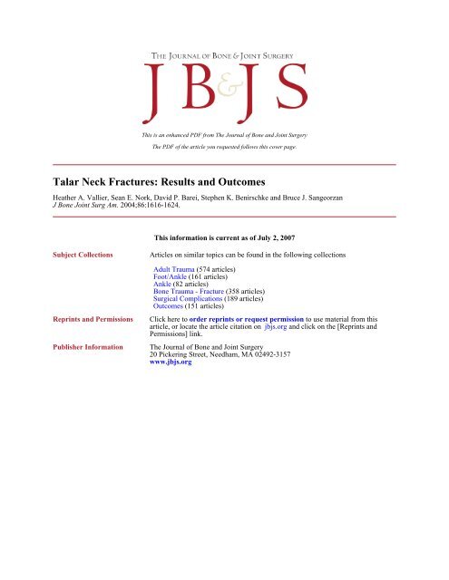
This is an enhanced PDF from The Journal of Bone <strong>and</strong> Joint SurgeryThe PDF of the article you requested follows this cover page.<strong>Talar</strong> <strong>Neck</strong> <strong>Fractures</strong>: <strong>Results</strong> <strong>and</strong> <strong>Outcomes</strong>Heather A. Vallier, Sean E. Nork, David P. Barei, Stephen K. Benirschke <strong>and</strong> Bruce J. SangeorzanJ Bone Joint Surg Am. 2004;86:1616-1624.This information is current as of July 2, 2007Subject CollectionsReprints <strong>and</strong> PermissionsPublisher InformationArticles on similar topics can be found in the following collectionsAdult Trauma (574 articles)Foot/Ankle (161 articles)Ankle (82 articles)Bone Trauma - Fracture (358 articles)Surgical Complications (189 articles)<strong>Outcomes</strong> (151 articles)Click here to order reprints or request permission to use material from thisarticle, or locate the article citation on jbjs.org <strong>and</strong> click on the [Reprints <strong>and</strong>Permissions] link.The Journal of Bone <strong>and</strong> Joint Surgery20 Pickering Street, Needham, MA 02492-3157www.jbjs.org
1616COPYRIGHT © 2004 BY THE JOURNAL OF BONE AND JOINT SURGERY, INCORPORATED<strong>Talar</strong> <strong>Neck</strong> <strong>Fractures</strong>:<strong>Results</strong> <strong>and</strong> <strong>Outcomes</strong>BY HEATHER A. VALLIER, MD, SEAN E. NORK, MD, DAVID P. BAREI, MD,STEPHEN K. BENIRSCHKE, MD, AND BRUCE J. SANGEORZAN, MDInvestigation performed at Harborview Medical Center, Seattle, WashingtonBackground: <strong>Talar</strong> neck fractures occur infrequently <strong>and</strong> have been associated with high complication rates. The purposesof the present study were to evaluate the rates of early <strong>and</strong> late complications after operative treatment of talarneck fractures, to ascertain the effect of surgical delay on the development of osteonecrosis, <strong>and</strong> to determinethe functional outcomes after operative treatment of such fractures.Methods: We retrospectively reviewed the records of 100 patients with 102 fractures of the talar neck who had beenmanaged at a level-1 trauma center. All fractures had been treated with open reduction <strong>and</strong> internal fixation. Sixtyfractures were evaluated at an average of thirty-six months (range, twelve to seventy-four months) after surgery. Complications<strong>and</strong> secondary procedures were reviewed, <strong>and</strong> radiographic evidence of osteonecrosis <strong>and</strong> posttraumaticarthritis was evaluated. The Foot Function Index <strong>and</strong> Musculoskeletal Function Assessment questionnaires were administered.<strong>Results</strong>: Radiographic evidence of osteonecrosis was seen in nineteen (49%) of the thirty-nine patients with completeradiographic data. However, seven (37%) of these nineteen patients demonstrated revascularization of the talardome without collapse. Overall, osteonecrosis with collapse of the dome occurred in twelve (31%) of thirty-nine patients.Osteonecrosis was seen in association with nine (39%) of twenty-three Hawkins group-II fractures <strong>and</strong> nine(64%) of fourteen Hawkins group-III fractures. The mean time to fixation was 3.4 days for patients who had developmentof osteonecrosis, compared with 5.0 days for patients who did not have development of osteonecrosis. Withthe numbers available, no correlation could be identified between surgical delay <strong>and</strong> the development of osteonecrosis.Osteonecrosis was associated with comminution of the talar neck (p < 0.03) <strong>and</strong> open fracture (p < 0.05).Twenty-one (54%) of thirty-nine patients had development of posttraumatic arthritis, which was more common aftercomminuted fractures (p < 0.07) <strong>and</strong> open fractures (p = 0.09). Patients with comminuted fractures also had worsefunctional outcome scores.Conclusions: <strong>Fractures</strong> of the talar neck are associated with high rates of morbidity <strong>and</strong> complications. Although thenumbers in the present series were small, no correlation was found between the timing of fixation <strong>and</strong> the developmentof osteonecrosis. Osteonecrosis was associated with talar neck comminution <strong>and</strong> open fractures, confirmingthat higher-energy injuries are associated with more complications <strong>and</strong> a worse prognosis. This finding was strengthenedby the poor Foot Function Index <strong>and</strong> Musculoskeletal Function Assessment scores in these patients. We recommendurgent reduction of dislocations <strong>and</strong> treatment of open injuries. Proceeding with definitive rigid internal fixationof talar neck fractures after soft-tissue swelling has subsided may minimize soft-tissue complications.Level of Evidence: Prognostic study, Level II-1 (retrospective study). See Instructions to Authors for a complete descriptionof levels of evidence.<strong>Talar</strong> neck fractures occur infrequently. Such fracturestypically are the result of high-energy trauma <strong>and</strong> arecharacterized by displacement, comminution, <strong>and</strong> softtissueinjury. These features have an impact on treatment <strong>and</strong>may have a negative effect on the result. In addition, associatednoncontiguous skeletal injuries <strong>and</strong>/or systemic traumamay delay the diagnosis or definitive care of the talar neckfracture. Urgent reduction <strong>and</strong> fixation of these fractures hasbeen advocated to protect any remaining blood supply to thetalar body <strong>and</strong> to promote revascularization 1-7 . The blood supplyto the talus has been well characterized 8-10 <strong>and</strong> is primarilylocalized to the talar neck <strong>and</strong> the medial part of the talarbody. Consequently, fractures of the talar neck that cause subluxationor dislocation of the talar body presumably compromiseits blood supply. It has been suggested that initialfracture displacement <strong>and</strong> delayed fracture fixation are associ-
1617THE JOURNAL OF BONE & JOINT SURGERY · JBJS.ORGVOLUME 86-A · NUMBER 8 · AUGUST 2004TALAR NECK FRACTURES:RESULTS AND OUTCOMESated with an increased incidence of osteonecrosis 1,2,4,5,7,8,11-21 .<strong>Talar</strong> neck fractures often disrupt the congruity of theperitalar joints 22,23 . Restoration <strong>and</strong> preservation of anatomicalignment may limit the development of posttraumatic arthritis.Sequelae of posttraumatic arthritis <strong>and</strong> osteonecrosishave been described in previous reports on talar neck fractures2,5,12,13,16,20,24-27 . These complications frequently have a devastatingimpact on overall function, cause chronic pain <strong>and</strong>stiffness, <strong>and</strong> often lead to secondary procedures 6,11,15,19-21 . Thepurposes of the present study were to characterize talar neckfractures, to evaluate the early <strong>and</strong> late complications of suchfractures, <strong>and</strong> to present the functional outcomes after surgicaltreatment. We also attempted to assess the impact ofsurgical delay on the development of osteonecrosis <strong>and</strong> posttraumaticarthritis.Materials <strong>and</strong> Methodse retrospectively reviewed the records of 100 consecu-patients with 102 fractures of the talar neck (classi-Wtivefication 72-A1 according to the system of the OrthopaedicTrauma Association 28 ) who had been managed operatively at alevel-1 trauma center over a sixty-seven-month period. Sixtymale patients <strong>and</strong> forty female patients with an average ageof 32.6 years (range, thirteen to seventy-seven years) <strong>and</strong> anaverage Injury Severity Score 29 of 15.8 points (range, 9 to 50points) were identified. The mechanism of injury was a motorvehicleaccident for sixty patients, a fall from a height fortwenty-seven, a motorcycle accident for seven, a pedestrianmotorvehicle accident for two, sports-related trauma for two,a plane crash for one, <strong>and</strong> an industrial accident for one.Initially, patients were assessed <strong>and</strong> resuscitated accordingto Advanced Trauma Life-Support guidelines (AmericanCollege of Surgeons, Chicago, Illinois). Sterile dressings wereapplied to open wounds, <strong>and</strong> intravenous antibiotics <strong>and</strong> tetanusprophylaxis were administered. Twenty-four fractures wereopen; of these, one was classified as type I, one was classifiedas type II, <strong>and</strong> twenty-two were classified as type IIIA accordingto the criteria of Gustilo <strong>and</strong> Anderson 30,31 . Plain radiographsof the foot <strong>and</strong> ankle were made in all cases. Thefractures were classified into groups as described by Hawkins 14<strong>and</strong> modified by Canale <strong>and</strong> Kelly 13 . Four fractures wereminimally displaced (group I), sixty-eight were associated withsubluxation or dislocation of the subtalar joint (group II),twenty-five were associated with dislocation of the tibiotalarjoint (group III), <strong>and</strong> five were associated with dislocation ofthe tibiotalar, subtalar, <strong>and</strong> talonavicular joints (group IV).Twenty-three patients had a contiguous talar body fracture<strong>and</strong>/or fracture extension, <strong>and</strong> twenty had an associated lateraltalar process fracture. Forty-four patients had additionalipsilateral foot <strong>and</strong> ankle injuries, <strong>and</strong> twenty-six had contralateralfoot <strong>and</strong> ankle injuries. Comminution was definedon plain radiographs as a talar neck fracture with more thanthree fracture fragments.Although closed, manipulative reduction was attemptedat the time of the initial assessment, all fractures subsequentlywere treated with open reduction <strong>and</strong> internal fixation withstainless steel small-fragment <strong>and</strong>/or mini-fragment implants.An attempt was made to perform surgery on an urgent basis.However, associated life-threatening injuries or a delay indiagnosis or presentation to our hospital precluded urgenttreatment in some cases. Dual anteromedial <strong>and</strong> anterolateralsurgical approaches were used for ninety-one fractures (Figs.1-A through 1-D) 17 . The remaining fractures were treatedthrough a single medial approach (eight fractures) or a singlelateral approach (three fractures). Eight patients also had an osteotomyof the medial malleolus to enhance the surgical exposure.Six of these osteotomies were performed in patients whohad associated fracture extensions involving the talar body.Postoperative management consisted of immobilizationof the ankle in neutral alignment until the wounds were sealed<strong>and</strong> the swelling had diminished. Range-of-motion exercisesfor the ankle <strong>and</strong> foot were then initiated. No weight-bearingon the affected limb was recommended for a total of twelveweeks after surgery or until fracture union had occurred.Progressive weight-bearing, however, was not delayed by radiographicevidence of osteonecrosis. Plain radiographs of thelateral aspects of the foot <strong>and</strong> ankle, ankle mortise radiographs,<strong>and</strong> Canale 13 radiographs were made after fixation<strong>and</strong> at approximately six-week, twelve-week, six-month, <strong>and</strong>twelve-month intervals postoperatively. Computerized tomographywas not used to assess fractures preoperatively or toevaluate alignment postoperatively. Osteonecrosis was definedon plain radiographs as any area of increased density of thetalar dome relative to the adjacent structures. Magnetic resonanceimaging was not routinely used to diagnose osteonecrosisor to follow its progression. Posttraumatic arthritiswas defined as any loss of joint space, formation of osteophytes,or development of subchondral sclerosis or cysts.Functional outcome measurements included the FootFunction Index (FFI) <strong>and</strong> the Musculoskeletal Function Assessment(MFA). These measurements were collected at themost recent clinic visit or by means of a questionnaire that wasadministered over the phone or by mail. The FFI is a specificlower-extremity outcome index consisting of scores for pain(81 points), disability (81 points), <strong>and</strong> activity (45 points) 32 .The total score is the average of these three scores, with higherscores indicating greater impairment of function. The MFA isa general-health-status outcome index consisting of ten categories,from which a total score can be calculated 33 . MFA valuesrange from 0 to 100, with higher scores indicating a lowerlevel of overall function. Both the FFI <strong>and</strong> MFA instrumentshave been determined to be valid, reliable, <strong>and</strong> consistent 32-36 .The FFI has been validated for the evaluation of individualswith systemic arthritis <strong>and</strong> measures activity specific to thefoot <strong>and</strong> ankle in individuals with a low level of function. TheMFA has been validated for the evaluation of trauma patientswith greater activity levels but includes the entire musculoskeletalsystem.Statistical analysis was performed with the SAS statisticalpackage (SAS Institute, Cary, North Carolina). The possiblepredictive variables included the type of fracture (open orclosed), fracture comminution, Hawkins’ classification, <strong>and</strong>
1618THE JOURNAL OF BONE & JOINT SURGERY · JBJS.ORGVOLUME 86-A · NUMBER 8 · AUGUST 2004TALAR NECK FRACTURES:RESULTS AND OUTCOMESage (less than forty years or forty years or more). The clinicaloutcomes included osteonecrosis, collapse of the talar dome,<strong>and</strong> arthritis of the tibiotalar or subtalar joint. Bivariate analysis<strong>and</strong> Fisher’s exact test were used to test the association betweenthe possible predictive variables <strong>and</strong> these clinicaloutcomes. With use of chi-square analysis, the timing of fixationwas analyzed as a continuous variable (within six hours,eight hours, twelve hours, twenty-four hours, or more thantwenty-four hours) to test the association between surgicaltiming <strong>and</strong> osteonecrosis. The Student t test was used to identifythe associations between the functional outcomes (as indicatedby the FFI <strong>and</strong> MFA scores) <strong>and</strong> the possible predictivevariables as well as to identify the associations between functional<strong>and</strong> clinical outcomes.<strong>Results</strong>orty-one patients with forty-two fractures were unavail-for follow-up. Two patients died of unrelated causes,Fabletwo patients had severe closed head injuries <strong>and</strong> were unableto walk or to communicate, one patient was in jail, one patientdid not speak English, <strong>and</strong> thirty-five patients could notbe located. Seventeen of the thirty-five patients who couldnot be located had been followed for six to twelve months,<strong>and</strong> none of them had had any clinical problems or symptomaticradiographic abnormalities at the time of the most recentfollow-up. The remaining fifty-nine patients (sixty fractures)were evaluated at an average of thirty-six months (range,TABLE I Incidence of Early <strong>and</strong> Late Complications AfterOpen Reduction <strong>and</strong> Internal Fixation of <strong>Talar</strong><strong>Neck</strong> <strong>Fractures</strong>IncidenceSuperficial infection 3.3% (2 of 60)Wound dehiscence 3.3% (2 of 60)Deep infection 5.0% (3 of 60)Delayed union 1.7% (1 of 60)Nonunion 3.3% (2 of 60)Osteonecrosis <strong>and</strong> dome collapse 31% (12 of 39)Ankle osteoarthritis 18% (7 of 39)Subtalar osteoarthritis 15% (6 of 39)twelve to seventy-four months) after surgery. Forty-five ofthese patients had complete functional outcome data, <strong>and</strong>thirty-nine had complete radiographic data. Six of the sixtyfractures were associated with early complications (Table I).Two patients had development of a superficial infection, <strong>and</strong>one had partial medial wound dehiscence. These three patientswere successfully managed with oral antibiotics <strong>and</strong>dressing changes. Another patient with wound dehiscence haddevelopment of a deep wound infection that required serialirrigation <strong>and</strong> débridement <strong>and</strong> intravenous administrationof antibiotics. After two years, there had been no recurrenceFig. 1-BFig. 1-AFigs. 1-A through 1-D A twenty-one-year-old man who sustained a Hawkins’ Group-III talar neck fracture with associated tibiotalar <strong>and</strong> subtalar dislocationsin a motor-vehicle accident. Figs. 1-A <strong>and</strong> 1-B Preoperative anteroposterior (Fig. 1-A) <strong>and</strong> lateral (Fig. 1-B) radiographs.
1619THE JOURNAL OF BONE & JOINT SURGERY · JBJS.ORGVOLUME 86-A · NUMBER 8 · AUGUST 2004TALAR NECK FRACTURES:RESULTS AND OUTCOMESFig. 1-DFig. 1-CFigs. 1-C <strong>and</strong> 1-D Anteroposterior (Fig. 1-C) <strong>and</strong> lateral (Fig. 1-D) radiographs made after open reduction <strong>and</strong> internal fixation through anteromedial<strong>and</strong> anterolateral approaches.of infection. Two other patients had development of a deepinfection within the first three months postoperatively. Bothof these patients underwent serial irrigation <strong>and</strong> débridementprocedures, <strong>and</strong> both were managed with intravenousadministration of antibiotics for several weeks. Neither patienthad had a recurrence of infection at the time of the mostrecent follow-up.Three of the sixty fractures did not demonstrate radiographicevidence of union within the first three months.One of the patients with a deep infection had developmentof a nonunion. This patient also had osteonecrosis of thetalar dome, which progressed to collapse. The talar fractureunited following a subtalar arthrodesis <strong>and</strong> revision of fixation.One other nonunion united successfully three monthsafter iliac-crest bone-grafting. One patient had a delayedunion <strong>and</strong> was followed with plain radiographs until fractureunion occurred at twenty-four weeks after surgery.Radiographic evidence of osteonecrosis was identifiedin nineteen (49%) of thirty-nine patients. Osteonecrosis occurredwithin the first ten months after the injury (at a meanof nineteen weeks) in all cases. Osteonecrosis was seen in associationwith nine (39%) of twenty-three Hawkins’ II fractures(five of which progressed to collapse of the talar dome), nine(64%) of fourteen Hawkins’ III fractures (six of which progressedto collapse), <strong>and</strong> one Hawkins’ IV fracture (whichprogressed to collapse). Overall, osteonecrosis with collapse ofthe talar dome occurred in twelve (31%) of thirty-nine patients.Collapse occurred at a mean of thirty-nine weeks(range, twenty-six to sixty-five weeks) after osteonecrosis wasdetected on plain radiographs. Seven (37%) of the nineteenpatients with osteonecrosis demonstrated revascularization ofthe talar dome on plain radiographs with no evidence of collapse.Revascularization occurred an average of thirty-fiveweeks (range, twenty-five to sixty-five weeks) postoperatively.All of these patients were followed for at least eighteen monthsafter the return of normal bone density. None of them demonstratedsubsequent evidence of osteonecrosis or collapse.The average time from injury to fixation was 3.7 days(range, four hours to forty-eight days). With the numbersavailable, chi-square analysis of fixation within six hours,eight hours, twelve hours, twenty-four hours, or more thantwenty-four hours did not demonstrate a correlation betweensurgical delay <strong>and</strong> the development of osteonecrosis orcollapse. The mean time from injury to fixation was 3.4 days(range, four hours to twenty days) for patients who had developmentof osteonecrosis <strong>and</strong> 5.0 days (range, four hours toforty-eight days) for those who did not (Figs. 2-A, 2-B, <strong>and</strong>2-C). The mean time between injury <strong>and</strong> fixation was 12.9hours for patients who had development of osteonecrosis <strong>and</strong>13.0 hours for those who did not. Time was also analyzed as acontinuous variable <strong>and</strong>, with the small numbers available,no correlation was observed between the timing of surgery
1620THE JOURNAL OF BONE & JOINT SURGERY · JBJS.ORGVOLUME 86-A · NUMBER 8 · AUGUST 2004TALAR NECK FRACTURES:RESULTS AND OUTCOMESFig. 2-AFig. 2-BFigs. 2-A, 2-B, <strong>and</strong> 2-C A thirty-eight-year-old man who sustained a talar neck fracture with dislocation of the subtalar joint in a motorcycle accident.The patient presented to our hospital more than twelve hours after the injury with severe soft-tissue swelling <strong>and</strong> fracture blisters. Fig. 2-ALateral radiograph made at the time of presentation. Fig. 2-B Lateral radiograph made after closed reduction, which was facilitated with percutaneousinsertion of a calcaneal pin. Seventeen days later, after some improvement in the soft-tissue injury, the patient underwent open reduction <strong>and</strong>internal fixation.<strong>and</strong> the development of osteonecrosis with or without collapse.This test was performed on the entire group of patients<strong>and</strong> was repeated for just those patients who had received fixationwithin twenty-four hours.Osteonecrosis occurred in eighteen of thirty-one patientswith comminution of the talar neck (p < 0.03). Twelveof these eighteen patients had collapse of the talar dome; a significantassociation was noted between comminution of thetalar neck <strong>and</strong> osteonecrosis with collapse (p = 0.02). Nine ofthirteen patients with an open fracture also had developmentof osteonecrosis; osteonecrosis occurred significantly morefrequently in patients with open fractures than in patientswith closed fractures (p < 0.05). Eight of nine patients whohad an open fracture with development of osteonecrosis hadprogression to collapse of the talar dome (p < 0.02). With thenumbers available, neither osteonecrosis nor collapse was associatedwith the age of the patient, Hawkins’ classification, orthe presence of an associated talar body fracture. However,our data showed a trend toward increased rates of osteonecrosis<strong>and</strong> collapse in association with greater initial fracture displacementaccording to Hawkins’ classification.Twenty-one (54%) of thirty-nine patients had radiographicfindings consistent with posttraumatic arthritis ofthe ankle joint <strong>and</strong>/or the subtalar joint. End-stage arthritiswith complete loss of the joint space was identified in thesubtalar joint of six patients <strong>and</strong> in the ankle joint of sevenpatients. Six of twenty-three patients with Hawkins’ II fractures<strong>and</strong> six of fourteen patients with Hawkins’ III fracturesFig. 2-CAnteroposterior radiograph, made nineteen months postoperatively,demonstrating a viable talar dome without evidence of osteonecrosisor collapse.
1621THE JOURNAL OF BONE & JOINT SURGERY · JBJS.ORGVOLUME 86-A · NUMBER 8 · AUGUST 2004TALAR NECK FRACTURES:RESULTS AND OUTCOMESTABLE II Mean Foot Function Index <strong>and</strong> Musculoskeletal Function Assessment ScoresFoot Function Index Scores*Pain Activity Disability TotalMusculoskeletalFunctionAssessment Score*ComminutionNo 13.3 11.4 25.3 16.7 11.2Yes 24.6 22.4 36.4 27.8 26.9P value 0.02†
1622THE JOURNAL OF BONE & JOINT SURGERY · JBJS.ORGVOLUME 86-A · NUMBER 8 · AUGUST 2004TALAR NECK FRACTURES:RESULTS AND OUTCOMESfracture into the talar body had no discernable effect on theMFA score.Discussionractures of the talar neck have been associated with highFratesof early <strong>and</strong> late complications 2,4-6,12,13,16,20,21,24-27,38 . Thehigh-energy nature of the majority of these injuries producesnot only fracture displacement <strong>and</strong> comminution but also severesoft-tissue damage, frequently in association with openwounds. These characteristics often portend a poor prognosis.Early complications such as skin necrosis, wound dehiscence,<strong>and</strong> infection have occurred in as many as 77% of cases 14,16,38 .Reports of late problems such as osteonecrosis, posttraumaticarthritis, <strong>and</strong> osteomyelitis with associated stiffness, pain, <strong>and</strong>loss of function also have been common 2,5,12,13,16,20,24-27 . Treatmentstrategies initially evolved from reduction <strong>and</strong> immobilization6,24,26,27to limited, often temporary, fixation 14,16,27,38 . Currently,open reduction <strong>and</strong> internal fixation is performed formost talar neck fractures 1,3-5,7,11,12,15,17,18,20,39 . The goals of surgicaltreatment have been to restore overall alignment of the talusin order to maximize function of the limb.Surgical treatment is necessary to restore <strong>and</strong> to maintainalignment after a displaced talar neck fracture. Mostfractures are associated with some subluxation or dislocationof the talar body. As direct exposure permits accurate reductionin these cases, we advocate dual anteromedial <strong>and</strong> anterolateralapproaches for most talar neck fractures 1,3,7,11,17 . Anadjunctive osteotomy of the medial malleolus may be performedto enhance visualization of the talar body 40 . The anterolateralexposure, along with intraoperative radiographs,greatly aids in establishing an anatomic reduction. The Canaleview 13is particularly helpful for assessing deformity.Medial comminution of the talar neck may otherwise be underestimated,<strong>and</strong> varus malalignment can result 22,23 . Once anaccurate reduction has been achieved, strategic, rigid internalfixation with plates <strong>and</strong>/or screws usually will provideadequate stability to permit early mobilization of the adjacentjoints 3 , which may reduce ankle <strong>and</strong> foot stiffness <strong>and</strong>may maximize overall function 2,5,7 . The effect of dual surgicalapproaches on the blood supply of the talus is unknown.It is possible that this technique could further compromisethe vascularity of fracture fragments or that of the talarbody. As 91% of the patients in the present study had bothanteromedial <strong>and</strong> anterolateral surgical approaches, wecould not assess whether this method has an advantage comparedwith other surgical techniques in terms of vascularcompromise.Osteonecrosis occurs frequently after talar neck fractures.It also has been proposed that surgical treatment maypromote revascularization of the talar body 1-7 . Hawkins developeda classification scheme that was based on the fracturedisplacement that is observed on the initial plain radiographs14 . He also provided treatment recommendations <strong>and</strong>presented prognostic information that was based on this classificationsystem. In his study, osteonecrosis was identified inno Group-I patients, in 42% of Group-II patients, <strong>and</strong> in91% of Group-III patients. Canale <strong>and</strong> Kelly later modifiedthis classification scheme, describing a fourth type of talarneck fracture with associated talonavicular dislocation 13 . Studiesin the literature to date have demonstrated the occurrenceof osteonecrosis in association with as many as 13% of Hawkins’I fractures, as many as 50% of Hawkins’ II fractures, asmany as 84% of Hawkins’ III fractures, <strong>and</strong> as many as 100%of Hawkins’ IV fractures 2-4,18,25,38 . Our data showed a trend towardan increased rate of osteonecrosis in association withgreater initial fracture displacement as described with Hawkins’classification. Comminution of the talar neck <strong>and</strong> openfractures were associated with an increased risk of osteonecrosisin our study.Some authors have advocated the use of protectedweight-bearing to reduce the possibility of collapse of thetalar dome in patients with osteonecrosis 1,6,13,16,18,24,27 . This recommendationremains controversial, however, <strong>and</strong> other authorshave recommended the progression of weight-bearingregardless of the presence of osteonecrosis 14,25,38 . We agree thatonce fracture union has been achieved, weight-bearing as toleratedmay be started. In the present study, 37% of the patientswith radiographic evidence of osteonecrosis showed a returnto normal talar dome density on plain radiographs. No collapseof the talar dome was detected in these patients despiteweight-bearing as tolerated without bracing. Thus, we believethat the effect of weight-bearing on the progression of osteonecrosisremains unknown. Additional studies are needed toresolve this issue.<strong>Talar</strong> neck fractures often are treated urgently to reducethe risk of osteonecrosis 1,4-7,20 because urgent reduction of dislocationsmay help to preserve any remaining blood supply tothe posterior portion of the talus. However, to our knowledge,surgical timing has not been previously shown to impact thedevelopment of osteonecrosis. Although the numbers in thepresent study were small, no correlation was found betweenthe timing of reduction <strong>and</strong> fixation <strong>and</strong> the development ofosteonecrosis. Because we did not have consistent informationabout the duration of dislocation in our patients, we were unableto determine the impact of prolonged dislocation on thedevelopment of osteonecrosis. Other characteristics of the initialinjury <strong>and</strong> its treatment, such as the presence of comminutionor an open fracture, may have a greater influence onwhether or not osteonecrosis will occur 7,12 .We advocate urgent fracture reduction through eitherclosed or percutaneous manipulation, <strong>and</strong> we recommendresorting to open reduction only when the fracture is notreducible through closed means. Once a reduction of the talarneck fracture has been achieved, we speculate that a delayin fixation of the fracture will not affect the developmentof osteonecrosis. In some cases, internal fixation cannot besafely undertaken on an urgent basis. This may be becauseof life-threatening trauma or because of severe soft-tissuedamage <strong>and</strong> swelling of the ankle <strong>and</strong> foot. It has been suggestedthat a surgical delay will allow improvement in theassociated soft-tissue injury <strong>and</strong> will decrease the rates ofwound complications <strong>and</strong> infection after fractures of the
1623THE JOURNAL OF BONE & JOINT SURGERY · JBJS.ORGVOLUME 86-A · NUMBER 8 · AUGUST 2004TALAR NECK FRACTURES:RESULTS AND OUTCOMEStibial plafond or calcaneus 41-46 . Previous reports have demonstratedskin necrosis, wound dehiscence, <strong>and</strong> infection inassociation with as many as 77% of talar neck fractures 14,16,38 .Perhaps these complications could be minimized by achievinga closed reduction but delaying definitive fixation inorder to allow for improvement in the soft-tissue injury beforeproceeding with the dual surgical approach that wefavor.In conclusion, it has been suggested that early operativeintervention protects the already tenuous blood supply tothe posterior portion of the talus after a fracture of the talarneck. Although the numbers in this series were small, no correlationwas found between the timing of fixation <strong>and</strong> thedevelopment of osteonecrosis. Osteonecrosis was associatedwith talar neck comminution <strong>and</strong> open fractures, confirmingthat higher-energy injuries are associated with more complications<strong>and</strong> a worse prognosis. This concept is furtherstrengthened by the poor FFI <strong>and</strong> MFA scores in patientswith comminuted fractures. We continue to recommend urgenttreatment of open injuries <strong>and</strong> reduction of dislocations.Proceeding with definitive fixation when there is minimalsoft-tissue swelling will provide rigid fracture stability to promotefracture-healing <strong>and</strong> to achieve an earlier return toweight-bearing function. Heather A. Vallier, MDDepartment of Orthopaedic Surgery, MetroHealth Medical Center,2500 MetroHealth Drive, Clevel<strong>and</strong>, OH 44109. E-mail address:heathervallier@yahoo.comSean E. Nork, MDDavid P. Barei, MDStephen K. Benirschke, MDBruce J. Sangeorzan, MDDepartment of Orthopaedic Surgery, Harborview Medical Center,Box 359798, 325 Ninth Avenue, Seattle, WA 98104-2499The authors did not receive grants or outside funding in support of theirresearch or preparation of this manuscript. They did not receive paymentsor other benefits or a commitment or agreement to provide suchbenefits from a commercial entity. No commercial entity paid ordirected, or agreed to pay or direct, any benefits to any research fund,foundation, educational institution, or other charitable or nonprofitorganization with which the authors are affiliated or associated.References1. Berlet GC, Lee TH, Massa EG. <strong>Talar</strong> neck fractures. Orthop Clin North Am.2001;32:53-64.2. Elgafy H, Ebraheim NA, Tile M, Stephen D, Kase J. <strong>Fractures</strong> of the talus:experience of two level 1 trauma centers. Foot Ankle Int. 2000;21:1023-9.3. Fleuriau Chateau PB, Brokaw DS, Jelen BA, Scheid DK, Weber TG. Platefixation of talar neck fractures: preliminary review of a new technique intwenty-three patients. J Orthop Trauma. 2002;16:213-9.4. Frawley PA, Hart JA, Young DA. Treatment outcome of major fractures of thetalus. Foot Ankle Int. 1995;16:339-45.5. Inokuchi S, Ogawa K, Usami N, Hashimoto T. Long-term follow up of talusfractures. Orthopedics. 1996;19:477-81.6. Mindell ER, Cisek EE, Kartalian G, Dziob JM. Late results of injuries to thetalus. Analysis of forty cases. J Bone Joint Surg Am. 1963;45:221-45.7. Smith PN, Ziran BH. <strong>Fractures</strong> of the talus. Operative Tech Orthop. 1999;9:229-38.8. Gelberman RH, Mortensen WW. The arterial anatomy of the talus. Foot Ankle.1983;4:64-72.9. Haliburton RA, Sullivan CR, Kelly PJ, Peterson LF. The extra-osseous <strong>and</strong>intra-osseous blood supply of the talus. J Bone Joint Surg Am. 1958;40:1115-20.10. Mulfinger GL, Trueta J. The blood supply of the talus. J Bone Joint Surg Br.1970;52:160-7.11. Archdeacon M, Wilber R. <strong>Fractures</strong> of the talar neck. Orthop Clin North Am.2002;33:247-62.12. Canale ST. <strong>Fractures</strong> of the neck of the talus. Orthopedics. 1990;13:1105-15.13. Canale ST, Kelly FB Jr. <strong>Fractures</strong> of the neck of the talus. Long-term evaluationof seventy-one cases. J Bone Joint Surg Am. 1978;60:143-56.14. Hawkins LG. <strong>Fractures</strong> of the neck of the talus. J Bone Joint Surg Am. 1970;52:991-1002.15. Higgins TF, Baumgaertner MR. Diagnosis <strong>and</strong> treatment of fractures of thetalus: a comprehensive review of the literature. Foot Ankle Int. 1999;20:595-605.16. Kenwright J, Taylor RG. Major injuries of the talus. J Bone Joint Surg Br.1970;52:36-48.17. Mayo KA. <strong>Fractures</strong> of the talus: principles of management <strong>and</strong> techniquesof treatment. Tech Orthop. 1987;2:42-54.18. Pajenda G, Vecsei V, Reddy B, Heinz T. Treatment of talar neck fractures:clinical results of 50 patients. J Foot Ankle Surg. 2000;39:365-75.19. Santavirta S, Seitsalo S, Kiviluoto O, Myllynen P. <strong>Fractures</strong> of the talus.JTrauma. 1984;24:986-9.20. Schulze W, Richter J, Russe O, Ingelfinger P, Muhr G. Surgical treatment oftalus fractures: a retrospective study of 80 cases followed for 1-15 years.Acta Orthop Sc<strong>and</strong>. 2002;73:344-51.21. Szyszkowitz R, Reschauer R, Seggl W. Eighty-five talus fractures treated byORIF with five to eight years of follow-up study of 69 patients. Clin Orthop.1985;199:97-107.22. Daniels TR, Smith JW, Ross TI. Varus malalignment of the talar neck. Its effecton the position of the foot <strong>and</strong> on subtalar motion. J Bone Joint Surg Am.1996;78:1559-67.23. Sangeorzan BJ, Wagner UA, Harrington RM, Tencer AF. Contact characteristicsof the subtalar joint: the effect of talar neck misalignment. J Orthop Res.1992;10:544-51.24. Coltart WD. Aviator’s astragalus. J Bone Joint Surg Br. 1952;34:545-66.25. Grob D, Simpson LA, Weber BG, Bray T. Operative treatment of displacedtalus fractures. Clin Orthop. 1985;199:88-96.26. Kleiger B. <strong>Fractures</strong> of the talus. J Bone Joint Surg Am. 1948;30:735-44.27. Pennal GF. <strong>Fractures</strong> of the talus. Clin Orthop. 1963;30:53-63.28. Orthopaedic Trauma Association Committee for Coding <strong>and</strong> Classification.Fracture <strong>and</strong> dislocation compendium. J Orthop Trauma. 1996;10(Suppl 1):v-ix,1-154.29. Baker SP, O’Neill B, Haddon W Jr, Long WB. The injury severity score: amethod for describing patients with multiple injuries <strong>and</strong> evaluating emergencycare. J Trauma. 1974;14:187-96.30. Gustilo RB, Anderson JT. Prevention of infection in the treatment of one thous<strong>and</strong><strong>and</strong> twenty-five open fractures of long bones: retrospective <strong>and</strong> prospectiveanalyses. J Bone Joint Surg Am. 1976;58:453-8.31. Gustilo RB, Mendoza RM, Williams DN. Problems in the management oftype III (severe) open fractures: a new classification of type III open fractures.J Trauma. 1984;24:742-6.32. Budiman-Mak E, Conrad KJ, Roach KE. The Foot Function Index: a measureof foot pain <strong>and</strong> disability. J Clin Epidemiol. 1991;44:561-70.33. Martin DP, Engelberg R, Agel J, Snapp D, Swiontkowski MF. Development ofa musculoskeletal extremity health status instrument: the MusculoskeletalFunction Assessment instrument. J Orthop Res. 1996;14:173-81.34. Coester LM, Saltzman CL, Leupold J, Pontarelli W. Long-term results follow-
1624THE JOURNAL OF BONE & JOINT SURGERY · JBJS.ORGVOLUME 86-A · NUMBER 8 · AUGUST 2004TALAR NECK FRACTURES:RESULTS AND OUTCOMESing ankle arthrodesis for post-traumatic arthritis. J Bone Joint Surg Am.2001;83:219-28.35. Domsic RT, Saltzman CL. Ankle osteoarthritis scale. Foot Ankle Int. 1998;19:466-71.36. Engelberg R, Martin DP, Agel J, Obremsky W, Coronado G, SwiontkowskiMF. Musculoskeletal Function Assessment instrument: criterion <strong>and</strong> constructvalidity. J Orthop Res. 1996;14:182-92.37. Engelberg R, Martin DP, Agel J, Swiontkowski MF. Musculoskeletal FunctionAssessment: reference values for patient <strong>and</strong> non-patient samples. J OrthopRes. 1999;17:101-9.38. Gilquist J, Oretop N, Stenstrom A, Rieger A, Wennberg E. Late results aftervertical fracture of the talus. Injury. 1974;6:173-9.39. Sangeorzan BJ, Mayo KA, Hansen ST. Intraarticular fractures of the foot. Talus<strong>and</strong> lesser tarsals. Clin Orthop. 1993;292:135-41.40. Ziran BH, Abidi NA, Scheel MJ. Medial malleolar osteotomy for exposure ofcomplex talar body fractures. J Orthop Trauma. 2001;15:513-8.41. Barei DP, Bellabarba C, Sangeorzan BJ, Benirschke SK. <strong>Fractures</strong> of the calcaneus.Orthop Clin North Am. 2002;33:263-85.42. Egol KA, Wolinsky P, Koval KJ. Open reduction <strong>and</strong> internal fixation of tibialpilon fractures. Foot Ankle Clin. 2000;5:873-85.43. Folk JW, Starr AJ, Early JS. Early wound complications of operative treatmentof calcaneus fractures: analysis of 190 fractures. J Orthop Trauma. 1999;13:369-72.44. Patterson MJ, Cole JD. Two-staged delayed open reduction <strong>and</strong> internal fixationof severe pilon fractures. J Orthop Trauma. 1999;13:85-91.45. Sirkin M, S<strong>and</strong>ers R, DiPasquale T, Herscovici D Jr. A staged protocol forsoft tissue management in the treatment of complex pilon fractures. J OrthopTrauma. 1999;13:78-84.46. Sirkin M, S<strong>and</strong>ers R. The treatment of pilon fractures. Orthop Clin North Am.2001;32:91-102.