Pelvic Disruption: Assessment and Classification
Pelvic Disruption: Assessment and Classification
Pelvic Disruption: Assessment and Classification

You also want an ePaper? Increase the reach of your titles
YUMPU automatically turns print PDFs into web optimized ePapers that Google loves.
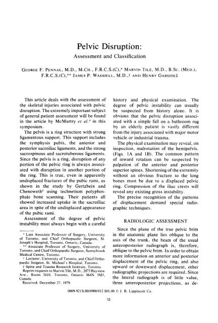
Clinical Orthopaedics14 Pennal, et al. <strong>and</strong> Related ResearchFIG. 3A. For the inlet projection, the beam isdirected from the head to the mid-pelvis at anangle of 40" to the X-ray plate.Let us consider the possible displacementsof one half of the pelvis <strong>and</strong> theirrecognition in the three projections. Theoretically,displacements may be anterior,posterior, lateral, medial, superior or inferior.Rotation may also occur about anyaxis parallel or perpendicular to the pelvicbrim. However, the perpendicular axis isusually through the sacroiliac joint.The inlet view is most helpful in disclosingtrue posterior displacement of the hemipelvis<strong>and</strong> inward or outward rotation of theanterior portion of the pelvis.The tangential view is helpful in disclosingsuperior displacement of the posterior halfof the pelvis <strong>and</strong> either superior or inferiordisplacement in the anterior portion of thepelvis. Thus, in a true rotational deformityof the bucket-h<strong>and</strong>le type, the combinedviews reveal the displacement to be inwardmedially <strong>and</strong> upward. These various rotatorydisplacements are more easily understoodfrom the motion picture "Fracturesof the Pelvis.""Tomograms may also be helpful in assessmentof the posterior fracture complex, <strong>and</strong>may show gross disruption through thesacral foramena (Fig. 5).Finally, in a ring-type structure, computed-assistedtomography (CT scan) maybe of some help in determining fracturepatterns <strong>and</strong> patterns of displacement.CLASSIFICATION<strong>Pelvic</strong> disruptions may be classified accordingto the direction of force producingthe injury: (1) anteroposterior compression;(2) lateral compression with or withoutrotation; <strong>and</strong> (3) vertical shear (Table I).This classification, based on the radiologictechniques previously described, shouldallow the surgeon to precisely define theinjury <strong>and</strong> its mechanism, <strong>and</strong> therefore,to apply counter forces to maintain stableFIGS. 3B <strong>and</strong> 3C. (Left) Anatomic appearance. (Right) Radiologic appearance in the inlet projection.
Number 151September, 1980 <strong>Pelvic</strong> <strong>Disruption</strong> <strong>Assessment</strong> 15reduction. In trauma as complicated aspelvic disruption, not all injuries can beclassified precisely, but nevertheless, apattern does evolve as described. In eachof the three major forces, the anterior lesionmay be through the symphysis pubis orthrough the pubic rami. The posteriorlesion may be through the sacrum, throughthe sacroiliac joint with a marginal fractureof the ilium, or through the ilium itself.Also, <strong>and</strong> this must be stressed, theanteroposterior compression <strong>and</strong> lateralcompression fractures may be associatedwith a stable or unstable hemipelvis; bydefinition, the vertical shear fractures areall grossly unstable (Fig. 6). The degree ofinstability, indicated best by the disruption<strong>and</strong> posterior displacement at the sacroiliacarea, is of extreme importance as aprognostic indicator for the general resuscitationof the patient.Those patients with gross posterior instabilityapparent on the original X-rayrequired 2% times as much blood replacement<strong>and</strong> had four times the m~rtality.~The patterns of injury are described inmore detail below.ANTEROPOSTERIOR COMPRESSIONOpen-book rype. Anterior compressionagainst the anterior superior spines pro-FIG. 4A. Tangential projection: the beam isdirected from the foot to the symphysis at anangle of 40" to the plate.duces a typical fracture pattern with separationat the symphysis pubis <strong>and</strong> a correspondingdegree of separation at bothsacroiliac joints anteriorly (Figs. 7A <strong>and</strong>7B). This has been produced experimentally;'the findings indicate that separationat the symphysis of more than 2.5 cm mustbe associated with a significant degree ofseparation at one or both sacroiliac joints.Experimental testing also revealed thatrupture of the sacrospinous ligament oravulsion of the ischial spine associatedwith pelvic ring disruption resulted inmarked instability. A ligamentous injuryFIGS, 4B <strong>and</strong> 4C. (Left) Anatomic appearance. (Right) Radiologic appearance in the tangentialprojection.
16 Pennal, et al.Clinical Orthopaedics<strong>and</strong> Related ResearchAFIG. 6. The posterior lesion may be unstablewhen these ligaments making up the so-called“tension b<strong>and</strong> of the pelvis” are ruptured.FIG. 5. Tomogram showing gross disruptionof the sacrum <strong>and</strong> an avulsion fracture of thetransverse process of L,, an important indicatorof pelvic instability. The patient sustained anL, nerve root avulsion.may be apparent on the initial X-ray as anavulsion of the ischial spine, <strong>and</strong> occasionallylater as calcification in the ligament.While, in the open-book type, the anteriordisruption is almost always a massivediastasis of the symphysis, the posteriorTABLE 1. <strong>Classification</strong> of <strong>Pelvic</strong><strong>Disruption</strong> Based on Directionof ForceAnteroposterior compressionLateral compressionA. Ipsilateral anterior <strong>and</strong> posterior lesionB. Contralateral anterior <strong>and</strong> posterior lesion(bucket-h<strong>and</strong>le)C. Four-rami <strong>and</strong> posterior lesionD. MiscellaneousVertical shearlesion may occasionally consist of a fracturethrough the ilium.Most fractures of the open-book typemaintain the integrity of the posterior aspectof the pelvic ring, that is, the posteriorsacroiliac ligaments. Occasionally, however,the posterior ligaments may rupture<strong>and</strong> render the hemipelvis unstable.Four-rami fracture. Isolated bilateral fracturesof the anterior rami can result fromeither anterior or lateral compressive forces.This so-called straddle fracture is usuallycaused by a lateral compressive force,described below. Rarely, it may be causedby a direct blow on the anterior pubic rami.The displacement of the floating fragmentis usually posterior <strong>and</strong> superior <strong>and</strong> maypersist by the pull of the rectus abdominismuscles. On one occasion, we were attemptingto reduce the pelvic disruptionwith a floating anterior segment, the patientcoughed on intubation. We were watchingthe image intensifier, <strong>and</strong> noted the floatingsegment migrate almost to the level of theumbilicus because of spasm in the rectusabdominis muscle. Rarely, displacement maypersist because of the unopposed pull of thismuscle, if the patient is not managed in
Number 151September, 1980 <strong>Pelvic</strong> <strong>Disruption</strong> <strong>Assessment</strong> 17nL 1;1FIGS. 7A <strong>and</strong> 7B. (Left) Diagram of a typical anteroposterior compression fracture (open-book type)showing a disruption of the symphysis pubis <strong>and</strong> the anterior sacroiliac ligaments. (Right) Radiographof a patient with this typical injury. The arrows indicate the widened sacroiliac joint anteriorly.some degree of flexion of the hip joints(Fig. 8).LATERAL COMPRESSIONThe greatest number of fractures of thepelvis occur from lateral compressive forces.Most are caused by simple falls <strong>and</strong> arerelatively undisplaced when first seen.Several types of lateral compressioninjury are identified (Table 1). As in theanteroposterior compression injury, theanterior disruption may be through thesacrum, sacroiliac joint or ilium. The patterns,however, are vastly different fromthose produced by anteroposterior compression<strong>and</strong> are marked by inward rotationof the hemipelvis. The lateral compressionforce may crush the sacrum anteriorly,leaving the posterior sacroiliac ligamentsintact. In these cases, the pelvis remainsstable <strong>and</strong> management is uncomplicated.In some cases, however, continuation of thelateral compression force ruptures theposterior sacroiliac ligaments, renderingthe pelvis unstable (Fig. 6).pelvis rotates inward. Usually the rami onthe site of impact break, <strong>and</strong> as the forcecontinues medially, the anterior sacrum iscrushed, (Figs. 9A <strong>and</strong> 9B). In some cases,continuation of the compressive force willrupture the posterior sacroiliac <strong>and</strong> iliolumbarligaments <strong>and</strong> render the hemipelvisunstable (Fig. 6). When the patient arrivessupine in the X-ray department, spontane-
18 Pennal, et al.Clinical Orthopaedics<strong>and</strong> Related ResearchFIGS. 9A <strong>and</strong> 9B. (Left) Diagram of a lateral compression injury, ipsilateral type, showing aposteriorfracture <strong>and</strong> the anterior disruption of the pubic rami with internal rotation of the hemipelvis. (Right)Radiograph of a typical lateral compression injury.ous reduction may have occurred <strong>and</strong> theposterior injury may be difficult to diagnose.The potential instability of an apparentlyinnocent fracture is often unrecognized.The use of a sling in this situation increasesthe deformity, <strong>and</strong> is therefore, contraindicated.Figures 10A <strong>and</strong> 10B show anexample of the degree of instability withthe sling in place. Persistence of this methodof treatment may cause a malrotation ofthe pelvis <strong>and</strong> permanent disability tothe bladder.Type B: Contralutrrml (bucket-h<strong>and</strong>le).When a lateral compressive force combinedwith an upward rotatory component occurs,the fracture pattern is surprisingly constantwith fractures of both pubic rami on the sideopposite the impact, <strong>and</strong> fracture in thesacroiliac area posteriorly on the side ofimpact (Figs. 11A <strong>and</strong> 11B). The resultantFIGS. 10A <strong>and</strong> 10B. (Left) The use of a pelvic sling increases the deformity by lateral compression.(Right) X-ray indicating displacement secondary to use of a sling.
Number 151September, 1980 <strong>Pelvic</strong> <strong>Disruption</strong> <strong>Assessment</strong> 19FIGS. 11A <strong>and</strong> 11B. (Left) Drawing of a typical lateral compression injury (contralateral or bucketh<strong>and</strong>letype) showing the right posterior lesion <strong>and</strong> the left pubic rami lesion. (Right) Radiograph of a19-year-old girl struck from the right side by a motor vehicle causing this typical injury. Note themarked impaction of the right sacroiliac fracture. Even with the patient under a general anesthetic, itwas virtually impossible to move this fracture five days following injury.displacement of the hemipelvis is superior<strong>and</strong> medial with rotation like the h<strong>and</strong>leof a bucket. A less common pattern from asimilar force is a diastasis of the symphysiswith a fracture through the posterior pelvicring. The leg is usually in the internallyrotated <strong>and</strong> shortened position (Figs. 1A<strong>and</strong> IB).Type C: Four-rumi with posterior disruption.Many of the violent lateral compressionforces may disrupt all four pubic ramianteriorly giving a picture of a so-calledstraddle injury, with disruption posteriorlythrough the sacrum, sacroiliac joint or ilium(Figs. 12A <strong>and</strong> 12B). Occasionally, disruptionof the symphysis <strong>and</strong> two pubicrami occur in this injury pattern. In somecases, the hemipelvis is crushed <strong>and</strong> stablein the deformed position; in others, theposterior ligaments tear, rendering thehemipelvis unstable. In our recent reviewof major pelvic trauma,6 this pattern wasFIGS. 12A <strong>and</strong> 12B. (Left) Diagram of a lateral compression injury (Type C in Table 1) showing theposterior disruption of the right sacroiliac complex <strong>and</strong> all four pubic rami fractured anteriorly. (Right)Inlet radiograph of this lesion.
20 Pennal, et al.the most common lateral compression injury.It was accompanied by the largestnumber of associated injuries <strong>and</strong> complications,indicating the severe violence causingthe injury.Type D: Miscellaneous. Two other patternsare recognized. In the first type, thesuperior pubic ramus buckles <strong>and</strong> the medialfragment rotates through the disruptedsymphysis (Figs. 13A <strong>and</strong> 13B). This causesrotation of the medial fragment into theperineum. The fragment may be felt infemale patients through the vagina. Thisinjury has been seen more commonly infemales than in males. Because of thedisplacement into the perineum, this fragmentrequires reduction.A rare type of lateral compression injuryoccurs when the anterior disruption isthrough the symphysis pubis <strong>and</strong> the forcecauses the symphysis to override <strong>and</strong> lock.Attempts at reduction may be extremelydifficult owing to the tremendous forcesnecessary to cause this injury.VERTICAL SHEARThe fracture pattern in this group, describedby Malgaigne (1855),3 consists of aClinical Orthopaedics<strong>and</strong> Related Researchfracture anteriorly through both rami or thesymphysis pubis in association with massiveposterior disruption either through thesacrum, the sacroiliac joint or the ilium(Figs. 14A <strong>and</strong> 14B). These fractures arealways the result of severe trauma, that is,falls from heights, motor vehicle accidents<strong>and</strong> crushing injuries in the mining or loggingindustries. The displacement suggests thatthe forces involved are in the vertical plane,<strong>and</strong> therefore, shearing in nature.The feature of the displacement in thisfracture dislocation which requires emphasis,in order to obtain correction, is theposterior displacement of the affected hemipelvisseen on the inlet view, <strong>and</strong> superiordisplacement as seen on the tangential view.Since the inclination of the pelvis to thevertical plane is 40", vertical shear mustresult in posterior as well as upwarddisplacement.Many types of fracture dislocations in themiscellaneous category defy precise classification<strong>and</strong> are, because of complex forceresolutions, usually the result of motorvehicle accidents. We have seen completesacroiliac bilateral dislocations <strong>and</strong> otherpatterns. However, the basic concept of theFIGS. 13A <strong>and</strong> 13B. (Left) Diagram of an unusual type of lateral compression injury characterizedby buckling of the superior pubic ramus <strong>and</strong> disruption of the symphysis. The medial fragment rotatesinferiorly <strong>and</strong> anteriorly. (Right) Anteroposterior projection of the same lesion.
Number 151September, 1980 <strong>Pelvic</strong> <strong>Disruption</strong> <strong>Assessment</strong> 21FIGS. 14A <strong>and</strong> 14B. (Left) Diagram of a vertical shear type injury with complete disruption of the lefthemipelvis. (Right) Anteroposterior radiograph of a 29-year-old patient who fell down a mine shaftsustaining a vertical shear injury with a fracture through the left ilium adjacent to the sacroiliac joint<strong>and</strong> disruption of the left pubic rami <strong>and</strong> symphysis pubis. This was an open fracture inferiorly.fracture pattern production described aboveis helpful in the analysis of the more complexcases <strong>and</strong> provides the necessary cluesfor the application of appropriate forces,if reduction is indicated.SUMMARYA precise radiologic technique for assessingthe forces producing pelvic disruptionhas been helpful in arriving at a logicalclassification of pelvic injury. The radiologicexamination should include anteroposterior,inlet <strong>and</strong> outlet views, as wellas tomograms <strong>and</strong> occasionally computedassistedtomographic evaluation (CT scanning).On the basis of this radiologic assessmentwith some biomechanical studies, aclassification of three major forces producinginjury is suggested. The anteroposterior<strong>and</strong> lateral compression types, while vastlydifferent, may both have stable <strong>and</strong> unstablesubtypes associated with them. The verticalshear fracture is always unstable. Anaccurate history <strong>and</strong> physical examinationin conjunction with the above radiologicprinciples will lead the surgeon to a precisedetermination of the fracture pattern. Aknowledge of the forces necessary toproduce this pattern is helpful in themanagement of the patient with this particulartraumatic lesion.REFERENCES1. Garside, H., <strong>and</strong> Pennal, G. F., Personal communication.2. Gertzbein, S. D., <strong>and</strong> Chenoweth, D. R.: Occultinjuries of the pelvic ring, Clin. Orthop. 128:202,1977.3. Malgaigne, .I. F.: Traite des Fractures et desLuxations, Paris, J. B. Bailliere, 1855.4. McMurtry, R., Walton, D., Dickinson, D., Kellarn,J., <strong>and</strong> Tile, M.: <strong>Pelvic</strong> disruption in the polytraumatizedpatient: A management protocol,Clin. Orthop. 151:22, 1980.5. Pennal, G. F., <strong>and</strong> Sutherl<strong>and</strong>, G. 0.: Fracturesof the Pelvis. Motion picture in AAOS filmlibrary, 1961.6. Tile, M., Lifeso, R., <strong>and</strong> Dickinson, D.: <strong>Pelvic</strong>disruption. Presented to the Canadian OrthopedicAssociation Convention, Calgary, Alberta, June1980.