Biomechanical Evaluation of Current Patella Fracture Fixation ...
Biomechanical Evaluation of Current Patella Fracture Fixation ...
Biomechanical Evaluation of Current Patella Fracture Fixation ...
- No tags were found...
Create successful ePaper yourself
Turn your PDF publications into a flip-book with our unique Google optimized e-Paper software.
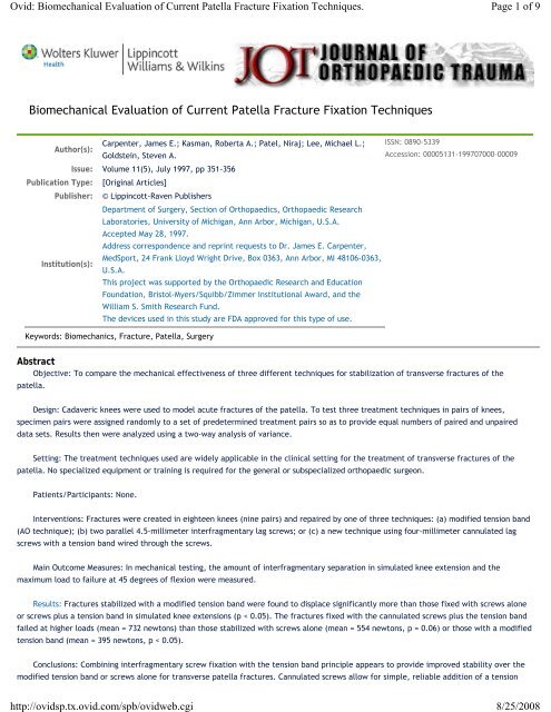
Ovid: <strong>Biomechanical</strong> <strong>Evaluation</strong> <strong>of</strong> <strong>Current</strong> <strong>Patella</strong> <strong>Fracture</strong> <strong>Fixation</strong> Techniques.http://ovidsp.tx.ovid.com/spb/ovidweb.cgiPage 1 <strong>of</strong> 98/25/2008<strong>Biomechanical</strong> <strong>Evaluation</strong> <strong>of</strong> <strong>Current</strong> <strong>Patella</strong> <strong>Fracture</strong> <strong>Fixation</strong> TechniquesCarpenter, James E.; Kasman, Roberta A.; Patel, Niraj; Lee, Michael L.; ISSN: 0890-5339Author(s):Goldstein, Steven A.Accession: 00005131-199707000-00009Issue: Volume 11(5), July 1997, pp 351-356Publication Type: [Original Articles]Publisher: © Lippincott-Raven PublishersDepartment <strong>of</strong> Surgery, Section <strong>of</strong> Orthopaedics, Orthopaedic ResearchLaboratories, University <strong>of</strong> Michigan, Ann Arbor, Michigan, U.S.A.Accepted May 28, 1997.Address correspondence and reprint requests to Dr. James E. Carpenter,MedSport, 24 Frank Lloyd Wright Drive, Box 0363, Ann Arbor, MI 48106-0363,Institution(s):U.S.A.This project was supported by the Orthopaedic Research and EducationFoundation, Bristol-Myers/Squibb/Zimmer Institutional Award, and theWilliam S. Smith Research Fund.The devices used in this study are FDA approved for this type <strong>of</strong> use.Keywords: Biomechanics, <strong>Fracture</strong>, <strong>Patella</strong>, SurgeryAbstractObjective: To compare the mechanical effectiveness <strong>of</strong> three different techniques for stabilization <strong>of</strong> transverse fractures <strong>of</strong> thepatella.Design: Cadaveric knees were used to model acute fractures <strong>of</strong> the patella. To test three treatment techniques in pairs <strong>of</strong> knees,specimen pairs were assigned randomly to a set <strong>of</strong> predetermined treatment pairs so as to provide equal numbers <strong>of</strong> paired and unpaireddata sets. Results then were analyzed using a two-way analysis <strong>of</strong> variance.Setting: The treatment techniques used are widely applicable in the clinical setting for the treatment <strong>of</strong> transverse fractures <strong>of</strong> thepatella. No specialized equipment or training is required for the general or subspecialized orthopaedic surgeon.Patients/Participants: None.Interventions: <strong>Fracture</strong>s were created in eighteen knees (nine pairs) and repaired by one <strong>of</strong> three techniques: (a) modified tension band(AO technique); (b) two parallel 4.5-millimeter interfragmentary lag screws; or (c) a new technique using four-millimeter cannulated lagscrews with a tension band wired through the screws.Main Outcome Measures: In mechanical testing, the amount <strong>of</strong> interfragmentary separation in simulated knee extension and themaximum load to failure at 45 degrees <strong>of</strong> flexion were measured.Results: <strong>Fracture</strong>s stabilized with a modified tension band were found to displace significantly more than those fixed with screws aloneor screws plus a tension band in simulated knee extensions (p < 0.05). The fractures fixed with the cannulated screws plus the tension bandfailed at higher loads (mean = 732 newtons) than those stabilized with screws alone (mean = 554 newtons, p = 0.06) or those with a modifiedtension band (mean = 395 newtons, p < 0.05).Conclusions: Combining interfragmentary screw fixation with the tension band principle appears to provide improved stability over themodified tension band or screws alone for transverse patella fractures. Cannulated screws allow for simple, reliable addition <strong>of</strong> a tension
Ovid: <strong>Biomechanical</strong> <strong>Evaluation</strong> <strong>of</strong> <strong>Current</strong> <strong>Patella</strong> <strong>Fracture</strong> <strong>Fixation</strong> Techniques.http://ovidsp.tx.ovid.com/spb/ovidweb.cgiPage 2 <strong>of</strong> 98/25/2008band to screw fixation<strong>Patella</strong> fractures are relatively uncommon, accounting for approximately 1 percent <strong>of</strong> all skeletal injuries. However, because the patellaplays such an important role in knee function, effective treatment <strong>of</strong> patella fractures is essential. Many patella fractures, especiallytransverse fractures, are associated with complete disruption <strong>of</strong> the extensor mechanism. When the extensor retinaculum is torn as well, thequadriceps muscle displaces the superior fragment proximally, making adequate reduction <strong>of</strong> the patella fragments impossible by closedmeans and necessitating operative treatment. In addition, fractures with articular incongruity <strong>of</strong> greater than two to three millimeters arebelieved to be at increased risk to develop post-traumatic osteoarthritis as a result <strong>of</strong> the high contact forces in the patell<strong>of</strong>emoral joint.For these reasons, operative treatment <strong>of</strong> displaced transverse patella fractures generally is indicated (8,16).Several different techniques for internal fixation have been employed. Cerclage wiring or wires placed through drill holes were the mostcommonly used techniques until the introduction <strong>of</strong> the modified tension band wiring method by the AO group (6,12). This technique,designed to resist the forces across the patella in bending, has been shown in some studies to be superior to circumferential wiring or screwfixation alone, but probably is not as effective in pure distraction when the knee is in full extension (1,18). Clinically, it can be difficult tosecure the tension band wire directly down against the patella, allowing the fragments to slip apart with quadriceps contraction. In addition,patient reports <strong>of</strong> skin irritation from prominent hardware are very common postoperatively.Some surgeons have used interfragmentary screws to prevent the fracture from sliding apart and to decrease the frequency <strong>of</strong> hardwareirritation to surrounding s<strong>of</strong>t tissues. The use <strong>of</strong> screws alone, however, may not provide enough stability to resist the bending forces thatoccur with knee flexion. Because <strong>of</strong> this, others have recommended using a tension band in combination with intrafragmentary screws (8).However, because the tension band wire cannot reliably engage the ends <strong>of</strong> the screws as they protrude from the bone, the wire may needto be drilled through the patella or just held in s<strong>of</strong>t tissue. With the introduction <strong>of</strong> the cannulated screw for fracture treatment, a newopportunity exists for combining screw fixation with tension band wiring <strong>of</strong> patella fractures. Wires can be inserted easily through thecannulated shaft <strong>of</strong> the screw and brought over the top <strong>of</strong> the patella, creating a secure tension band construct with the added advantage <strong>of</strong>intrafragmentary compression from the screw fixation. The objective in this study was to characterize the structural properties <strong>of</strong> threetechniques for repairing transverse patella fractures: modified tension band, interfragmentary screw fixation alone, and cannulated screwfixation combined with a tension band.MATERIALS AND METHODSSpecimen PreparationNine pairs <strong>of</strong> fresh cadaver knees were obtained from the Department <strong>of</strong> Anatomy at the University <strong>of</strong> Michigan (Ann Arbor) for a total <strong>of</strong>eighteen specimens. The average patient age at time <strong>of</strong> death was seventy-one years (range, fifty-three to ninety-three years; standarddeviation, fourteen years). None <strong>of</strong> the specimens had any evidence <strong>of</strong> significant joint pathology or trauma. All knees were stripped <strong>of</strong> skin,subcutaneous tissue, and muscle, carefully preserving the knee capsule, ligaments, and the patellar extensor mechanism. Approximately tento fifteen centimeters <strong>of</strong> distal femur and proximal tibia were preserved. In each specimen, a transverse patellar osteotomy was created bydrilling two-millimeter holes across the patella at five-millimeter intervals. An osteotome was used to complete the osteotomy, simulating atransverse patellar fracture without comminution. This technique provided consistent, reproducible fractures. The medial and lateralpatellar retinacula were divided in line with the patellar fracture. Then the patellar fracture was reduced anatomically and instrumentedwith one <strong>of</strong> the three fixation techniques. The irregularities at the surfaces approximated fracture line interdigitation when reduced. Sixspecimens were fixed by each technique. The two specimens in each pair were fixed by different techniques using a randomization protocol(Table 1).
Ovid: <strong>Biomechanical</strong> <strong>Evaluation</strong> <strong>of</strong> <strong>Current</strong> <strong>Patella</strong> <strong>Fracture</strong> <strong>Fixation</strong> Techniques.http://ovidsp.tx.ovid.com/spb/ovidweb.cgiPage 3 <strong>of</strong> 98/25/2008TABLE 1. Randomization scheme for testing the three techniques <strong>of</strong> fracture fixation using nine pairs (eighteen knees) <strong>of</strong> cadaveric lowerextremitiesThe three fracture fixation techniques are illustrated in Fig. 1. For the modified tension band technique, two two-millimeter Kirschnerwires were placed longitudinally across the fracture site, and an eighteen-gauge wire was secured around the K-wires anteriorly in a figure<strong>of</strong>-eightpattern. The wire bands were tensioned by twisting at two points to provide even tension. The quadriceps and patellar tendonswere left intact, and the wire was secured as close to the patella as possible. The second group was fixed using two parallel 4.5-millimetercortical screws (Synthes, Paoli, PA, U.S.A.). The distal pole was overdrilled so that the screws acted as lag screws, providing compressionacross the fracture site. The third technique combined features <strong>of</strong> the first two techniques. The fracture was first fixed with two parallelfour-millimeter cannulated screws (Smith & Nephew Richards, Inc., Memphis, TN, U.S.A.) lagged across the fracture site. An eighteen-gaugewire was then threaded through the cannulated screws to create a figure-<strong>of</strong>-eight pattern tension band across the anterior surface <strong>of</strong> thepatella, as illustrated in Fig. 1. The patellar retinaculum was not repaired for any <strong>of</strong> the specimens so that the strength <strong>of</strong> the fixationmethod alone could be assessed. Anatomic reduction was confirmed by visual inspection and radiographically. Threaded steel rods werecemented into the intramedullary canals <strong>of</strong> the distal femur and proximal tibia using polymethyl methacrylate to allow attachment <strong>of</strong> thespecimens into the testing fixtures. Specimens were then stored frozen at -30°C until ready for testing.
Ovid: <strong>Biomechanical</strong> <strong>Evaluation</strong> <strong>of</strong> <strong>Current</strong> <strong>Patella</strong> <strong>Fracture</strong> <strong>Fixation</strong> Techniques.http://ovidsp.tx.ovid.com/spb/ovidweb.cgiPage 4 <strong>of</strong> 98/25/2008FIG. 1. Techniques <strong>of</strong> internal fixation studied. A: Modified tension band. B: Interfragmentary 4.5-millimeter screws. C: Cannulated fourmillimeterscrews plus tension band.Testing ProtocolEach specimen was thawed at room temperature in a water bath and kept moist throughout testing. The specimens were mounted in aspecially designed servohydraulic testing apparatus, as illustrated in Fig. 2. This apparatus secured the femur with its shaft parallel to thefloor. The tibia could move freely through a range <strong>of</strong> motion at the knee <strong>of</strong> 0 to 90 degrees or be locked in any one position. The quadricepstendon was gripped in a sinusoidal shaped clamp attached in series with a load cell (Interface, Inc., Scottsdale, AZ, U.S.A.) and a hydraulicactuator (Continental Hydraulics, Savage, MN, U.S.A.). This allowed loads to be applied to the knee via the quadriceps tendon in line withthe femur, simulating the pull <strong>of</strong> the quadriceps muscle. To simulate the weight <strong>of</strong> the intact foot and leg, a 4.5-kilogram weight wasattached to the tibial rod 16.5 centimeters distal to the joint line. Previous studies have demonstrated that this construct provides theidentical moment at the knee as an average 70-kilogram individual (3). A clip gage was fabricated from a strip <strong>of</strong> copper bent into aninverted "U" shape. Strain gages were fastened to the inner and outer surfaces at the top <strong>of</strong> the "U," and metal pins were soldered to theends. Displacement between these pins deformed the copper strip, was measured by the strain gages, and was calibrated so that its outputwas proportional to displacement. The device was accurate to ± 0.01 millimeter over a range <strong>of</strong> zero to ten millimeters. This clip gage wasattached to the patella across the fracture site to measure displacement between fragments. An electrogoniometer was attached to thefemur and tibia with its center <strong>of</strong> rotation at the knee joint. We therefore had the capacity to record the following variables: load on thequadriceps tendon, displacement at the fracture site, and knee flexion angle.
Ovid: <strong>Biomechanical</strong> <strong>Evaluation</strong> <strong>of</strong> <strong>Current</strong> <strong>Patella</strong> <strong>Fracture</strong> <strong>Fixation</strong> Techniques.http://ovidsp.tx.ovid.com/spb/ovidweb.cgiPage 5 <strong>of</strong> 98/25/2008FIG. 2. Testing apparatus. Hydraulic cylinder applies load to quadriceps tendon, which is measured by the load cell. Tibia can move freely orbe locked into place at any position from 0 to 90 degrees. Clip gauge measures displacement at the fracture site.Each knee was then tested in two modes. First, it was tested in simulated active extension <strong>of</strong> the knee against gravity in a seatedposition. In a theoretical analysis <strong>of</strong> patell<strong>of</strong>emoral joint mechanics, Hungerford showed that twenty-eight kilograms <strong>of</strong> quadriceps tensionwas required to extend the knee (7). In experimental studies, Kaufer showed that full knee extension required seventeen to twenty-sixkilograms <strong>of</strong> force on the quadriceps tendon (9). Therefore, in our first set <strong>of</strong> tests, cyclic loads <strong>of</strong> zero to thirty kilograms were applied tothe quadriceps tendon with the tibia free to allow the knee to extend at a rate <strong>of</strong> fifty newtons/second. Purposefully, no preloading orpreconditioning was used so the displacement that occurs with initial loading, simulating the clinical scenario, could be recorded. Tenflexion/extension cycles were performed for each specimen based on pilot data that showed no significant changes after the initial tencycles. The quadriceps load, fracture site displacement, and knee flexion angle were acquired directly by a Macintosh microcomputerequipped with National Instruments A/D boards and Lab VIEW s<strong>of</strong>tware (Lab VIEW, National Instruments, Austin, TX, U.S.A.).In the second testing mode, the ultimate strength <strong>of</strong> fixation for each method was measured. Goodfellow demonstrated that thepatell<strong>of</strong>emoral contact area is located in the center <strong>of</strong> the patella (at our fracture site) at approximately 45 degrees <strong>of</strong> knee flexion (5).Because others have demonstrated that maximum disruptive forces occur near 45 degrees <strong>of</strong> knee flexion, we chose this position to load thefractures to failure (7,14). We fixed the tibia with the knee at 45 degrees flexion, and applied a load on the quadriceps tendon at a rate <strong>of</strong>fifty newtons/second until failure occurred. We defined failure as three-millimeter displacement at the fracture site, as in previous studies(1,18). Quadriceps load and fracture site displacement were acquired directly using the microcomputer system.Statistical AnalysisBecause three fracture fixation techniques were used and the knees all were matched pairs, we had some paired and some unpairedtrials for each technique. The statistical analysis was performed using a two-way analysis <strong>of</strong> variance with a balanced incomplete blockdesign. Dependent variables were displacement at the fracture site and load to failure; the subject variables were specimen pair andtreatment group. Significance level was set at p < 0.05.RESULTSIn cyclic tests, we analyzed the maximum fracture site displacement over ten knee flexion/extension cycles and computed the meanmaximum displacement for each technique. <strong>Fracture</strong>s fixed with a modified tension band were found to displace an average <strong>of</strong> 4.37millimeters [standard deviation (SD) = 0.86 millimeter] with cyclic knee flexion/extension, which was significantly (p < 0.05) more thanthose fixed with screws alone (1.52 millimeters, SD = 1.89 millimeters) or screws plus tension band (1.04 millimeters, SD = 1.95 millimeters).This is shown graphically in Fig. 3. The patellar fragments tended to slide apart on the K-wires when the knee was initially loaded and thenstabilized in this distracted position. The actual tension band wire did not fail. This displacement generally occurred in the first few cyclesand then there was no further distraction. Screws alone or screws plus tension band held the fractures together better under this cyclicloading, with less than one-millimeter fracture site displacement in four <strong>of</strong> six specimens fixed with screws and in five <strong>of</strong> six specimens fixedwith screws plus tension band. There was no statistically significant difference between the screws alone and the screws with tension bandgroups.
Ovid: <strong>Biomechanical</strong> <strong>Evaluation</strong> <strong>of</strong> <strong>Current</strong> <strong>Patella</strong> <strong>Fracture</strong> <strong>Fixation</strong> Techniques.http://ovidsp.tx.ovid.com/spb/ovidweb.cgiPage 6 <strong>of</strong> 98/25/2008FIG. 3. Displacement <strong>of</strong> fracture site with knee extension. Mean displacement measured at the fracture site (± standard deviation) as theknee is extended and flexed with zero to 300 newtons <strong>of</strong> force on the quadriceps tendon. Displacement for screws and tenstion band withscrews were both statistically significantly lower than for modified tension band (p < 0.05).In the static tests, we determined the load to failure, defined as the point where three-millimeter displacement occurred at thefracture site. In four specimens, fixation failure occurred during cyclic testing (two specimens fixed with modified tension band, onespecimen fixed with screws alone, and one specimen fixed with screws plus tension band). The fractures fixed with the screws plus tensionband generally failed at higher loads (mean = 732 newtons, SD = 316 newtons) than the screws alone (mean = 554 newtons, SD = 306newtons) or the modified tension band (mean = 395 newtons, SD = 147 newtons), as shown in Fig. 4. These differences were statisticallysignificant between the screws plus tension band technique and the modified tension band technique (p < 0.05). However, because <strong>of</strong> thevariation between specimens and the relatively small sample size, the differences between the loads to failure using screws alone comparedwith screws plus a tension band were not found to be statistically significant (p = 0.06) despite the large differences in the means. Aspreviously stated, those fractures fixed with the modified tension band failed initially by sliding apart along the K-wires, followed by actualfailure <strong>of</strong> the tension band wire. All specimens fixed with screws alone failed catastrophically, with significant fragmentation <strong>of</strong> the distalpole <strong>of</strong> the patella as the heads <strong>of</strong> the screws pulled through. For those specimens fixed with screws plus tension band, two <strong>of</strong> six pulled out<strong>of</strong> the distal pole with distal pole fragmentation, one <strong>of</strong> six pulled out <strong>of</strong> the proximal pole without fragmentation, and three <strong>of</strong> six slid apartalong the screws without bony comminution.
Ovid: <strong>Biomechanical</strong> <strong>Evaluation</strong> <strong>of</strong> <strong>Current</strong> <strong>Patella</strong> <strong>Fracture</strong> <strong>Fixation</strong> Techniques.http://ovidsp.tx.ovid.com/spb/ovidweb.cgiPage 7 <strong>of</strong> 98/25/2008FIG. 4. Mean loads to failure at 45 degrees <strong>of</strong> flexion. With the knee locked at 45 degrees, the mean load (± standard deviation) required todisplace the fracture more than three millimeters was measured. The difference between screws and tension band with screws wassignificant at p = 0.05. Other differences were not statistically significant.DISCUSSIONIn this study, we evaluated three techniques for stabilizing transverse fractures <strong>of</strong> the patella. We hypothesized that combininginterfragmentary screw fixation with a tension band would provide the most stable fixation. In biomechanical testing, this construct wassuperior to screws alone or a modified tension band. Because successful clinical results depend in part on stable fixation, it is hoped thatusing this construct in the clinical setting might provide improved results.Because <strong>of</strong> the important mechanical function <strong>of</strong> the patella, attempts should be made to preserve the patella after fracture if at allpossible. Kaufer was one <strong>of</strong> the first to quantitatively study this function <strong>of</strong> the patella and found a loss <strong>of</strong> extension torque in the knee <strong>of</strong>approximately 30 percent in cadaver knees after patellectomy (9). In a similar study, Wendt reported a 40 percent decrease in knee torquefrom 0 to 40 degrees <strong>of</strong> knee flexion (18). In patients with patella fractures, good results have been reported in approximately 80 percent <strong>of</strong>patients treated with open reduction and internal fixation versus 50 percent in patients with patellectomy (4,10,11). <strong>Patella</strong> fractures shouldbe treated with patella preservation if at all possible. If not possible because <strong>of</strong> comminution, partial patellectomy with removal <strong>of</strong> thecomminuted poles also has been used effectively (15).The objectives <strong>of</strong> operative treatment are anatomic reduction <strong>of</strong> the articular surface and restoration <strong>of</strong> the extensor mechanism whilepreserving the patella. Internal fixation is used to maintain reduction until the fracture is healed. Early range <strong>of</strong> motion <strong>of</strong> the knee willreduce the incidence <strong>of</strong> postoperative knee stiffness and shorten disability after patella fractures. Internal fixation techniques must to bestrong enough to resist the bending and distraction forces across the patella during this postoperative period to allow for early motion.Cadaveric studies can simulate accurately the structural properties <strong>of</strong> transverse patella fractures after internal fixation in theimmediate postoperative period. On this basis, several biomechanical studies have examined these properties in cadaveric knees. Weber etal. compared four wiring techniques in their ability to resist fragment separation through simulated knee extension from 0 to 90 degrees <strong>of</strong>flexion (17). They found the modified tension band technique as described by the AO group to allow the least interfragmentarydisplacement. Specimens were not tested to failure. In contrast, Curtis compared the AO modified tension band with a technique combininga circumferential wiring with a tension band (2). He reported no difference in fracture displacement with loading, but a significantly lowerload to failure with the modified tension band.
Ovid: <strong>Biomechanical</strong> <strong>Evaluation</strong> <strong>of</strong> <strong>Current</strong> <strong>Patella</strong> <strong>Fracture</strong> <strong>Fixation</strong> Techniques.http://ovidsp.tx.ovid.com/spb/ovidweb.cgiPage 8 <strong>of</strong> 98/25/2008Interfragmentary screw fixation also has been studied as a fixation technique. The use <strong>of</strong> screws has the potential to improveinterfragmentary stability and reduce the frequency <strong>of</strong> painful skin irritation from subcutaneous wires. In cadaveric knees, Benjamin et al.compared screw fixation with modified tension band, Lotke longitudinal anterior band, and Magnusson wiring (1). They reported that themost consistent results were achieved with the modified tension band followed by screw fixation. They recommended screw fixation forpatients with good bone stock and modified tension band for patients with osteopenia or comminution. Wiring alone did not provide secureenough fixation to allow early range <strong>of</strong> motion. In an analysis <strong>of</strong> using a load sharing cable from the superior patella fragment to the tibia,Perry et al. improved the stability <strong>of</strong> fractures treated with either interfragmentary screws or a modified tension band (13). This techniquewould be most helpful in fractures in which secure internal fixation could not be obtained otherwise. They did not compare directly screwfixation with the modified tension band.In contrast to Benjamin et al., we have found interfragmentary screw fixation to provide more rigid and strong fixation than themodified tension band. This might be explained by the differences in testing protocol, cadaveric bone quality, or details <strong>of</strong> surgicaltechniques, such as insertion technique and position <strong>of</strong> placement. In addition, we also demonstrated that fixation can be improved furtherby the addition <strong>of</strong> an anterior tension band to screw fixation. The fact that 4.5-millimeter lag screws used in this study have a larger headand thread size with greater pullout strength than the four-millimeter cannulated screws used adds some additional weight to this finding.Unfortunately, at the time <strong>of</strong> this study, 4.5-millimeter cannulated screws were not available so as to allow a more direct comparison. Thereare several reasons that may help explain why the addition <strong>of</strong> a anterior tension band provides superior fixation.The principle behind tension band wiring <strong>of</strong> patellar fractures is to resist bending loads across the fracture as the knee is flexed. Withthe knee flexed, there is a bending load across the patella caused by the angle between the quadriceps and the patellar tendon. If thetension on the anterior surface is carried by the tension band, then there are compressive loads at the articular surface that improvefracture stability and presumably healing. However, with the knee in extension, the quadriceps and patellar tendon are almost parallel,applying a more uniform tensile load across the patella. In this position, the tension band does not resist displacement at the articular side<strong>of</strong> the fracture site as effectively as it can in bending. Therefore, quadriceps contraction with the knee extended may pull apart thearticular surface at the fracture site as the fragments slide on the K-wires. This is the position usually employed for initial immobilizationafter fixation <strong>of</strong> patellar fractures. Screw fixation might better resist this displacement.Use <strong>of</strong> lag screws across the fracture site theoretically can apply compression across the fracture site in any knee position if adequatescrew fixation can be obtained. Addition <strong>of</strong> an anterior tension band, as described in this article, may help to resist some <strong>of</strong> the tensileforces across the fracture site with the knee in a flexed position, and actually convert them to compressive forces at the articular surface asthe knee flexes. When using cannulated screws, this tension band can be placed easily and securely in the clinical setting. This is difficult toachieve with noncannulated screws. Also, because the tension band is threaded through the cannulated screws rather than around K-wires,it can be laid down closer to the poles <strong>of</strong> the patella with less interposition <strong>of</strong> s<strong>of</strong>t tissue, leaving less initial slack in the system than withthe modified tension band technique. As the wires exit the ends <strong>of</strong> the screws, they can be subjected to sharp corners that might increasethe risk <strong>of</strong> wire breakage. This can be minimized by leaving the screws flush with or short <strong>of</strong> the end <strong>of</strong> the patella.We found that combining lag screw and tension band principles provided more secure fixation <strong>of</strong> transverse patellar fractures thaneither method alone, with less displacement at the fracture site as the knee is extended and a higher load to failure. This should allow forpreservation <strong>of</strong> anatomic reduction <strong>of</strong> the articular surface and early mobilization to prevent knee stiffness. Although we cannot concludefrom our study that clinical results necessarily will be improved over those achieved with conventional techniques, we believe that ourresults support a prospective clinical trial <strong>of</strong> this technique <strong>of</strong> patella fracture fixation.Acknowledgment: We thank John Warner, Center for Statistical Consultation and Research, University <strong>of</strong> Michigan, for assistance withthe statistical analysis.REFERENCES1. Benjamin J, Bried J, Dohm M, McMurtry M. <strong>Biomechanical</strong> evaluation <strong>of</strong> various forms <strong>of</strong> fixation <strong>of</strong> transverse patellar fractures. J OrthopTrauma 1987;1:219-222. Bibliographic Links [Context Link]2. Curtis MJ. Internal fixation for fracture <strong>of</strong> the patella; a comparison <strong>of</strong> two methods. J Bone Joint Surg 1990;72B:280-282. [Context Link]3. Dempster WT. Free body diagrams as an approach to the mechanics <strong>of</strong> human posture and locomotion. In: Evans FG, ed. <strong>Biomechanical</strong>studies <strong>of</strong> the musculoskeletal system. Springfield, IL, Charles C. Thomas, 1961, pp 81-93. [Context Link]
Ovid: <strong>Biomechanical</strong> <strong>Evaluation</strong> <strong>of</strong> <strong>Current</strong> <strong>Patella</strong> <strong>Fracture</strong> <strong>Fixation</strong> Techniques.http://ovidsp.tx.ovid.com/spb/ovidweb.cgiPage 9 <strong>of</strong> 98/25/20084. Edwards B, Johnell O, Redlund-Johnell I. <strong>Patella</strong>r fractures: a 30-year follow-up. Acta Orthop Scand 1989;60:712-714. [Context Link]5. Goodfellow J, Hungerford DS, Zindel M. Patell<strong>of</strong>emoral joint mechanics and pathology: I. Functional anatomy <strong>of</strong> the patell<strong>of</strong>emoral joint.J Bone Joint Surg 1976;58B:287-290. [Context Link]6. Hung LK, Chan KM, Chow YN, Leung PC. <strong>Fracture</strong>d patella: operative treatment using the tension band principle. Injury 1985;16:343-347.Bibliographic Links [Context Link]7. Hungerford DS, Barry M. Biomechanics <strong>of</strong> the patell<strong>of</strong>emoral joint. Clin Orthop 1979;144:9-15. Ovid Full Text Bibliographic Links[Context Link]8. Johnson EE. <strong>Fracture</strong>s <strong>of</strong> the knee, In: Rockwood CA, Green DP, Bucholz RW, eds. <strong>Fracture</strong>s in adults, 3rd ed. Philadelphia, JB Lippincott,1991:1762-1777. [Context Link]9. Kaufer H. Mechanical function <strong>of</strong> the patella. J Bone Joint Surg 1971;53A:1551-1560. [Context Link]10. Levack B, Flannagan JP, Hobbs S. Results <strong>of</strong> surgical treatment <strong>of</strong> patellar fractures. J Bone Joint Surg 1985;67BL:416-419. [Context Link]11. Marya SKS, Bhan S, Dave PK. Comparative study <strong>of</strong> knee function after pallectomy and osteosynthesis with a tension band wire followingpatellar fractures. Int Surg 1987;72:211-213. Bibliographic Links [Context Link]12. Muller ME, Allgower M, Schneider R, Willeneger H. Manual <strong>of</strong> internal fixation: techniques recommended by the AO group. Berlin,Springer-Verlag, 1979, pp 248-253. [Context Link]13. Perry CR, McCarty JA, Kain CC, Pearson RL., <strong>Patella</strong>r fixation protected with a load-sharing cable: a mechanical and clinical study. JOrthop Trauma 1988;2:234-240. [Context Link]14. Reilly DT, Martens M. Experimental analysis <strong>of</strong> the quadriceps muscle force and patell<strong>of</strong>emoral reaction force for various activities. ActaOrthop Scand 1972;43:126-137. [Context Link]15. Saltzman CL, Goulet JA, McClellan RT, Schneider LA, Matthews LS. Results <strong>of</strong> treatment <strong>of</strong> displaced patellar fractures by partialpatellectomy. J Bone Joint Surg 1990;72-A:1279-1285. [Context Link]16. Saunders R. <strong>Patella</strong> fractures and extensor mechanism injuries. In: Browner BD, Jupiter JB, Levine AM, Trafton PG, eds. Skeletal trauma.Philadelphia, WB Saunders, 1992, pp 1685-1715. [Context Link]17. Weber MJ, Janecki CJ, McLeod P, Nelson CL, Thompson JA. Efficacy <strong>of</strong> various forms <strong>of</strong> fixation <strong>of</strong> transverse fractures <strong>of</strong> the patella. JBone Joint Surg 1980;62A:215-220. [Context Link]18. Wendt PP, Johnson RP. A study <strong>of</strong> quadriceps excursion, torque, and the effect <strong>of</strong> pallectomy on cadaver knees. J Bone Joint Surg1985;67-A:726-732. [Context Link]Key Words: Biomechanics; <strong>Fracture</strong>; <strong>Patella</strong>; SurgeryCopyright (c) 2000-2008 Ovid Technologies, Inc.Version: OvidSP_UI01.02.01, SourceID 36463


