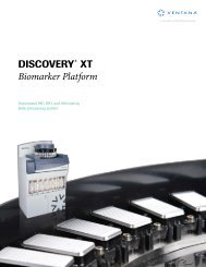
BlueMap Detection Kit Application Notes - Ventana Medical Systems
BlueMap Detection Kit Application Notes - Ventana Medical Systems
BlueMap Detection Kit Application Notes - Ventana Medical Systems

Create successful ePaper yourself
Turn your PDF publications into a flip-book with our unique Google optimized e-Paper software.
<strong>BlueMap</strong> <strong>Kit</strong>Chromogenic <strong>Detection</strong> kit for the <strong>Ventana</strong> Discovery ® Series of InstrumentsCatalog Number: 760-120125 TestsIntended UseFor research use only. Not for use in diagnostic procedures.IntroductionThe Discovery <strong>BlueMap</strong> <strong>Kit</strong> was designed for mRNA and antigen localization in frozen sections, formalin/paraformaldehyde-fixed,paraffin-embedded tissue sections, or IHC applications requiring added sensitivity. The <strong>BlueMap</strong> <strong>Kit</strong> was designed to be used inconjunction with the Discovery series of instruments and <strong>Ventana</strong> <strong>Medical</strong> <strong>Systems</strong>’ ancillary reagents for optimal performance.<strong>Kit</strong> ComponentsStorage<strong>BlueMap</strong> Package InsertPAb-Block(For reducing background staining)SA-Alk Phos(Streptavidin alkaline phosphatase conjugate)Activator(For increasing signal intensity)<strong>BlueMap</strong> NBT(Hue enhancer)<strong>BlueMap</strong> BCIP(Substrate for alkaline phosphatase)1 copy125 tests125 tests125 tests125 tests125 tests2-8°C(Do not freeze)2-8°C(Do not freeze)2-8°C(Do not freeze)2-8°C(Do not freeze)2-8°C(Do not freeze)Instructions for UseRegister the kit on your Discovery series instrument as described in the on screen directions. Open the package, and take out thedispensers. Remove the cap from the nozzle of each dispenser, and place it on the nozzle cap holder on the rear of the dispenser.While holding the dispenser upright, remove the yellow shipping key by pulling the key tab to disengage it from each end. Do notcover the nozzle tip or depress the dispenser while removing key. Place the dispensers on the reagent tray, along with theappropriate accessory reagents.The Discovery series of instruments and the <strong>BlueMap</strong> <strong>Kit</strong> form an integrated system. ALL KIT COMPONENTS MUST BE USEDTOGETHER in order to obtain high-quality and consistent results. Omitting or changing any of the solutions may compromise thefinal outcome.Bulk reagents should be prepared using quality reagent-grade water, not tap water. Carboys for storing bulk reagents should berinsed thoroughly between fillings.ControlsA known positive tissue should be run with every antibody being used. This tissue serves as a reagent control to demonstrate all ofthe reagents used in the assay are functional. The staining of the positive control also serves as a baseline for evaluating run-to-runand/or day-to-day consistency. For IHC, in addition to the positive control, a non-immune Ig control should be run on every tissuetested. Thus, the specificity of the staining reaction can be documented by comparing the non-immune Ig staining to the primaryantibody staining. For ISH applications, in addition to the positive control, a sense probe control on non-immune Ig should be run onevery tissue tested. The sense control should be used to set appropriate stringency conditions for each probe by increasingstringency until staining with the sense probe is “negative”. When this is done, the specificity of the staining reaction can bedocumented by comparing the anti-sense probe staining to sense staining.<strong>Ventana</strong> Molecular Discovery <strong>Systems</strong> March 2006 1http://www.ventanadiscovery.com
ProtocolsAlthough general tissue processing protocols are similar among laboratories, a single universal protocol is not in place, thus no twolaboratories prepare tissue samples in exactly the same way. Tissue processing has the greatest single impact on the end result,and different tissue types often require slightly different pretreatments for optimum results. It is not surprising then that there is not asingle protocol that is optimal for all cases. However, a general protocol (see below) on the Discovery series instruments can serveas a starting point and guideline for optimizing the <strong>BlueMap</strong> <strong>Kit</strong> protocols. The ISH and FISH general protocols below include theuse of other products such as RiboPrep, RiboClear, and RiboFix reagents available from <strong>Ventana</strong> <strong>Medical</strong> <strong>Systems</strong>.Sample protocolsSelectable StepISH(Dig-labeled riboprobe)<strong>Application</strong>FISH(Biotin-labeled DNA probe)Tissue Sample √ √ √Non-ParaffinWet Slide LoadPre-FixativeParaffin √ √ √Deparaffinization √ √ √Fixative RiboPrep 32’ @37ºC RiboPrep 28’ @37ºCPretreatment #1 √ √Use Reaction Buffer for PT1Disable Heat for PT1Heat Slides for PT1-RBUse EZ Buffer for PT1 √ √Disable Heat for PT1-EZHeat Slides for PT1-EZ RiboClear 12’ @37ºC RiboClear 8’ @37ºCCell Conditioning √ √ √Conditioner #1Mild CC1Standard CC1Extended CC1Conditioner #2 √ √Mild CC2 √ √Standard CC2Extended CC2Pretreatment #2 √ √Use Reaction Buffer for PT2 √ √Disable Heat for PT2Heat Slides for PT2-RB Protease 3 for 16’ Protease 3 for 20’Use EZ Buffer for PT2Disable Heat for PT2-EZHeat Slides for PT2-EZPretreatment #3Use Reaction Buffer for PT3Disable Heat for PT3Heat Slides for PT3-RBUse EZ Buffer for PT3Disable Heat for PT3-EZHeat Slides for PT3-EZProbe √ √Auto-DispenseTitration De: 70ºC 8’ Hyb:65ºC 6h De: 90ºC 8’ Hyb:50ºC 6hStringency Wash #1 √ √High Temp Stringency 0.1X @75ºC for 4’ 2.0X @50ºC for 4’Stringency Wash #2 √ √High Temp Stringency 0.1X @75ºC for 4’ 2.0X @50ºC for 4’Stringency Wash #3High Temp StringencyIHC√√√<strong>Ventana</strong> Molecular Discovery <strong>Systems</strong> March 2006 2http://www.ventanadiscovery.com
<strong>Application</strong>Selectable StepISHFISH(Dig-labeled riboprobe) (Biotin-labeled DNA probe)IHCEnzymeOptionPost Fixative (ISH) RiboFix 20’ RiboFix 4’A/B BlockNo HeatAntibody √ √Auto Dispense√Antibody BlockingStandard Ab Incubation FITC/Anti-biotin 20’Extended Ab IncubationTitration√Antibody BlockingStandard Ab Incubation√Extended Ab IncubationPost Fixative (IHC)AmplifyPolyclonalMonoclonal2 nd Antibody √ √ √2 nd Antibody Auto Dispense Anti-DIG biotin Anti-mouse/FITC 20’ √Antibody Blocking2 nd Antibody TitrationAntibody BlockingTyramide RunTSADisable Blocker (Pab, D, or R)Substrate Incubation (<strong>BlueMap</strong> only) 6 hr 30 minCounterstain√Post Counterstain√Slide Cleaning√Note: If the mouse monoclonal biotin-labeled anti-DIG antibody (clone DI-22) from Sigma is being used as the secondaryantibody, it is highly recommended that it be used in conjunction with DISCOVERY Antibody Block.NexES ® Protocol Editor Window1. Open the NexES software.2. To create a protocol, click on the “Protocols” button on the main screen. A window will appear on the screen with “Create/EditProtocol” and “Manage Protocol”. Click on “Create/Edit Protocol” to open the “NexES Protocol Editor” window.3. Select the “Research IHC <strong>BlueMap</strong>” or “Research ISH <strong>BlueMap</strong>” procedure under the “Procedure” field.Saving the Protocol1. After all options have been selected (i.e. no more yellow boxes), click on the “Save As” button. Fields will appear for a protocolname and protocol number. Type in an unused name for the protocol, and select an unused number in the appropriate boxes.Click on the “Save” button, and the protocol will be saved.Preparing Labels and Loading Slides1. From the toolbar on the bottom of the main screen, click on the bar code symbol. Click on the “Protocols” button. Highlight theprotocol number and name desired in the protocol field. Click on the “Add>>” button once for each protocol bar code label youwant to print. Click on the “Close/Print” button. Enter any additional information you want to appear on the label in the "UserPrompt" fields. Click the “Print” button. When the last bar code has been printed, click on the “Exit” button.2. Place the bar code(s) on the slide(s), load them carefully onto the instrument, close the instrument, and click on the “Run”button. Click on the “Reagents/Reagent Tray Loaded” box and “Reagent Caps Removed” box. Enter the number of slidesloaded, and click on “Start Run”.End of Run Instructions1. All runs will go into a hold step before Counterstain/Slide Cleaning. To complete the run, press the logo button on theinstrument. If Counterstain and/or Slide Cleaning were selected in the protocol, the instrument will perform these functionsthen stop. If Counterstain and/or Slide Cleaning were not selected, the instrument will “home,” and then the run will end.<strong>Ventana</strong> Molecular Discovery <strong>Systems</strong> March 2006 3http://www.ventanadiscovery.com
IHC TroubleshootingProblem Possible Cause Next StepNo SignalGood Tissue MorphologyNo SignalPoor Tissue Morphology1. Insufficient cell conditioning2. Inadequate protease digestion1. Over cell conditioned2. Over digested1. Insufficient cell conditioning1. Increase cell conditioning time2. Use stronger proteaseORExtend protease digestion time1. Decrease or remove cell conditioning steps2. Use weaker protease or shorter digestion time1. Increase cell conditioning timeWeak SignalLow BackgroundWeak SignalHigh BackgroundSignal Too StrongLow BackgroundSignal Too StrongHigh Background2. Inadequate protease digestion3. Antibody too dilute1. Non-specific binding of the primary2. Antibody concentration too high2. Use stronger proteaseORExtend protease digestion time3. Increase antibody concentrationORExtend antibody incubation time1. Use blocking reagent during the primaryantibody incubation step2. Decrease the antibody concentration andincrease the incubation time1. Antibody concentration too high 1. Decrease antibody concentrationAND/ORDecrease antibody incubation time1. Antibody concentration too high2. Over cell conditioned1. Decrease antibody concentrationand/or decrease antibody incubation time2. Decrease or remove cell conditioning stepsNOTE: For ISH Troubleshooting recommendations, see the RiboMap ® <strong>Kit</strong> package insert.Intellectual Property<strong>BlueMap</strong>, RiboPrep, RiboClear, and RiboFix are trademarks of <strong>Ventana</strong> <strong>Medical</strong> <strong>Systems</strong>, Inc. Discovery ® , NexES ® ,RiboMap ® , and <strong>Ventana</strong> ® are registered trademarks of <strong>Ventana</strong> <strong>Medical</strong> <strong>Systems</strong>, Inc.<strong>Ventana</strong> grants to Purchaser a single-use only license under the following patents:U.S. Pat. Nos. 6045 759, 6192 945 B1, 6416 713 B1, and foreign counterparts.Technical ConsultationAdditional technical information can be obtained from our Molecular Discovery <strong>Systems</strong> Technical Center at:U.S.A. : (800) 227-2155<strong>Ventana</strong> <strong>Medical</strong> <strong>Systems</strong>, Inc.1910 E. Innovation Park DriveTucson, AZ 85755USAJapan : (+81) 3 5408 5071<strong>Ventana</strong> Japan K.K.Landmark Tower, 35F2-2-1, Minato Mirai, Nishi-kuYokohama, Kanagawa 220-8135JapanEurope : (+33) 3 90 40 52 00<strong>Ventana</strong> <strong>Medical</strong> <strong>Systems</strong> S.A.Parc d’InnovationRue G. de KaysersbergBP 30144F-67404 Illkirch CEDEXFranceAustralia : +61 (0) 3 9817 3077<strong>Ventana</strong> <strong>Medical</strong> <strong>Systems</strong>, Pty. Ltd.5/39 Grand BoulevardMontmorency, Victoria 3094Australia<strong>Ventana</strong> Molecular Discovery <strong>Systems</strong> March 2006 4http://www.ventanadiscovery.com