

Nasolabial flap for oral cavity reconstruction - Vula - University of ...
Nasolabial flap for oral cavity reconstruction - Vula - University of ...
Nasolabial flap for oral cavity reconstruction - Vula - University of ...

Create successful ePaper yourself
Turn your PDF publications into a flip-book with our unique Google optimized e-Paper software.
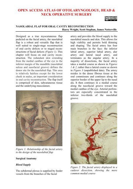
OPEN ACCESS ATLAS OF OTOLARYNGOLOGY, HEAD &NECK OPERATIVE SURGERYNASOLABIAL FLAP FOR ORAL CAVITY RECONSTRUCTIONHarry Wright, Scott Stephan, James NettervilleDesigned as a true myocutaneous <strong>flap</strong>pedicled on the facial artery, the nasolabial<strong>flap</strong> is a robust and versatile <strong>flap</strong> that iswell suited to single-stage <strong>reconstruction</strong><strong>of</strong> <strong>oral</strong> <strong>cavity</strong> defects or to staged <strong>reconstruction</strong>s<strong>of</strong> facial defects (Figure 1). Thischapter will focus on <strong>oral</strong> <strong>cavity</strong> <strong>reconstruction</strong>.The redundant skin extendingfrom the medial canthus <strong>of</strong> the eye to theinferior margin <strong>of</strong> the mandible (nasolabialsulcus and nas<strong>of</strong>acial groove) defines thedonor site <strong>for</strong> the nasolabial <strong>flap</strong>. This areais relatively hairless except <strong>for</strong> the lowercheek in males, an important considerationin <strong>oral</strong> <strong>cavity</strong> <strong>reconstruction</strong>. The <strong>flap</strong> itselfis comprised <strong>of</strong> skin, subcutaneous tissueand the underlying musculature.artery and provides the blood supply to thenasolabial muscle and skin. This allows <strong>for</strong>high viability and permits bold thinningand shaping. The facial artery has fourmain branches in the face: the inferiorlabial artery, superior labial artery, alarartery and lateral nasal artery, andterminates as the angular artery. In amajority <strong>of</strong> dissections, the facial arterytakes a medial course as shown in Figures1 & 2, rather than a lateral course as shownin Figure 3 (unpublished data). The arteryresides in the dense fibrous tissue at the<strong>oral</strong> commissure and continues along thesuperior border <strong>of</strong> the upper lip to the nasalala. It then continues in a medial coursealong the nas<strong>of</strong>acial groove toward themedial canthus <strong>of</strong> the eye. Arterial per<strong>for</strong>atorsare especially concentrated in theinferior two-thirds <strong>of</strong> the nasolabialgroove.Figure 1: Relationship <strong>of</strong> the facial arteryto the design <strong>of</strong> the nasolabial <strong>flap</strong>Surgical AnatomyBlood SupplyThe subdermal plexus is supplied by feedervessels from the branches <strong>of</strong> the facialFigure 2: The facial artery displayed in acadaver dissection, showing the morecommon medial course
Figure 12: Wound closure and final resultFigure 14: Outline <strong>of</strong> <strong>flap</strong>Case #2: Mouth floor <strong>reconstruction</strong>This floor <strong>of</strong> mouth cancer resectionresulted in an alveolar ridge, gingiva, andfloor <strong>of</strong> mouth defect; the inferior border<strong>of</strong> the defect is at the level <strong>of</strong> the superiorborder <strong>of</strong> the mandible (Figure 13). Thedefect was reconstructed with a nasolabial<strong>flap</strong> (Figures 14-16).Though the <strong>flap</strong> is hairless in the imageshown, the surgeon must anticipate theimplications <strong>of</strong> transferring hair‐bearingskin intra<strong>oral</strong>ly.Figure 15: Facial artery is bluntly dissecttedFigure 16: Flap inset into floor <strong>of</strong> mouthFigure 13: Defect following resection5
Case #3: Mouth floor <strong>reconstruction</strong>Figures 17-20 illustrate the postoperativeresults <strong>of</strong> <strong>reconstruction</strong> <strong>of</strong> an anteriorfloor <strong>of</strong> mouth defect crossing the midline.Figure 20: Though the right upper lip isparalysed, the concomitant lip lift providedby the harvest <strong>of</strong> nasolabial donor skinaids with symmetry at restFigure 17: The robust nasolabial <strong>flap</strong>minimises the risk <strong>of</strong> ankyloglossia.Case #4: Buccal <strong>cavity</strong> <strong>reconstruction</strong>The robust blood supply to the nasolabial<strong>flap</strong> permits shaping <strong>of</strong> the <strong>flap</strong> toprecisely fill the defect (Figures 21-24).Figure 18: Early postoperative imageshows aesthetically acceptable appearance<strong>of</strong> donor siteFigure 21: Buccal defectFigure 19: Later image shows a wellhealedfloor <strong>of</strong> mouth <strong>reconstruction</strong>6
Figure 21: Flap inset into buccal defectFigure 22: Final cosmetic resultCase #5: Palate <strong>reconstruction</strong>(postoperative)Figure 23 demonstrates a healed palatal<strong>reconstruction</strong>, with an implant visible <strong>for</strong>a dental prosthesis.Figure 21: Healed <strong>flap</strong> in buccal defectFigure 23: Healed palatal defect7
Reconstruction <strong>of</strong> palatal defects requiresthat the inferior border <strong>of</strong> the incision beapproximately at the level <strong>of</strong> the <strong>oral</strong>commissure. Note the excellent camouflaging<strong>of</strong> the scar and the facial symmetryat rest (Figure 24). When smiling thepatient demonstrates the expected paralysis<strong>of</strong> the ipsilateral upper lip which isgenerally well-tolerated (Figure 25); thisadverse outcome is further discussedbelow.Case 6: Bilateral nasolabial <strong>flap</strong>sThis patient has a large defect <strong>of</strong> the hardpalate, alveolus and gingivobuccal sulcus(Figure 26). Bilateral nasolabial <strong>flap</strong>s wereraised (Figure 27). In Figure 28 the terminalbranch <strong>of</strong> the facial artery i.e. the angularartery, is demonstrated.Figure 26: Defect <strong>of</strong> hard palate, alveolusand gingivobuccal sulcusFigure 24: Excellent camouflage <strong>of</strong> scarand facial symmetry at restFigure 25: Asymmetry with smilingFigure 27: Bilateral nasolabial <strong>flap</strong>s8
Figure 31: Healed facial scarsAdverse OutcomesFigure 28: Angular artery visibleUpper lip weaknessUpper lip weakness may result if theterminal buccal branches <strong>of</strong> the facialnerve supplying lip elevators are lysed or ifthe principle lip elevators (zygomaticusmajor/minor, levator labii alique nasi,levator anguli oris) are completelydivided. Muscle disruption may occurwhen extensive mobilisation and rotation<strong>of</strong> the <strong>flap</strong> is required.Figure 29: Flap being inset in a side-bysideconfigurationPostoperative images show well-healeddonor sites and intact and symmetricelevation <strong>of</strong> the corners <strong>of</strong> the mouth i.e.intact cranial nerve VII (Figures 30. 31).Figure 30: Healed palatal <strong>reconstruction</strong>A unilateral nasolabial <strong>flap</strong> typically doesnot result in any appreciable ipsilateral lipweakness. As a correlation, deinnervatedAbbe <strong>flap</strong> <strong>reconstruction</strong> <strong>of</strong> the upper liphas been shown on electromyography (1and 5 years postop) to regain 80 and 100%reinnervation <strong>of</strong> the transposed musclesegment. 1 However, extensive disruption<strong>of</strong> innervation and mimetic muscleintegrity during harvesting <strong>of</strong> bilateralnasolabial <strong>flap</strong>s may result in clinicallyappreciable weakness <strong>of</strong> the upper lip.Though permanent, it is usually welltolerated. As shown in the selected casesabove, the resulting upper lip weakness ismitigated by the mild lip lift provided byclosure <strong>of</strong> the donor site.9
Oral competenceBilateral upper lip weakness and numbnesscould significantly impair <strong>oral</strong> competence,especially lip-pursing, and must beconsidered if bilateral nasolabial <strong>flap</strong>s areplanned. Special care should be taken toavoid disruption <strong>of</strong> the distal facial nervebranches in such cases.Summary<strong>Nasolabial</strong> <strong>flap</strong> <strong>reconstruction</strong> <strong>of</strong> intra<strong>oral</strong>defects is a well-recognised technique.Because the <strong>flap</strong> is pedicled on the facialartery, single-stage closure with a smallerpedicle may be achieved if the proximalportion <strong>of</strong> the <strong>flap</strong> is de-epithelialised. Arobust blood supply helps to ensure <strong>flap</strong>viability and prevents <strong>flap</strong> breakdown andfistula <strong>for</strong>mation even in adverse conditions<strong>of</strong> excess tension or mild compression<strong>of</strong> the transbuccal pedicle. The bulkprovided by the facial musculature helps t<strong>of</strong>ill larger defects. It does not impair speechand causes only minimal donor sitemorbidity. The <strong>flap</strong> is not overly timeconsumingor technically difficult tomaster; however knowledge <strong>of</strong> the relevantanatomy is essential.ReferencesBurget G, Menick F. Aesthetic restoration<strong>of</strong> one-half <strong>of</strong> the upper lip. Plast ReconstrSurg. 1986;78(5):583–93AcknowledgementsIllustrations by Paul GrossCadaver dissections by David Haynes,MD FACSAuthorsHarry V. Wright MD, MSFellowFacial Plastic and Reconstructive SurgeryOtolaryngology and Head & Neck Surgery<strong>University</strong> <strong>of</strong> South FloridaUSAdr.hvwright@gmail.comScott Stephan MDAssistant Pr<strong>of</strong>essorFacial and Plastic SurgeryVanderbilt Bill Wilkerson CenterNashville, TEUSAstephan@vanderbilt.eduJames L. Netterville MDDirector <strong>of</strong> Head and Neck SurgeryVanderbilt Bill Wilkerson CenterNashville, TEUSAjames.netterville@vanderbilt.eduEditorJohan Fagan MBChB, FCORL, MMedPr<strong>of</strong>essor and ChairmanDivision <strong>of</strong> Otolaryngology<strong>University</strong> <strong>of</strong> Cape TownCape Town, South Africajohannes.fagan@uct.ac.zaTHE OPEN ACCESS ATLAS OFOTOLARYNGOLOGY, HEAD &NECK OPERATIVE SURGERYwww.entdev.uct.ac.zaThe Open Access Atlas <strong>of</strong> Otolaryngology, Head &Neck Operative Surgery by Johan Fagan (Editor)johannes.fagan@uct.ac.za is licensed under a CreativeCommons Attribution - Non-Commercial 3.0 UnportedLicense10


