Iowa Section of AADR - The University of Iowa College of Dentistry
Iowa Section of AADR - The University of Iowa College of Dentistry
Iowa Section of AADR - The University of Iowa College of Dentistry

You also want an ePaper? Increase the reach of your titles
YUMPU automatically turns print PDFs into web optimized ePapers that Google loves.
1<br />
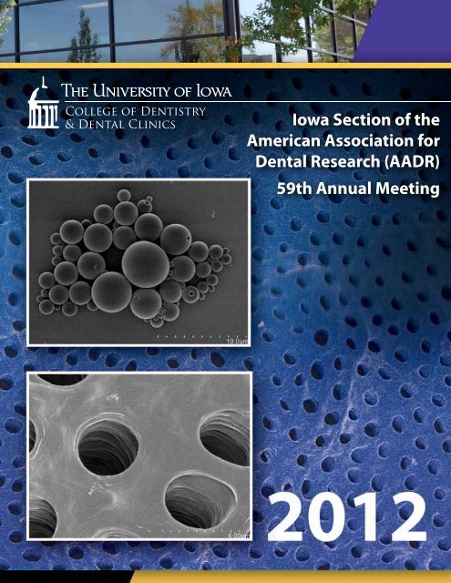
<strong>Iowa</strong> <strong>Section</strong> <strong>of</strong> the<br />
American Association for<br />
Dental Research (<strong>AADR</strong>)<br />
59th Annual Meeting
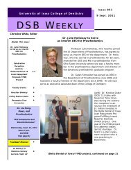
Our Keynote Speaker:<br />
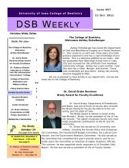
Isabel Garcia, D.D.S., M.P.H.<br />
Deputy Director<br />
National Institute <strong>of</strong> Dental and Crani<strong>of</strong>acial Research<br />
Dr. Isabel Garcia is the Deputy Director <strong>of</strong> the National Institute <strong>of</strong> Dental and Crani<strong>of</strong>acial Research.<br />
NIDCR is the federal government’s largest funder <strong>of</strong> research and research training focusing on oral,<br />
dental and crani<strong>of</strong>acial diseases and disorders. As deputy director, Dr. Garcia shares the responsibility<br />
with the director to lead and manage <strong>of</strong> all NIDCR’s programs and activities. Dr. Garcia is a career-<strong>of</strong>ficer<br />
with the U.S. Public Health Service and holds the rank <strong>of</strong> Rear Admiral - Lower Half. She has been<br />
NIDCR’s Deputy Director since January 2007.<br />
Dr. Garcia received her Bachelor degree in Science from the <strong>University</strong><br />
<strong>of</strong> Mary Washington, a Doctor in Dental Surgery from the Medical <strong>College</strong><br />
<strong>of</strong> Virginia, and a Master in Public Health from the <strong>University</strong> <strong>of</strong> Michigan<br />
where she also completed a Residency in Dental Public Health. Her experience<br />
spans over 30 years <strong>of</strong> work in dental public health, research and<br />
administration at the local, state and national levels. Since joining NIDCR<br />
in 1995 Dr. Garcia has held several positions within the Institute. In addition<br />
to serving as acting institute director from August 2010-2011, Dr. Garcia<br />
has led NIDCR’s science transfer efforts, directed the Institute’s science<br />
planning, health policy, legislative, and evaluation programs and led the<br />
development <strong>of</strong> NIDCR’s last two strategic plans. Prior to her career in the<br />
Public Health Service Dr. Garcia worked in the private sector as a clinician<br />
and later held health management positions in Virginia and Ohio.<br />
Dr. Garcia is a Diplomate <strong>of</strong> the American Board <strong>of</strong> Dental Public Health. She has served as an <strong>of</strong>ficer<br />
in the American Association <strong>of</strong> Public Health <strong>Dentistry</strong>’s Executive Council, the Oral Health <strong>Section</strong> <strong>of</strong><br />
the American Public Health Association and the American Board <strong>of</strong> Dental Public Health. In addition<br />
to her duties as Deputy, Dr. Garcia directs NIDCR’s Residency Program in Dental Public Health and<br />
serves as her Institutes’ coordinator for International Health. In addition to numerous honors and<br />
awards Dr. Garcia has been recognized for her role in a Presidential and Secretarial health diplomacy<br />
mission which provided health care to people in 12 countries in Latin America.<br />
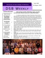
Dental research images for the cover were provided<br />
by Jessica Grabouski, Jaxon Hoopes, David Thuet,<br />
Na Wei, and Min Zhu.<br />
2
Table <strong>of</strong> Contents<br />
Letter from Dean David Johnsen ........................................................................................................ 2<br />
Letter from Associate Dean for Research Clark Stanford ................................................................. 3<br />
Letter from Officers <strong>of</strong> the <strong>Iowa</strong> Chapter <strong>of</strong> the <strong>AADR</strong> .................................................................... 4<br />
Program ................................................................................................................................................ 5<br />
Presentation Assignments.................................................................................................................... 6<br />
Abstracts ............................................................................................................................................... 9<br />
Author-Abstract Index ....................................................................................................................... 32<br />
<strong>Iowa</strong> <strong>Section</strong> <strong>of</strong> <strong>AADR</strong> — Past Presidents ......................................................................................... 33<br />
Acknowledgments .............................................................................................................................. 34<br />
1
Dear Colleagues:<br />
Thank you for your participation in the <strong>University</strong> <strong>of</strong> <strong>Iowa</strong> <strong>College</strong> <strong>of</strong> <strong>Dentistry</strong>’s Local Research<br />
Day on February 14, 2012. Research is central to our mission. Research is important in itself<br />
and for the culture <strong>of</strong> inquiry that it supports. This day is one <strong>of</strong> the highlights <strong>of</strong> our life as an<br />
academic community. <strong>The</strong> event’s planning committee and research presenters are to be heartily<br />
commended for their hard work.<br />
We are honored to host Dr. Isabel Garcia, Deputy Director <strong>of</strong> NIDCR, as our keynote speaker.<br />
Dr. Garcia has worked tirelessly and effectively, not only to lead NIDCR as Acting Director, but to<br />
raise the visibility <strong>of</strong> dental research at the NIH. This critical visibility for dental research at the<br />
NIH was enhanced through her work on the NIH Roadmap and in assisting Dr. Larry Tabak in<br />
his post as Interim Deputy Director <strong>of</strong> the larger NIH.<br />
Since our last Research Day, half <strong>of</strong> our fourth floor research space has been renovated and occupied.<br />
This state-<strong>of</strong>-the-art transformation was possible with the support <strong>of</strong> the Roy J. Carver<br />
Charitable Trust. <strong>The</strong> Carver Charitable Trust will be represented by Troy Ross, Ph.D. and<br />
Lynne Sasmazer, Ph.D.<br />
Local Research Day shows the people and the spirit <strong>of</strong> discovery that have always made possible<br />
outstanding education, service, research, and patient care within our <strong>College</strong>.<br />
Local Research Day and this research abstract book <strong>of</strong>fer many opportunities to learn about fascinating<br />
research within our <strong>College</strong>. Thank you for being a part <strong>of</strong> this important event.<br />
Best wishes,<br />
David C. Johnsen, D.D.S., M.S.<br />
Dean<br />
2
Dental Research participants and <strong>Iowa</strong> <strong>Section</strong> <strong>of</strong> the <strong>AADR</strong><br />
I would like to join in welcoming everyone to the <strong>Iowa</strong> section <strong>of</strong> the <strong>AADR</strong> annual research day.<br />
It brings special meaning this year. This year we have a special guest, Dr. Isabel Garcia DDS,<br />
MPH who was recently the acting director <strong>of</strong> NIDCR. Dr. Garcia brings insights and perspectives<br />
which are truly inspirational.<br />
This is a day that celebrates the range <strong>of</strong> research interests across the <strong>College</strong> and shows the<br />
multiple links that we have with the entire campus at the <strong>University</strong> <strong>of</strong> <strong>Iowa</strong>. <strong>The</strong> research presented<br />
is by undergraduates, dental students, resident, graduate students and faculty. <strong>The</strong> work<br />
spans the areas <strong>of</strong> basic, translational, clinical and health services research all within one forum.<br />
This is an unusual event.<br />
Of value during this day is the chance to discuss, critique and learn from each other. This is a<br />
critical event in the life <strong>of</strong> the <strong>College</strong> and I hope you enjoy the day!<br />
Warmest Regards,<br />
Clark M. Stanford DDS, Ph.D.<br />
Associate Dean for Research<br />
Centennial Fund Pr<strong>of</strong>essor<br />
<strong>College</strong> <strong>of</strong> <strong>Dentistry</strong><br />
<strong>University</strong> <strong>of</strong> <strong>Iowa</strong><br />
3
Dear Colleagues,<br />
On behalf <strong>of</strong> the <strong>Iowa</strong> <strong>Section</strong> <strong>of</strong> the American Association for Dental Research (<strong>AADR</strong>), we are<br />
very pleased to welcome you to the <strong>University</strong> <strong>of</strong> <strong>Iowa</strong>, <strong>College</strong> <strong>of</strong> <strong>Dentistry</strong> Research Day. We<br />
would like to thank everyone, including the presenters and participants, for your cooperation<br />
and support in making this event successful.<br />
This year we are honored to have Dr. Isabel Garcia from the National Institute <strong>of</strong> Dental and<br />
Crani<strong>of</strong>acial Research (NIDCR) as the keynote speaker. Dr. Garcia is the Deputy Director <strong>of</strong><br />
NIDCR and is responsible for leading and managing all <strong>of</strong> NIDCR’s programs and activities.<br />
<strong>The</strong> quality, diversity, and breadth <strong>of</strong> research at the <strong>University</strong> <strong>of</strong> <strong>Iowa</strong>, <strong>College</strong> <strong>of</strong> <strong>Dentistry</strong> is<br />
extraordinary. This “Local <strong>AADR</strong> Research Day” allows all <strong>of</strong> us the opportunity to share and<br />
learn about the research endeavors conducted at the <strong>College</strong> <strong>of</strong> <strong>Dentistry</strong>.<br />
Todays activities demonstrate the research accomplishments <strong>of</strong> dental students, graduate students,<br />
post-graduate students, faculty and staff through posters, table clinics and oral presentations.<br />
We are excited about the active participation in this research day.<br />
Lastly, we would like to extend a special thank you to all <strong>of</strong> our sponsors as well as all <strong>of</strong> you who<br />
have contributed to make this day possible.<br />
THANK YOU FOR YOUR SUPPORT AND PARTICIPATION!<br />
Sincerely,<br />
Justine Kolker, DDS, MS, PhD Sherry Timmons, DDS, PhD<br />
Associate Pr<strong>of</strong>essor, Assistant Pr<strong>of</strong>essor<br />
Department <strong>of</strong> Operative <strong>Dentistry</strong> Department <strong>of</strong> Oral Pathology, Radiology<br />
and Medicine Vice-President, <strong>Iowa</strong> <strong>Section</strong> <strong>of</strong> <strong>AADR</strong><br />
President, <strong>Iowa</strong> <strong>Section</strong> <strong>of</strong> <strong>AADR</strong><br />
Sharon Seydel<br />
Department Administrative Manager<br />
Dows Institute <strong>of</strong> Research<br />
Secretary/Treasurer <strong>Iowa</strong> <strong>Section</strong> <strong>of</strong> <strong>AADR</strong><br />
4
Program<br />
<strong>Iowa</strong> <strong>Section</strong> <strong>of</strong> the American Association for Dental Research (<strong>AADR</strong>)<br />
59th Annual Meeting, Tuesday February 14th, 2012<br />
8:00 a.m. Reception with C<strong>of</strong>fee and Rolls (1st floor link)<br />
8: 30 a.m. Dean’s Welcome (Galagan Auditoria)<br />
Dr. David Johnsen<br />
Keynote Speaker Introduction<br />
Drs. Clark Stanford and Steve Levy<br />
8:45 a.m. Keynote Address (Galagan Auditoria)<br />
Dr. Isabel Garcia<br />
9:45 a.m. Break<br />
10:00 a.m. - 11:15 a.m. Oral Presentations (Galagan Auditoria)<br />
Pre-doctoral (Gal. A)<br />
Graduate & Post-doctoral (Gal. B)<br />
11:30 a.m. – 12:40 p.m. Poster & Table Clinic Presentations (W220 A&B)<br />
5:30 p.m. Awards Banquet Reception with Cash Bar (Coralville Holiday Inn)<br />
6:30 p.m. Awards Banquet Dinner & Awards (Coralville Holiday Inn)<br />
5
Presentation Assignments<br />
Oral Session 1: Pre-Doctoral<br />
10:00 a.m. - 11:15 a.m., Galagan A<br />
(a) Max Smith Pre-Doctoral Competition<br />
(b) <strong>Iowa</strong> Society for Periodontology Pre-Doctoral Award<br />
▼<br />
1. R. Walton<br />
Incidence <strong>of</strong> Signs and Symptoms <strong>of</strong> Vertical Root Fracture<br />
2. a B. Smith, L.M. Moreno, S.F. Miller, G. Wehby, M. Dunnwald<br />
Digital Image Analysis for Reliable Characterization <strong>of</strong> Cleft Wound Phenotypes<br />
3. a,b L. Harvey, K. Brogden, A. Progulske-Fox<br />
HBD3-Enhanced Porphyromonas gingivalis rHagB Cytokine Responses in Dendritic Cells<br />
4. a,b J. Van Hemert , E. Recker, K. Walters, A. Progulske-Fox, K.A. Brogden<br />
HBD3 Inhibits Porphyromonas gingivalis rHagB Binding to Dendritic Cells<br />
Oral Session 2: Graduate & Post-Doctoral<br />
10:00 a.m. - 11:15 a.m., Galagan B<br />
(c) Max Smith Graduate and Post-Doctoral Competition<br />
(d) Dental Specialty Award Competition: Pediatric<br />
(k) Dental Specialty Award Competition: Operative<br />
▼<br />
5. c,d S.E. Swenson, K. Weber-Gasparoni, F. Qian, R.L. Ettinger<br />
Effectiveness <strong>of</strong> Group Home Caregivers’ Training Regarding Oral Care Delivery<br />
6. c C.L. Fischer, K. Walters, D.V. Dawson, D.R. Drake, K.A. Brogden, P.W. Wertz<br />
7. c,k<br />
Sphingolipids and Fatty Acids Alter Porphyromonas gingivalis Protein/Lipid Compositions<br />
L. St-Pierre, C. Bergeron, F. Qian, M.M. Hernandez, J.L. Kolker, D.S. Cobb, M.A. Vargas<br />
Effect <strong>of</strong> Finishing and Polishing Direction on the Marginal Adaptation <strong>of</strong> Resin-Based Composite<br />
Restorations In Vitro<br />
6<br />
Presenters are underlined.<br />
Mentors are italicized.
Pre-Doctoral, Graduate, and Post-Doctoral Posters & Table Clinics<br />
11:30 a.m. - 12:40 p.m., W220 A & B<br />
(b) <strong>Iowa</strong> Society for Periodontology Pre-Doctoral Award<br />
(d) Dental Specialty Award Competition: Pediatric<br />
(e) Proctor and Gamble Pre-Doctoral Award<br />
(f) ADA Pre-Doctoral Competition<br />
(g) ADA Graduate and Post-Doctoral Competition<br />
(h) <strong>Iowa</strong> Society for Periodontology Post-Doctoral Award<br />
(i) Endodontic Michael Fuller Post-Doctoral Award<br />
(j) Dental Specialty Award Competition: Preventive & Community<br />
(k) Dental Specialty Award Competition: Operative<br />
(l) Dental Specialty Award Competition: Basic Science<br />
▼<br />
8. e,f<br />
9. e,f<br />
10. e,f<br />
11. e,f<br />
E.K. Wang, D.R. Blanchette, C.M. Kummet, S.F. Miller, L.M. Moreno, D.V. Dawson<br />
Three-dimensional S<strong>of</strong>t Tissue Asymmetry in Unaffected Relatives <strong>of</strong> NSCL/P Individuals<br />
C.M. Schiltz, J.L. Kolker, J.S. Wefel, C.M. Kummet, M.M. Hogan, J.D. Harless, D.V. Dawson<br />
S.J. Christensen, D.V. Dawson, D.R. Blanchette, C.M. Stanford<br />
Evaluating the Validity <strong>of</strong> the Pink Esthetic Score<br />
C. Shao, M.R. McQuistan, C.L. Straub-Morarend, M.D. Macek<br />
Oral Health Knowledge among Eldery Dental Patients<br />
12. e,f A. Shimek, M. Zhu, J.A. Banas<br />
Streptococcus mutans Glucan-Binding Proteins<br />
13. e,f A. Kang, D.G. Gratton, C.M. Stanford<br />
Prospective, Comparative Assessment <strong>of</strong> Single-Tooth Replacement in Different Implant-Abutment<br />
Interface Settings: Digital Scanning Analysis <strong>of</strong> Alveolar Ridge Architecture Alterations<br />
14. e,f C.R. Allen, M.A. Mansilla, J.C. Murray<br />
GWAS Follow-up Studies: NTN1 and NOG, New Candidates for NSCL/P<br />
15. e,f<br />
16. e,f<br />
17. e,f<br />
A.J. Brasser, K.A. Brogden, P.W. Wertz<br />
Salivary Lipid Binding by SPLUNC1.<br />
H.V. Guenther, J.J. Warren, D.R. Drake, F. Qian<br />
Assessment <strong>of</strong> Convenience <strong>of</strong> Glycyrrhiza uralensis Lollipops for Caries Prevention<br />
J. Grabouski, J.J. Grabouski, R.N. Staley, C.M. Kummet<br />
Cephalometric Measurements in Mixed Dentitions with Class I Normal Occlusion<br />
18. e,f C. Gleichman, M.A. Vargas<br />
<strong>The</strong> Use <strong>of</strong> NaOCl to Increase Bond Strength<br />
19. e,f T.N. Kieu, K. Weber-Gasparoni, J.J. Warren, F. Qian<br />
Evaluation <strong>of</strong> a Caries Risk Assessment Tool — A Pilot Study<br />
20. e,f D.G. Meier, R.N. Staley, C.M. Kummet<br />
Growth Relationships between the Mandible and Standing Height by Occlusion<br />
7
21. e,f M. Popowski, M.R. McQuistan, C.L. Straub-Morarend, F. Qian<br />
Changes in Fourth Year Dental Students’ Presentation <strong>of</strong> Treatment Plans<br />
22. e,f N. Benassi, Y. Yu, V. Joshi, N. Wei, A.K. Salem, L. Hong<br />
Enhancement <strong>of</strong> Rat MSC Capabilities by Intracelluar Release <strong>of</strong> Estradiol<br />
23. e,f J. Kelly, N.E. Holton, S.D. Marshall, R. Franciscus, T.E. Southard<br />
<strong>The</strong> Effect <strong>of</strong> Maxillary Restriction on Mandibular Fossa Morphology<br />
24. e,f A. Murray, M.A. Vargas, S. Geraldeli, F. Qian, L. Chen<br />
Effect <strong>of</strong> Benzalkonium Chloride Adhesive System on Resin-Dentin Bond Durability.<br />
25. e,f J.J. Grabouski, R.N. Staley, C.M. Kummet<br />
Cephalometric Measurements in Adolescents with Class I Normal Occlusion<br />
26. A.D. Figueroa<br />
e,f , N.E. Holton , S.L. Kane, T.E. Southard<br />
Analysis <strong>of</strong> Cortical Bone Distribution in the Human Mandible<br />
27. E. Recker, C. Barwacz, L. Thomann, D.R. Blanchette, C.M. Kummet, D.V. Dawson, K.A. Brogden ,<br />
C.M. Stanford<br />
Bone Mediators in the Implant Sulcus Fluid Around Implant Abutments<br />
28. g,h B.T. Tingey, S.H. Clark, L.A. Humbert, J.D. Tingey, C.M. Kummet<br />
29. g,i<br />
30. g,i<br />
Use <strong>of</strong> IV Sedation in Periodontal Practice: A National Survey<br />
D. Thuet, A.E. Williamson, F. Qian<br />
TM<br />
Effect <strong>of</strong> Ultrasonic Activation Using ProUltra PiezoFlow in Curved Canals on Smear Layer<br />
Removal Using 17% EDTA<br />
J. Hoopes, A.E. Williamson, D.R. Drake, F. Qian<br />
Effect <strong>of</strong> Blood Contamination on Sealing Properties <strong>of</strong> EndoSequence Root Repair Material TM<br />
31. i,g J.M. Clark, W.T. Johnson, K.V. Krell, D.R. Drake, F. Qian, O. Maktabi<br />
Retrospective Analysis <strong>of</strong> the Survival <strong>of</strong> Teeth Treated with Endodontics and Crown Lengthening<br />
32. H.A. Reynolds, K. Weber-Gasparoni, F. Qian, S.E. Swenson, R.L. Ettinger<br />
Caregivers’ Perceived Comfort Regarding Oral Care Delivery in Group Homes.<br />
33. g,j G. Kavand S.M. Levy, J.J. Warren<br />
Longitudinal Changes in Dental Esthetic Perceptions <strong>of</strong> Adolescents and <strong>The</strong>ir Parents<br />
34. d,g M. Zhu, H. Zhang, Y. Ou, J.A. Banas<br />
35. g,i<br />
36. g,l<br />
R. Beasley, A.E. Williamson, B.C. Justman, D.R. Drake, F. Qian<br />
Percha From Moderately Curved Root Canals with ProTaper Retreatment Files<br />
N. Wei, L. Hong, N. Kim, Y. Yu, V. Joshi, A.K. Salem<br />
Effects <strong>of</strong> GR siRNA on Proliferation <strong>of</strong> MSCs In vitro<br />
8
Abstracts<br />
1. Incidence <strong>of</strong> Signs and Symptoms <strong>of</strong> Vertical Root Fracture<br />
R. Walton 1<br />
1 <strong>University</strong> <strong>of</strong> <strong>Iowa</strong><br />
Objectives:<br />
Materials and Methods:<br />
Results: Signs and symptoms had no consistent diagnostic patterns. All fractured roots had RCT. Reported pain<br />
was none-mild; swelling (history or presence at 77%) or sinus tract (31%) were more common. Probings showed<br />
different patterns (narrow-rectangular was prevalent at 66%), including no defect (21%). Radiographic patterns<br />
ranged from no change to extensive bone loss (most common was apical+lateral+crestal at 45%); the fracture was<br />
on most roots.<br />
Conclusions:<br />
9
2. Digital Image Analysis for Reliable Characterization <strong>of</strong> Cleft Wound Phenotypes<br />
B. Smith 1 , L.M. Moreno 1 , S.F. Miller 1 , G. Wehby 1 , M. Dunnwald 1<br />
1 <strong>University</strong> <strong>of</strong> <strong>Iowa</strong><br />
Cleft lip is a common birth defect with major physical and psychological impacts on affected individuals’ lives.<br />
Satisfaction with facial appearance, which is partly affected by success/quality <strong>of</strong> surgical cleft repair, is widely<br />
IRF6 or other clefting candidate<br />
genes. However, understanding the genetic basis <strong>of</strong> wound healing among affected individuals requires accurate<br />
characterization <strong>of</strong> the wound healing phenotype.<br />
Objectives: To develop wound healing phenotypes measured from digital 2D and 3D images <strong>of</strong> cleft lip wounds for<br />
genotype-phenotype correlation studies.<br />
Methods: We used 3-D images <strong>of</strong> 68 surgically repaired unilateral cleft lip individuals captured using the 3DMD<br />
image system. Size and RGB color histogram data <strong>of</strong> affected and control regions <strong>of</strong> the philtrum and upper lip<br />
were acquired using 3DMD and Image J s<strong>of</strong>tware. Reliability was determined from repeated measurements <strong>of</strong><br />
color similarity <strong>of</strong> several non-affected control points <strong>of</strong> the philtrum. We compared the color <strong>of</strong> affected versus<br />
unaffected control areas using t-test analysis.<br />
Results: Our method demonstrated high repeatability from ICC scores >8.0 for all repeated area measurements<br />
Pearson correlation (p
phenotypes are currently being evaluated and will be presented.<br />
Supported by: CDC grant 5R01DD000295; R03-AR055313; NIDCR Training Grant T32 DE014678-09<br />
3. HBD3-enhanced Porphyromonas gingivalis rHagB Cytokine Responses in Dendritic Cells<br />
L. Harvey 1 , K.A. Brogden 1 , A. Progulske-Fox 6<br />
1 <strong>University</strong> <strong>of</strong> <strong>Iowa</strong>; 6 <strong>University</strong> <strong>of</strong> Florida, Gainesville, FL<br />
Previously, we found that HBD3, co-administered with recombinant hemagglutinin B (rHagB) attenuates a<br />
Porphyromonas gingivalis rHagB in human myeloid dendritic<br />
cells.<br />
Objectives: <strong>The</strong> objective <strong>of</strong> this study was to determine if the chemokine and cytokine response <strong>of</strong> dendritic cells<br />
induced by rHagB is still attenuated when HBD3 is administered prior to exposure with rHagB; when administered<br />
simultaneously with rHagB; or when administered after exposure to rHagB.<br />
Methods:<br />
post exposure, culture supernatants were removed for the determination <strong>of</strong> 22 chemokines and cytokines using<br />
Instrument (Luminex, Austin, TX).<br />
Results: rHagB alone induced a robust chemokine and cytokine response. <strong>The</strong> timing <strong>of</strong> HBD3 administration<br />
was important. HBD3 co-incubated with rHagB (for 30 min prior to exposure) attenuated a chemokine and<br />
simultaneously, but not together or if HBD3 was administered 1 hour after, then rHagB induces a modestly robust<br />
Conclusions: <strong>The</strong> results show that the timing <strong>of</strong> HBD3 administration is important and HBD3 has the capacity to<br />
modulate the cytokine and chemokine response <strong>of</strong> dendritic cells to rHagB. Supported by funds from NIH, NIDCR<br />
R01 DEO14390.<br />
10<br />
Supported by: <strong>University</strong> <strong>of</strong> <strong>Iowa</strong>, <strong>College</strong> <strong>of</strong> <strong>Dentistry</strong>, <strong>Iowa</strong> Dental Research Grant<br />
4. HBD3 Inhibits Porphyromonas gingivalis rHagB Binding to Dendritic Cells<br />
J. Van Hemert 5 , E. Recker 5 , K. Walters 2 , A. Progulske-Fox 6 , K.A. Brogden 5<br />
2 5 6 <strong>University</strong> <strong>of</strong> <strong>Iowa</strong>, <strong>Iowa</strong> City, IA; <strong>University</strong> <strong>of</strong> <strong>Iowa</strong> <strong>College</strong> <strong>of</strong> <strong>Dentistry</strong>, <strong>Iowa</strong> City, IA; <strong>University</strong> <strong>of</strong> Florida,<br />
Gainesville, FL<br />
Human β-defensin 3 (HBD3) is a small, well-characterized peptide with broad antimicrobial activities and<br />
diverse innate immune functions. Previously, we found that HBD3 binds to recombinant Porphyromonas gingivalis<br />
dendritic cells.<br />
Objectives: Our objective was to determine if HBD3 binding to rHagB alters the binding <strong>of</strong> rHagB to the surface <strong>of</strong><br />
human myeloid dendritic cells.<br />
Methods: To test this, human myeloid dendritic cells and mouse JAWS II cells were incubated with 0.1 μM<br />
rHagB, 1.0 μM HBD3+0.1 μM rHagB (10:1 molar ratio), 1.0 μM HBD3, or 0.1 M PBS, pH 7.2. After 5 minutes,<br />
with monoclonal MoAb 1858 to rHagB, polyclonal rabbit to rHagB, and polyclonal 500-P241 rabbit antibody to<br />
microscopy.<br />
Results: For confocal microscopy, 6.3 x 10⁴ dendritic cells in chamber slides were incubated with rHagB and had<br />
Conclusion: Overall, these results strongly suggest that HBD3 binding to rHagB alters the binding <strong>of</strong> rHagB to the<br />
response <strong>of</strong> rHagB in dendritic cells. This work was supported by NIH, NIDCR grant R01 DEO14390.<br />
Supported by: NIH, NIDCR grant R01 DEO14390.<br />
11
R01 DEO14390.<br />
Supported by: <strong>University</strong> <strong>of</strong> <strong>Iowa</strong>, <strong>College</strong> <strong>of</strong> <strong>Dentistry</strong>, <strong>Iowa</strong> Dental Research Grant<br />
4. HBD3 Inhibits Porphyromonas gingivalis rHagB Binding to Dendritic Cells<br />
J. Van Hemert 5 , E. Recker 5 , K. Walters 2 , A. Progulske-Fox 6 , K.A. Brogden 5<br />
2 <strong>University</strong> <strong>of</strong> <strong>Iowa</strong>, <strong>Iowa</strong> City, IA; 5 <strong>University</strong> <strong>of</strong> <strong>Iowa</strong> <strong>College</strong> <strong>of</strong> <strong>Dentistry</strong>, <strong>Iowa</strong> City, IA; 6 <strong>University</strong> <strong>of</strong> Florida,<br />
Gainesville, FL<br />
Human β-defensin 3 (HBD3) is a small, well-characterized peptide with broad antimicrobial activities and<br />
diverse innate immune functions. Previously, we found that HBD3 binds to recombinant Porphyromonas gingivalis<br />
dendritic cells.<br />
Objectives: Our objective was to determine if HBD3 binding to rHagB alters the binding <strong>of</strong> rHagB to the surface <strong>of</strong><br />
human myeloid dendritic cells.<br />
Methods: To test this, human myeloid dendritic cells and mouse JAWS II cells were incubated with 0.1 μM<br />
rHagB, 1.0 μM HBD3+0.1 μM rHagB (10:1 molar ratio), 1.0 μM HBD3, or 0.1 M PBS, pH 7.2. After 5 minutes,<br />
with monoclonal MoAb 1858 to rHagB, polyclonal rabbit to rHagB, and polyclonal 500-P241 rabbit antibody to<br />
microscopy.<br />
Results: For confocal microscopy, 6.3 x 10⁴ dendritic cells in chamber slides were incubated with rHagB and had<br />
Conclusion: Overall, these results strongly suggest that HBD3 binding to rHagB alters the binding <strong>of</strong> rHagB to the<br />
response <strong>of</strong> rHagB in dendritic cells. This work was supported by NIH, NIDCR grant R01 DEO14390.<br />
Supported by: NIH, NIDCR grant R01 DEO14390.<br />
11<br />
12
5. Effectiveness <strong>of</strong> Group Home Caregivers’ Training Regarding Oral Care Delivery<br />
S.E. Swenson 1 , K. Weber-Gasparoni 1 , F. Qian 1 , R.L. Ettinger 1<br />
1 <strong>University</strong> <strong>of</strong> <strong>Iowa</strong><br />
Purpose: <strong>The</strong> purpose <strong>of</strong> this study was to investigate the effectiveness <strong>of</strong> training on the behaviors <strong>of</strong> group home<br />
caregivers who provided oral hygiene care for individuals with special health care needs (SHCN).<br />
Methods: A 24-item survey was distributed to 884 caregivers employed at six care facilities in <strong>Iowa</strong>. Bivariate analyses<br />
and logistic regression models were used to analyze the data (alpha=.05).<br />
Results:<br />
that caregivers who reported receiving training at their current facility (yes versus no) on how to provide oral<br />
hygiene care for individuals with SHCN were more likely to brush the teeth <strong>of</strong> uncooperative consumers more<br />
frequently (P=.003), and feel more comfortable providing oral hygiene care for those who verbally and physically<br />
resisted oral care (P=.0003). Multivariable logistic regression analyses indicated that individuals who reported<br />
receiving training were more likely to brush (P P=.03) the teeth <strong>of</strong> cooperative consumers,<br />
continue oral care delivery when consumers physically refused the care (P=.0041), provide direct care to a higher<br />
number <strong>of</strong> individuals with SHCN on a weekly basis (P=.0209), and disagree that lack <strong>of</strong> training was a barrier to<br />
providing oral hygiene care (P<br />
<strong>of</strong> consumers perceived as uncooperative.<br />
Conclusions: Caregivers who reported receiving training on how to provide oral hygiene care to individuals with<br />
SHCN displayed more positive behaviors when compared to those who did not receive training.<br />
Supported by: Delta Dental <strong>of</strong> <strong>Iowa</strong> Foundation<br />
6. Sphingolipids and Fatty Acids Alter Porphyromonas gingivalis Protein/Lipid Compositions<br />
C.L. Fischer 2 , K. Walters2 , D.V. Dawson 2 , D.R. Drake 2 , K.A. Brogden 2 , P.W. Wertz2 2 <strong>University</strong> <strong>of</strong> <strong>Iowa</strong>, <strong>Iowa</strong> City, IA<br />
Long chain bases: sphingosine, phytosphingosine, and dihydrosphingosine, and fatty acids: lauric acid and sapienic<br />
acid, commonly found on oral mucosa and on skin have potent antimicrobial activity for a variety <strong>of</strong> Gram-positive<br />
and Gram-negative bacteria including Porphyromonas gingivalis. Treatment with these lipids also induces striking<br />
ultrastructural damage to the overall bacterial cell and formation <strong>of</strong> unique intracellular inclusions in some<br />
bacteria. However, little is known about the mechanisms <strong>of</strong> antimicrobial action as well as the nature and the origin<br />
<strong>of</strong> the bacterial inclusions.<br />
Objectives:<br />
<strong>of</strong> bacteria following treatment with lipids; 2) We determined if bacteria exhibited cell-association <strong>of</strong> test lipids<br />
following exposure.<br />
Methods: P. gingivalis was treated with long chain bases or fatty acids and pelleted by centrifugation. For one<br />
treatment, whole-cell protein extracts were reduced and separated by SDS polyacrylamide gel electrophoresis<br />
and stained. For another treatment, lipids were extracted and separated by thin layer chromatography. Controls<br />
bases and fatty acids.<br />
Results: Of the long chain bases and fatty acids examined, sapienic acid induced the greatest alterations in the<br />
Conclusions:<br />
Mechanisms for these alterations are not yet known and are being explored. Long chain bases and fatty acids are<br />
present in cell pellets suggesting they may be associating with the bacterial membrane or accumulating in the cell,<br />
perhaps in the forming inclusions.<br />
Supported by: NIH/NIDCR RO1 DEO18032 and R01 DEO14390.<br />
13 12
7. Effect <strong>of</strong> Finishing and Polishing Direction on the Marginal Adaptation <strong>of</strong> Resin-Based<br />
Composite Restorations In Vitro<br />
L. St-Pierre 1 , C. Bergeron 1 , F. Qian 1 , M.M. Hernandez 1 , J.L. Kolker 1 , D.S. Cobb 1 , M.A. Vargas 1<br />
1 <strong>University</strong> <strong>of</strong> <strong>Iowa</strong><br />
Objective: To assess the effect <strong>of</strong> finishing and polishing direction on the marginal adaptation <strong>of</strong> resin-based<br />
composite restorations.<br />
Methods: Forty human molars were collected and sectioned along their mesio-distal axis. Buccal and lingual<br />
enamel surfaces were flattened and a triangular preparation (0.87mm deep and 3mm wide) representing two<br />
30° bevels was achieved. Specimens (n=20/per group) were randomly assigned in groups and restored with two<br />
resin-based composite materials: a nan<strong>of</strong>illed (Filtek Supreme Ultra)(FSU) and a microhybrid (Point4)(PT4) and<br />
two finishing/polishing techniques: a series <strong>of</strong> S<strong>of</strong>-Lex discs (SL) and a sequence <strong>of</strong> diamond bur/dark-orange<br />
SL/rubber polishers (HiLuster). On each specimen, both margins were finished and polished with the same<br />
technique, one from the resin-based composite to the tooth structure (C-T) and the other from the tooth structure<br />
to the resin-based composite (T-C). Replicas were made for FeSEM observation (200X) and quantitative margin<br />
analysis was performed based on four defined marginal quality criteria. Comparisons were made between polishing<br />
directions (paired-samples t-test, Wilcoxon signed-rank test), between resin-based composites and between<br />
polishing techniques (two-sample t-test and Wilcoxon rank sum test)<br />
Results: Significant differences were found between polishing directions (p
8. Three-Dimensional S<strong>of</strong>t Tissue Asymmetry in Unaffected Relatives <strong>of</strong> NSCL/P Individuals<br />
E.K. Wang 1 , D.R. Blanchette 1 , C.M. Kummet 1 , S. Miller 1 , L.M. Moreno 1 , D.V. Dawson 1<br />
1 <strong>University</strong> <strong>of</strong> <strong>Iowa</strong><br />
Objectives: To compare patterns <strong>of</strong> three-dimensional facial asymmetry in unaffected siblings <strong>of</strong> individuals with<br />
nonsyndromic cleft lip with or without palate (NSCL/P) to those <strong>of</strong> controls, including assessment <strong>of</strong> feasibility,<br />
developing a diagnostic system for crani<strong>of</strong>acial phenotyping.<br />
Methods: Existing 3D facial scans <strong>of</strong> 51 unaffected siblings (30M/21F) and 29 controls (15M/14F) with no family<br />
history <strong>of</strong> clefting were landmarked by a calibrated rater using 3dMD s<strong>of</strong>tware (Atlanta, GA) for 8 midline and 8<br />
pairs <strong>of</strong> bilateral sites, for a total <strong>of</strong> 24 standard anthropometric s<strong>of</strong>t tissue landmarks . Subject ages ranged from 5<br />
to 16 years (median 10 years). 3D landmark coordinates were extracted and standardized through translation and<br />
were used as measures <strong>of</strong> facial asymmetry. Differences in asymmetry between NSCL/P siblings vs. controls were<br />
assessed using the Wilcoxon Rank Sum test and via multivariate logistic regression to adjust for age and gender.<br />
Analyses were carried out with and without centroid scaling to adjust for facial size.<br />
Results: Feasibility <strong>of</strong> the method was established, with excellent intra-rater reliability (ICCs <strong>of</strong> 0.83 — 0.99 in all<br />
three coordinates <strong>of</strong> 24 landmarks). Evidence was found for an effect <strong>of</strong> age (p=0.037) on crista philtri asymmetry<br />
which strengthened (p=0.0085) after facial size adjustment via centroid scaling; a gender effect was also suggested<br />
measures in relatives <strong>of</strong> NSCL/P cases vs. controls.<br />
Conclusion: S<strong>of</strong>t tissue asymmetry studies are feasible and results from this pilot study will provide useful<br />
information for future well-powered studies, which should consider adjustment for the effects <strong>of</strong> age, facial size, and<br />
gender.<br />
Supported by: NIDCR Training Grant T32 DE014678-09<br />
9. faces.<br />
C.M. Schiltz 1 , J.L. Kolker 1 , J.S. Wefel 1 , C.M. Kummet 1 , M.M. Hogan 1 , J.D. Harless 1 , D.V. Dawson 1<br />
1 <strong>University</strong> <strong>of</strong> <strong>Iowa</strong><br />
Objective: This study aimed to evaluate the effectiveness <strong>of</strong> commercially available, and pr<strong>of</strong>essionally prescribed or<br />
Methods: On 121 non-carious root surfaces a 1mm X 6mm window was created and exposed to a simulated oral<br />
environment. Each surface was randomly assigned to 1 <strong>of</strong> 5 groups: Control, 3M Cavity Shield 5% NaF Varnish<br />
(22,000 ppm — applied once), Oral-B® Neutra-Foam® 2% NaF (9,000 ppm — applied once), PreviDent 5000<br />
Plus TM (5,000 ppm — applied daily), or Crest® Cavity Protection (1,100 ppm — applied daily). For 17 days,<br />
every 24 hours the teeth were exposed cyclically to demineralizing solution (acetic acid - pH 4.3 — 2 hours),<br />
(control, varnish, foam) for 1 hours/min. Teeth were then sectioned, exposing the internal surface <strong>of</strong> the root<br />
windows. Lesion depths were measured using polarized light microscopy and Image Pro Plus computer s<strong>of</strong>tware.<br />
Descriptive statistics, ANOVA, and pairwise comparisons were performed using SAS 9.2 and R statistical s<strong>of</strong>tware.<br />
Results:<br />
highest in the control group (144.52 µm) followed by Neutra-foam (128.63 µm), Varnish (120.18 µm), Crest (85.37<br />
each other after adjustment for multiple comparisons to preserve an overall Type I error <strong>of</strong> 0.05 (Tukey-Kramer<br />
adjusted p=0.0492 for 3M Cavity Shield Varnish and Neutra-Foam comparison and adjusted p
10. Evaluating the Validity <strong>of</strong> the Pink Esthetic Score<br />
S.J. Christensen 1 , D.V. Dawson 1 , D.R. Blanchette 1 , C.M. Stanford 1<br />
1 <strong>University</strong> <strong>of</strong> <strong>Iowa</strong><br />
Objectives: <strong>The</strong>re is a need for an objective system to evaluate esthetics <strong>of</strong> the peri-implant s<strong>of</strong>t tissue. <strong>The</strong> Pink<br />
Esthetic Score (PES), proposed by Furhauser et al., is one metric although it relies on a subjective ordinal set <strong>of</strong><br />
scales. <strong>The</strong> goal <strong>of</strong> this study is to develop calibrated examiners who can have a higher agreement than 70.5% as<br />
reported by Gehrke et al in 2008 using the PES.<br />
Methods: Five dentistry faculty used the PES to evaluate 10 digital photographs using the PES variables: mesial<br />
papilla, distal papilla, s<strong>of</strong>t tissue level, s<strong>of</strong>t tissue contour, s<strong>of</strong>t tissue color, s<strong>of</strong>t tissue texture, and the alveolar<br />
process. <strong>The</strong> participants rated the tooth in question using a 0 (poor), 1 (acceptable), 2 (best) ordinal scale to<br />
evaluate the PES variables compared with the contralateral tooth. Participants were than retested two weeks later.<br />
Eight additional dentists <strong>of</strong> different specialties were also given the survey to test differences between specialties<br />
(total 13 subjects). Qualtrics a secure, online data collection service was used for the survey.<br />
Results: 94% <strong>of</strong> the images were graded within 6 points <strong>of</strong> each other. <strong>The</strong> median difference for a given<br />
participant ranged from 0.5 to 2.5 points. <strong>The</strong> participants showed an inter-rater agreement <strong>of</strong> 0.53 which is<br />
dental students showed the highest agreement (0.77) based on Kendall’s Concordance.<br />
Conclusions: A web-based survey can be used for measurement <strong>of</strong> implant esthetics. Based on the results <strong>of</strong> this<br />
study the ordinal scale used to assess the s<strong>of</strong>t tissue esthetics surrounding a single tooth implant is highly variable.<br />
Calibration training prior to conducting the survey is critical but scoring drift still occurred.<br />
Supported by: Astra Tech AB<br />
11. Oral Health Knowledge Among Eldery Dental Patients<br />
C. Shao 1 , M.R. McQuistan 1 , C.L. Straub-Morarend 1 , M.D. Macek 3<br />
1 <strong>University</strong> <strong>of</strong> <strong>Iowa</strong>; 3 <strong>University</strong> <strong>of</strong> Maryland<br />
Objective: To assess the oral health knowledge <strong>of</strong> an elderly population.<br />
Methods:<br />
study. Participants completed an oral and written survey based on the Comprehensive Measure <strong>of</strong> Oral Health<br />
Knowledge (CMOHK), which was developed at the <strong>University</strong> <strong>of</strong> Maryland. Knowledge scores were summed and<br />
then dichotomized into poor/fair (0-14 points) vs. good (15-23 points). Data were entered into Excel. Descriptive<br />
and bivariate analyses were conducted utilizing SAS 9.3.<br />
Results: 40 subjects completed surveys. <strong>The</strong> median knowledge score was 17 points. Subjects were least likely to<br />
understand concepts related to children’s dentition, periodontal disease and oral cancer. <strong>The</strong> following subjects<br />
were most likely to receive “poor/fair” knowledge scores (p
12. Streptococcus mutans Glucan-Binding Proteins<br />
A. Shimek 1 , M. Zhu 1 , J.A. Banas 1<br />
1 <strong>University</strong> <strong>of</strong> <strong>Iowa</strong><br />
<strong>The</strong> aim <strong>of</strong> this study was to isolate individual glucan-binding proteins (Gbps A, B, C, and D) and determine their<br />
<strong>of</strong> Streptococcus mutans<br />
Biotin-tagged fusions were engineered for Gbps A, B, C, and D and expressed in E. coli. Cell lysates containing<br />
the eluted samples were run on a 12% polyacrylamide minigel, transferred onto a nitrocellulose membrane, and<br />
run compared to migration in gels without dextran. Samples were visualized by western blotting using Strepavidin-<br />
Alkaline Phosphatase.<br />
demonstrated to have a retardation rate <strong>of</strong> 10-14%. In one successful trial GbpA was demonstrated to have a 100%<br />
on native gels.<br />
This information could provide insight into the relative importance <strong>of</strong> each Gbp and whether preventative<br />
measures could be developed that target Gbps in the effort to diminish caries risk.<br />
Supported by: NIDCR Training Grant T32 DE014678-09<br />
13. Prospective, Comparative Assessment <strong>of</strong> Single-Tooth Replacement in Different<br />
Implant-Abutment Interface Settings: Digital Scanning Analysis <strong>of</strong> Alveolar Ridge<br />
Architecture Alterations<br />
A. Kang 1 , D.G. Gratton 1 , C.M. Stanford 1<br />
1 <strong>University</strong> <strong>of</strong> <strong>Iowa</strong><br />
Repeated investigations suggest that implants with connections that possess micromotion are associated with<br />
reactionary crestal bone loss. <strong>The</strong>refore, we aim to compare the peri-implant tissue responses to three different<br />
144 subjects are included at four centers and each subject being randomized to receive one <strong>of</strong> the three implant<br />
designs. <strong>The</strong> individuals received implant and abutment installation in a one-stage procedure with immediate<br />
provisionalization within region 14 to 24. Poly-vinyl siloxane (PVS) impressions were taken from each center at the<br />
screening visit, implant installation, permanent crown delivery, and follow up visits (6, 12, 24 and 36 months after<br />
implant placement). PVS impressions were sent to the <strong>University</strong> <strong>of</strong> <strong>Iowa</strong> <strong>College</strong> <strong>of</strong> <strong>Dentistry</strong> and are being poured<br />
in a type IV dental stone. We are in the process <strong>of</strong> successfully scanning dental casts utilizing a Kavo Everest ScanPro<br />
non-contact 3D striplight projection scanner with a scan accuracy <strong>of</strong> 20 µm. For each subject, the baseline data from<br />
screening visit will be aligned and pinned in 3D space. <strong>The</strong>n the 3D data for each <strong>of</strong> the subsequent time points<br />
will be aligned to the baseline surface using a global alignment tool <strong>of</strong> the Qualify s<strong>of</strong>tware. Once all data have<br />
been aligned, standardized measurements <strong>of</strong> gingival zenith, papilla, and alveolar ridge architecture alterations will<br />
be compared. Completion <strong>of</strong> the project will provide statistically robust evidence that different implant-abutment<br />
interface designs are associated with different peri-implant tissue related outcomes.<br />
Supported by: <strong>University</strong> <strong>of</strong> <strong>Iowa</strong>, <strong>College</strong> <strong>of</strong> <strong>Dentistry</strong>, <strong>Iowa</strong> Dental Research Grant<br />
17 16
14. GWAS Follow-Up Studies: NTN1 and NOG, New Candidates for NSCL/P<br />
C.R. Allen 5 , M.A. Mansilla 2 , J.C. Murray 2<br />
2 <strong>University</strong> <strong>of</strong> <strong>Iowa</strong>, <strong>Iowa</strong> City, IA; 5 <strong>University</strong> <strong>of</strong> <strong>Iowa</strong> <strong>College</strong> <strong>of</strong> <strong>Dentistry</strong>, <strong>Iowa</strong> City, IA<br />
Objectives: <strong>The</strong> or<strong>of</strong>acial defect cleft lip with or without palate (CL/P), is a common congenital anomaly with<br />
environmental and genetic contributors. Several new non-syndromic CL/P (NSCL/P) candidate genes, including<br />
NTN1<br />
NTN1<br />
Methods: NTN1 and NOG were genotyped in DNA from 275 <strong>Iowa</strong>n NSCL/P case-parent-trios<br />
and 220 Filipino NSCL/P multiplex-families, and analyzed by the Transmission-Disequilibrium Test (TDT) using a<br />
family-based association test (FBAT). Direct sequencing was performed for the coding exons <strong>of</strong> both genes in 304<br />
individuals from <strong>Iowa</strong> (217 cases, 84 controls) and 180 individuals from the Philippines (89 cases, 91 controls).<br />
Results: Initial FBAT statistics replicated previous results. A p-value <strong>of</strong> 6 x 10⁻³ was obtained for rs9788972 (NTN1)<br />
(NOG NTN1<br />
(5 missense, 2 synonymous, and 7 non-coding). Eight new mutations were found in NOG, all non-coding. Of the<br />
missense mutations in NTN1, Y162C and G440S are predicted to be probably damaging by PolyPhen-2 prediction<br />
algorithm.<br />
Conclusions:<br />
discovery <strong>of</strong> rare variants in these two genes within affected individuals, suggests their potential role in the etiology<br />
<strong>of</strong> NSCL/P, especially the two new missense mutations in NTN1 predicted to have damaging functional effects.<br />
Additional investigations are needed to further analyze these genes and the role <strong>of</strong> their variants.<br />
Supported by: NIH Grant: DE 08559; NIDCR Grant: T32 DE014678-09<br />
15. Salivary Lipid Binding by SPLUNC1.<br />
A.J. Brasser 1 , K.A. Brogden 1 , P.W. Wertz 1<br />
1 <strong>University</strong> <strong>of</strong> <strong>Iowa</strong><br />
Objectives: <strong>The</strong> purpose <strong>of</strong> this study was to determine whether short palate lung and nasal epithelial clone 1<br />
protein (SPLUNC1) binds salivary lipids. Previous work has demonstrated that salivary lipids consist <strong>of</strong> a mixture<br />
<strong>of</strong> cholesterol (3.9%), fatty acids (27.6%), triglycerides (32.1%), wax esters ((26.3%), cholesterol esters (1.9%) and<br />
squalene (10.3%) (Brasser et al. Archs Oral Biol 56:588-591, 2011).<br />
Methods: Five ml portions <strong>of</strong> unstimulated whole saliva were collected from volunteers. To each sample 15 ml<br />
<strong>of</strong> absolute ethanol was added. This precipitates all protein except SPLUNC1 (Campos et al. Am J Respir Cell Mol<br />
Biol 30:184-192, 2004). After centrifugation at 16,000 x g for 15 min, supernatants containing SPLUNC1 were<br />
concentrated to dryness via rotary evaporator. A synthetic lipid mixture approximating the composition <strong>of</strong> human<br />
salivary lipids (see above) was added to each <strong>of</strong> two tubes. SPLUNC1 was reconstituted in 5 ml distilled water<br />
and added to one tube. Five ml distilled water was added to the second tube. After sonication, the tubes were<br />
incubated at 37o for 1 hr. Samples were again centrifuged at 16,000 x g for 15 min. Supernatants were lyophilized,<br />
and the dried residues were extracted with chlor<strong>of</strong>orm:methanol. Recovered lipids were analyzed by thin-layer<br />
chromatography.<br />
Results: Lipids suspended in distilled water completely sedimented during centrifugation. All <strong>of</strong> the lipids in the<br />
cholesterol and cholesterol esters.<br />
Conclusions: SPLUNC1 can bind all <strong>of</strong> the neutral salivary lipids. It preferentially binds cholesterol and<br />
cholesterol esters.<br />
Supported by: RO1 DEO18032; NIDCR Training Grant T32 DE014678-09<br />
17 18
16. Assessment <strong>of</strong> Convenience <strong>of</strong> Glycyrrhiza uralensis Lollipops for Caries Prevention<br />
H.V. Guenther 1 , J.J. Warren 1 , D.R. Drake 1 , F. Qian 1<br />
1 <strong>University</strong> <strong>of</strong> <strong>Iowa</strong><br />
Objectives: Lollipops containing licorice root extract, Glycyrrhizol A, have been developed to reduce salivary levels<br />
<strong>of</strong> S. mutans, thus providing parents with another mode <strong>of</strong> caries prevention for their children. This pilot study<br />
sought to evaluate compliance and attitudes toward the “Lollipop Regimen.”<br />
Methods: 35 Head Start children were instructed to use lollipops twice per day for ten days (20 lollipops). Subjects<br />
were randomly assigned to either a control sugar-free lollipop group or to an herbal lollipop group (Dr. John’s<br />
Candies). Plaque samples were acquired from their teeth with a sterile cotton swab on Day 1 and Day 14, and were<br />
spiral-plated onto Mitis-Salivarius-Kanamycin-Bacitracin agar plates. On Day 14, parents were asked to complete a<br />
survey which assessed actual compliance, the children’s opinions on taste, and their attitudes on convenience <strong>of</strong> the<br />
“Lollipop Regimen” (scale <strong>of</strong> 1-5). Written responses elaborated reasons for these ratings.<br />
Results: 18 children (51%) completed the study. Of those, 5 consumed all 20 lollipops (28%), and 9 (50%)<br />
completed as many as 15 <strong>of</strong> 20. A slight majority (53%) who completed the survey rated the convenience as average<br />
or worse (score <strong>of</strong> 3 or less). Almost 75% <strong>of</strong> the children rated the taste highly, as either a 4 or 5. Compliance<br />
and ratings for both convenience and taste did not differ between the sugar-free or herbal group. <strong>The</strong>re were no<br />
differences in bacterial counts between groups at either Day 1 or Day 14.<br />
Conclusion: While children may approve <strong>of</strong> the taste <strong>of</strong> the herbal lollipops, actual compliance <strong>of</strong> the complete 10<br />
day “Lollipop Regimen” was low. Due to inconvenience, parents may not accept the herbal lollipops as an effective<br />
vehicle to use as a caries-preventive regimen. Further research, using larger samples sizes, should be conducted to<br />
further assess compliance and effectiveness <strong>of</strong> these lollipops.<br />
Supported by: NIDCR Training Grant, T32 DE014678-09<br />
17. Cephalometric Measurements in Mixed Dentitions with Class I Normal Occlusion<br />
J. Grabouski1 , J.J. Grabouski1 , R.N. Staley1 , C.M. Kummet1 1 <strong>University</strong> <strong>of</strong> <strong>Iowa</strong><br />
Objective: Using cephalometric (C) radiographs <strong>of</strong> mixed dentition (MD) subjects who had Class I normal<br />
occlusion in adolescence, (1) describe C norms for 8 year old children, (2) test hypothesis that C variables are<br />
similar in genders, and (3) test hypothesis that 8 and 12 year old C measurements taken by JJG are similar.<br />
Method: Longitudinal radiographs from 19 males (M) and 19 females (F) in the <strong>Iowa</strong> Growth Study [IRB<br />
permission] with Class I normal occlusion in permanent dentition (PD) were measured in MD at 8 years (Jessica)<br />
and 12 years in PD (Jeremy). C variables (11 angular, 3 linear) were measured with protractor and ruler. Intrarater<br />
reliability correlations (r = .9700-.9978, p< 0.0001) and inter-rater reliability correlations (r=.9548-.9986, p<<br />
0.0001) were acceptable. Student’s t-tests/Wilcoxon compared genders and ages. Histograms <strong>of</strong> MD variables<br />
were generated.<br />
Results: One variable, N-Me mm, differed between MD genders (M > F) (p=0.0152). Comparison <strong>of</strong> MD and<br />
p=0.0085), SNPog∞ (F: p=0.0000; M: p=0.0005), N-Me mm (F: p=0.0000; M: p=0.0000), MP : SN∞ (F: p=0.0053;<br />
M: p=0.0003), - : - ∞ (F: p=0.0052; M: p=0.0012), - : FH∞ (F: p=0.0221; M: p=0.0406), and - : NB mm (F: p=0.0023;<br />
M: p=0.0040). Other MD-PD differences were males, - : SN∞ (p=0.0080) and females, SNA∞ (p=0.0209), - : MP∞<br />
(p=0.0434), and - : APog mm (p=0.0098).<br />
Conclusions: <strong>The</strong> null hypothesis for differences between MD genders was accepted for all variables except N-Me<br />
mm. <strong>The</strong> null hypothesis for differences between MD and PD variables was rejected for 10 F variables and 8 M<br />
variables. Norms for age 8 are recommended.<br />
Supported by: <strong>University</strong> <strong>of</strong> <strong>Iowa</strong>, <strong>College</strong> <strong>of</strong> <strong>Dentistry</strong>, Dows Research Award<br />
19 18
18. <strong>The</strong> Use <strong>of</strong> NaOCl to Increase Bond Strength<br />
C. Gleichman 1 , M.A. Vargas 1<br />
1 <strong>University</strong> <strong>of</strong> <strong>Iowa</strong><br />
Objective: To evaluate if the addition <strong>of</strong> 5.25% NaOCl followed by 37% H 3 PO 4 etch will increase the bond strength<br />
by increasing the overall etched surface using a three-step etch-and-rinse adhesive system.<br />
Method:<br />
with a low-speed diamond saw. Each tooth will represent one <strong>of</strong> 6 groups.<br />
A. 37% H 3 PO 4 etch for 20 seconds, let sit as is<br />
B. 37% H 3 PO 4 etch rubbed into the enamel for 20 seconds<br />
C. 5.25% NaOCl for 30 seconds and 37% H 3 PO 4 etch for 20 seconds, let sit as is<br />
D. 5.25% NaOCl for 30 seconds and 37% H 3 PO 4 etch rubbed into the enamel for 20 seconds<br />
E. 5.25% NaOCl for 60 seconds and 37% H 3 PO 4 etch for 20 seconds, let sit as is<br />
F. 5.25% NaOCl for 60 seconds and 37% H 3 PO 4 etch rubbed into the enamel for 20 seconds<br />
Etchant and Optibond FL adhesive were applied according to manufacturer and instructions <strong>of</strong> group. A<br />
composite build-up <strong>of</strong> approximately 5 mm was made using Filtek Supreme, applied in 2 increments. After 24-hour<br />
storage in water, the resin-enamel bonded specimens were sectioned with a water-cooled diamond saw in both the<br />
x and y directions. Each rectangular stick had a cross section area <strong>of</strong> approximately 0.8mm. At random, 2 sticks<br />
from each tooth were subjected to tension at a crosshead speed <strong>of</strong> 1.0 mm/minute using a Zwick testing machine<br />
Results:<br />
Conclusion: <strong>The</strong> addition <strong>of</strong> NaOCl did not increase the long term bond strength <strong>of</strong> composite to enamel.<br />
Supported by: <strong>University</strong> <strong>of</strong> <strong>Iowa</strong>, <strong>College</strong> <strong>of</strong> <strong>Dentistry</strong>, <strong>Iowa</strong> Dental Research Grant<br />
19. Evaluation <strong>of</strong> a Caries Risk Assessment Tool — A Pilot Study<br />
T.N. Kieu 1 , K. Weber-Gasparoni 1 , J.J. Warren 1 , F. Qian 1<br />
1 <strong>University</strong> <strong>of</strong> <strong>Iowa</strong><br />
Objectives: <strong>The</strong> purpose <strong>of</strong> this pilot study was to determine the accuracy <strong>of</strong> a caries risk assessment tool used at the<br />
enrolled in the program and later seen at the <strong>University</strong> <strong>of</strong> <strong>Iowa</strong> <strong>College</strong> <strong>of</strong> <strong>Dentistry</strong> (COD). An additional goal<br />
was to identify the strongest caries-risk indicators collected at the IOHP for the children who eventually developed<br />
caries.<br />
Methods: Data were collected from 30 children aged 12 to 48 months. Fisher’s exact test was used to evaluate<br />
Results: Among the 30 children, 16 were girls and 14 were boys; 60% were 1 or 2 years at the baseline IOHP<br />
P
20. Growth Relationships between the Mandible and Standing Height by Occlusion<br />
D.G. Meier 1 , R.N. Staley 1 , C.M. Kummet 1<br />
1 <strong>University</strong> <strong>of</strong> <strong>Iowa</strong><br />
Objectives: To measure longitudinal growth <strong>of</strong> the mandible and standing height from 6-13 years in Class I normal<br />
occlusions (CINO) and Class II malocclusions (CIIM), and correlate the variables to describe the strength <strong>of</strong> the<br />
associations in the occlusion groups.<br />
Methods: Cephalograms <strong>of</strong> 30 male and 28 female participants in the <strong>Iowa</strong> Growth Study were used [IRB approval].<br />
Dial calipers were used to measure mandibular length between articulare-pogonion (Ar-Pg). Standing height<br />
measurements were obtained from the <strong>Iowa</strong> Growth Study. Spearman correlations were used to correlate growth <strong>of</strong><br />
standing height and articulare-pogonion in subjects with CINO (15 males and 15 females) and CIIM (15 males and<br />
Results:<br />
two occlusion groups occurred at age 9 in females (r=0.7235 [CINO]; r= -0.0070 [CIIM], p=0.0448). Growth curves<br />
for mandibular length and for standing height were similar but key differences occurred in growth acceleration in<br />
both genders in both occlusion groups.<br />
Conclusions: Correlations between growth in standing height and length <strong>of</strong> mandible in CINO and CIIM subjects<br />
were low to moderate positive. <strong>The</strong> correlations have minimal clinical application.<br />
Supported by: <strong>University</strong> <strong>of</strong> <strong>Iowa</strong>, <strong>College</strong> <strong>of</strong> <strong>Dentistry</strong>, Dows Research Award<br />
21. Changes in Fourth-Year Dental Students’ Presentation <strong>of</strong> Treatment Plans<br />
M. Popowski1 , M.R. McQuistan 1 , C.L. Straub-Morarend 1 , F. Qian1 1 <strong>University</strong> <strong>of</strong> <strong>Iowa</strong><br />
Objective: To assess whether dental students change their methods <strong>of</strong> presenting treatment plans to patients as<br />
they progress through the fourth year.<br />
Methods: A survey was developed and administered to all <strong>University</strong> <strong>of</strong> <strong>Iowa</strong> <strong>College</strong> <strong>of</strong> <strong>Dentistry</strong> fourth-year dental<br />
students at two points in time (N=77; July and October 2010). <strong>The</strong> survey assessed the duration and content <strong>of</strong><br />
students’ treatment plan presentations. Univariate analyses were performed. <strong>The</strong> nonparametric Wilcoxon signed<br />
the two time points. IRB approval was obtained prior to beginning the study.<br />
Results: 46 students completed the survey at two points in time for a matched response rate <strong>of</strong> 59.7%. On average,<br />
students spent 11-20 minutes presenting a treatment plan to a new patient, with 37% spending more time as<br />
the semester progressed. Students spent 1-10 minutes reviewing an accepted treatment plan with a returning<br />
patient, with 24% spending more time as the semester progressed. 47% <strong>of</strong> students frequently presented multiple<br />
treatment plans to a patient, but this decreased over time. During the initial presentation <strong>of</strong> the treatment plan,<br />
students were most likely to discuss cost <strong>of</strong> treatment (98%), number <strong>of</strong> visits to complete treatment (91%),<br />
etiology <strong>of</strong> treatment needs (80%), and maintenance (78%). Students were least likely to discuss evidence relevant<br />
to the planned treatment (13%), case prognosis (67%), and material selection (67%). With the exception <strong>of</strong> cost,<br />
the number <strong>of</strong> students who discussed the aforementioned aspects <strong>of</strong> the treatment decreased over time.<br />
Conclusion: As the semester progressed, students changed the amount <strong>of</strong> time they spent presenting treatment<br />
plans to patients and the content they presented to their patients.<br />
Supported by: <strong>University</strong> <strong>of</strong> <strong>Iowa</strong>, <strong>College</strong> <strong>of</strong> <strong>Dentistry</strong>, <strong>Iowa</strong> Dental Research Grant<br />
21 20
22. Enhancement <strong>of</strong> Rat MSC Capabilities by Intracelluar Release <strong>of</strong> Estradiol<br />
N. Benassi 1 , Y. Yu 1 , V. Joshi 1 , N. Wei 1 , A.K. Salem 1 , L. Hong 1<br />
1 <strong>University</strong> <strong>of</strong> <strong>Iowa</strong><br />
Stem cell-based bone tissue engineering has emerged as an increasingly promising alternative approach for the<br />
bone marrow-derived MSCs are considered a potentially attractive cell source for this approach, the natural<br />
population <strong>of</strong> MSC is very limited and their differentiation capabilities are easily reduced following in vitro cell<br />
expansion. Thus, it is essential to develop strategies that can be used to improve the potential utilization <strong>of</strong> MSCs<br />
for clinical application. Estrogen effectively improves MSC capabilities and has strong potential as a regulator <strong>of</strong><br />
can serve as a tool for intracellular delivery <strong>of</strong> estradiol (E2) to effectively execute estrogen regulation <strong>of</strong> MSCs.<br />
Objectives: This study is to evaluate in vitro and in vivo bone regenerative capability <strong>of</strong> MSCs with intracellular<br />
release <strong>of</strong> E2.<br />
Methods: After rat bone marrow MSCs took up E2-loaded PLGA microparticles, their proliferation and in vitro<br />
In vivo bone<br />
regeneration <strong>of</strong> these MSCs was investigated using a rat calvarial bone defect model.<br />
Results:<br />
proliferation rate compared to the controls. mRNA expression <strong>of</strong> osteocalcin, an osteogenic differentiation marker,<br />
differentiation medium in vitro. Bone regeneration at calvarial defects treated by MSCs with uptake <strong>of</strong> E2-loaded<br />
PLGA particles are faster than controls.<br />
Conclusions: Intracellular release <strong>of</strong> E2 delivered by PLGA microparticles effectively enhanced proliferation and<br />
bone regenerative capabilities <strong>of</strong> MSCs. This approach can be potentially used to improve the capabilities <strong>of</strong> MSCs<br />
for MSC-based bone tissue engineering.<br />
Supported by: <strong>University</strong> <strong>of</strong> <strong>Iowa</strong>, <strong>College</strong> <strong>of</strong> <strong>Dentistry</strong>, <strong>Iowa</strong> Dental Research Grant<br />
21 22
23. <strong>The</strong> Effect <strong>of</strong> Maxillary Restriction on Mandibular Fossa Morphology<br />
J. Kelly 1 , N.E. Holton 1 , S.D. Marshall 1 , R. Franciscus 1 , T.E. Southard 1<br />
1 <strong>University</strong> <strong>of</strong> <strong>Iowa</strong><br />
Objectives: In previous analyses, we found that experimentally-induced maxillary restriction in a pig model<br />
resulted in predictable changes in mandibular condylar morphology. In the present study we extend our analysis to<br />
assess the potential effects <strong>of</strong> maxillary restriction on the morphology <strong>of</strong> the mandibular fossa.<br />
Methods: Ten female domestic pig sibship cohorts, each consisting <strong>of</strong> three individuals, were allocated to one <strong>of</strong><br />
months. <strong>The</strong> sham group underwent surgery, receiving only screw implantation, and the control group underwent<br />
no surgical procedure. Pigs were euthanized at six months <strong>of</strong> age. Mandibular fossa length and breadth were<br />
assessed using the Mann-Whitney U-test. Additionally, we assessed variation in mandibular fossa size and shape<br />
via geometric morphometric analysis <strong>of</strong> coordinate landmark data describing the fossa. To assess these data,<br />
we used Procrustes analysis, which distills a form, represented by coordinate landmark data, into size and shape<br />
information. Shape differences were quantitatively assessed using principal components (PC) <strong>of</strong> Procrustes scaled<br />
coordinate landmarks.<br />
Results:<br />
differences in shape. <strong>The</strong> results <strong>of</strong> our principal components indicated that the experimental group, relative to<br />
the control and sham groups, had a more superiorly positioned joint surface with lateral displacement relative to<br />
(from our previous analysis) describing differences in mandibular condylar morphology (r=0.49; p=0.012).<br />
Conclusions:<br />
This suggests that altering maxillary growth may have important consequences on masticatory function and joint<br />
biomechanics.<br />
Supported by: <strong>University</strong> <strong>of</strong> <strong>Iowa</strong>, <strong>College</strong> <strong>of</strong> <strong>Dentistry</strong>, <strong>Iowa</strong> Dental Research Grant<br />
24. Effect <strong>of</strong> Benzalkonium Chloride Adhesive System on Resin-Dentin Bond Durability<br />
A. Murray 5 , M.A. Vargas 5 , S. Geraldeli 9 , F. Qian 8 , L. Chen 7<br />
5 <strong>University</strong> <strong>of</strong> <strong>Iowa</strong> <strong>College</strong> <strong>of</strong> <strong>Dentistry</strong>, <strong>Iowa</strong> City, IA; 7 Research and Development, Bisco, Schaumburg, IL;<br />
8 Department <strong>of</strong> Preventive and Community <strong>Dentistry</strong>, <strong>University</strong> <strong>of</strong> <strong>Iowa</strong>, <strong>Iowa</strong> City, IA,; 9 Restorative Dental<br />
Sciences, <strong>University</strong> <strong>of</strong> Florida, Gainesville, FL<br />
Objectives: <strong>The</strong> resin-dentin bond is degraded by endogenous dentinal proteinases activated by acidic compounds<br />
contained in dental adhesive systems. Several compounds have been suggested to neutralize these proteinases,<br />
including benzalkonium chloride (BAC). <strong>The</strong> objective is to evaluate the effect <strong>of</strong> incorporating various<br />
concentrations <strong>of</strong> BAC on the short and intermediate bond strength <strong>of</strong> a one-step self-etch adhesive.<br />
Methods: Forty-eight human molars stored in 0.5% chloramine solution were used within 3mo <strong>of</strong> extraction<br />
and randomly assigned to 4 groups (n = 12). Flat dentin surfaces were prepared with a water-cooled, low-speed<br />
diamond saw (Isomet1000, Buehler). A standardized smear layer was prepared using 600-grit wet sandpaper.<br />
Three experimental one-step self-etching adhesives (Bisco Inc.) were formulated with 0.1%, 0.2% and 0.5% BAC<br />
following manufacturer’s guidelines. A three increment build-up <strong>of</strong> approximately 5mm was made using resin<br />
composite Estelite Sigma Quick (Tokuyama). Each increment was light cured (690-730 mW/cm 2 ) for 20sec with<br />
sectioned to obtain six rectangular sticks from the central part <strong>of</strong> the bonded surface with a cross section area <strong>of</strong><br />
approximately 0.8mm. At random, two sticks from each group were subjected to tension at a crosshead speed <strong>of</strong><br />
1.0mm/min using a Zwick testing machine at 1wk and two sticks at 10wks.<br />
Results: A two-way repeated measures ANOVA was used to evaluate differences among the groups by adhesive and<br />
23 22
iomechanics.<br />
Supported by: <strong>University</strong> <strong>of</strong> <strong>Iowa</strong>, <strong>College</strong> <strong>of</strong> <strong>Dentistry</strong>, <strong>Iowa</strong> Dental Research Grant<br />
24. Effect <strong>of</strong> Benzalkonium Chloride Adhesive System on Resin-Dentin Bond Durability<br />
A. Murray 5 , M.A. Vargas 5 , S. Geraldeli 9 , F. Qian 8 , L. Chen 7<br />
5 <strong>University</strong> <strong>of</strong> <strong>Iowa</strong> <strong>College</strong> <strong>of</strong> <strong>Dentistry</strong>, <strong>Iowa</strong> City, IA; 7 Research and Development, Bisco, Schaumburg, IL;<br />
8 Department <strong>of</strong> Preventive and Community <strong>Dentistry</strong>, <strong>University</strong> <strong>of</strong> <strong>Iowa</strong>, <strong>Iowa</strong> City, IA,; 9 Restorative Dental<br />
Sciences, <strong>University</strong> <strong>of</strong> Florida, Gainesville, FL<br />
Objectives: <strong>The</strong> resin-dentin bond is degraded by endogenous dentinal proteinases activated by acidic compounds<br />
contained in dental adhesive systems. Several compounds have been suggested to neutralize these proteinases,<br />
including benzalkonium chloride (BAC). <strong>The</strong> objective is to evaluate the effect <strong>of</strong> incorporating various<br />
concentrations <strong>of</strong> BAC on the short and intermediate bond strength <strong>of</strong> a one-step self-etch adhesive.<br />
Methods: Forty-eight human molars stored in 0.5% chloramine solution were used within 3mo <strong>of</strong> extraction<br />
and randomly assigned to 4 groups (n = 12). Flat dentin surfaces were prepared with a water-cooled, low-speed<br />
diamond saw (Isomet1000, Buehler). A standardized smear layer was prepared using 600-grit wet sandpaper.<br />
Three experimental one-step self-etching adhesives (Bisco Inc.) were formulated with 0.1%, 0.2% and 0.5% BAC<br />
following manufacturer’s guidelines. A three increment build-up <strong>of</strong> approximately 5mm was made using resin<br />
composite Estelite Sigma Quick (Tokuyama). Each increment was light cured (690-730 mW/cm 2 ) for 20sec with<br />
sectioned to obtain six rectangular sticks from the central part <strong>of</strong> the bonded surface with a cross section area <strong>of</strong><br />
approximately 0.8mm. At random, two sticks from each group were subjected to tension at a crosshead speed <strong>of</strong><br />
1.0mm/min using a Zwick testing machine at 1wk and two sticks at 10wks.<br />
Results: A two-way repeated measures ANOVA was used to evaluate differences among the groups by adhesive and<br />
and BAC concentrations did not affect microtensile bond strength.<br />
Conclusions: <strong>The</strong> incorporation <strong>of</strong> BAC did not affect the bond strength <strong>of</strong> the experimental one-step self-etch<br />
22<br />
adhesive.<br />
Supported by: NIDCR Training Grant T32 DE014678-09<br />
25. Cephalometric Measurements in Adolescents with Class I Normal Occlusion<br />
J.J. Grabouski 1 , R.N. Staley 1 , C.M. Kummet 1<br />
1 <strong>University</strong> <strong>of</strong> <strong>Iowa</strong><br />
Objectives:<br />
by Angle Class. <strong>The</strong> objectives <strong>of</strong> this study were to (1) describe CN for a random sample <strong>of</strong> adolescents with Class I<br />
normal occlusion (CIN), (2) develop adolescent CN, and (3) test the hypothesis that CN <strong>of</strong> genders are similar.<br />
Methods: Radiographs <strong>of</strong> adolescents (19 male, 19 female, mean age=12.0 years) were taken from the <strong>Iowa</strong> Facial<br />
Growth Study [IRB permission]. Fourteen cephalometric measurements (11 angular, 3 linear) were recorded<br />
with a cephalometric protractor and digital calipers. Intra-rater reliability correlations (r = .9774-.9981, p< 0.0001)<br />
showed excellent agreement between duplicate measurements. Gender differences were analyzed using Student’s<br />
t-test. Visual comparisons <strong>of</strong> genders were generated using histograms <strong>of</strong> measurement variables.<br />
Results:<br />
between genders (p > 0.05). Non-statistical comparisons <strong>of</strong> means in this study (G) with previous means: Downs<br />
(D), Riedel(R), Michigan (M), Taylor & Hitchcock (TH), Harris (H), Saksena (S) yielded mostly similarity.<br />
Dissimilarities included FH/N-Pog 4.8° (1sd) higher in (D); MP/FH 7.2° (1sd) lower in (D); Inter-incisal angle 7.4°<br />
(1sd) higher in (D); upper incisor/A-Pog 3 mm (1sd) lower in (D); and male N-Me distance 6mm (1sd) higher in<br />
(M).<br />
Conclusions: <strong>The</strong> null hypothesis predicting no differences between adolescent males and females was accepted for<br />
all cephalometric variables. <strong>The</strong> differences between the present study means and those <strong>of</strong> previous studies may be<br />
explained by (1) sample differences, selection <strong>of</strong> subjects, and perhaps growth maturation in (D). Most previous<br />
CN observed appear satisfactory for adolescents.<br />
Supported by: <strong>University</strong> <strong>of</strong> <strong>Iowa</strong>, <strong>College</strong> <strong>of</strong> <strong>Dentistry</strong>, Dows Research Award<br />
24
adhesive.<br />
Supported by: NIDCR Training Grant T32 DE014678-09<br />
25. Cephalometric Measurements in Adolescents with Class I Normal Occlusion<br />
J.J. Grabouski 1 , R.N. Staley 1 , C.M. Kummet 1<br />
1 <strong>University</strong> <strong>of</strong> <strong>Iowa</strong><br />
Objectives:<br />
by Angle Class. <strong>The</strong> objectives <strong>of</strong> this study were to (1) describe CN for a random sample <strong>of</strong> adolescents with Class I<br />
normal occlusion (CIN), (2) develop adolescent CN, and (3) test the hypothesis that CN <strong>of</strong> genders are similar.<br />
Methods: Radiographs <strong>of</strong> adolescents (19 male, 19 female, mean age=12.0 years) were taken from the <strong>Iowa</strong> Facial<br />
Growth Study [IRB permission]. Fourteen cephalometric measurements (11 angular, 3 linear) were recorded<br />
with a cephalometric protractor and digital calipers. Intra-rater reliability correlations (r = .9774-.9981, p< 0.0001)<br />
showed excellent agreement between duplicate measurements. Gender differences were analyzed using Student’s<br />
t-test. Visual comparisons <strong>of</strong> genders were generated using histograms <strong>of</strong> measurement variables.<br />
Results:<br />
between genders (p > 0.05). Non-statistical comparisons <strong>of</strong> means in this study (G) with previous means: Downs<br />
(D), Riedel(R), Michigan (M), Taylor & Hitchcock (TH), Harris (H), Saksena (S) yielded mostly similarity.<br />
Dissimilarities included FH/N-Pog 4.8° (1sd) higher in (D); MP/FH 7.2° (1sd) lower in (D); Inter-incisal angle 7.4°<br />
(1sd) higher in (D); upper incisor/A-Pog 3 mm (1sd) lower in (D); and male N-Me distance 6mm (1sd) higher in<br />
(M).<br />
Conclusions: <strong>The</strong> null hypothesis predicting no differences between adolescent males and females was accepted for<br />
all cephalometric variables. <strong>The</strong> differences between the present study means and those <strong>of</strong> previous studies may be<br />
explained by (1) sample differences, selection <strong>of</strong> subjects, and perhaps growth maturation in (D). Most previous<br />
CN observed appear satisfactory for adolescents.<br />
Supported by: <strong>University</strong> <strong>of</strong> <strong>Iowa</strong>, <strong>College</strong> <strong>of</strong> <strong>Dentistry</strong>, Dows Research Award<br />
23<br />
25
26. Analysis <strong>of</strong> Cortical Bone Distribution in the Human Mandible<br />
1 1 1 1<br />
A.D. Figueroa, N.E. Holton , S.L. Kane , T.E. Southard<br />
1 <strong>University</strong> <strong>of</strong> <strong>Iowa</strong><br />
Objective: During mastication, the mandible experiences complex patterns <strong>of</strong> stresses and strains. Experimental<br />
studies have documented that variation in masticatory function (i.e., hard- vs. s<strong>of</strong>t-diets) results in predictable<br />
differences in mandibular cortical bone properties in response to differences in strain magnitudes. It is unclear,<br />
however, how the results <strong>of</strong> these studies relate to masticatory function and mandibular cortical bone properties<br />
in humans. We therefore tested the null hypothesis that there is no correlation between masticatory function and<br />
mandibular cortical bone properties in a human sample.<br />
Methods: We assessed mandibular cortical bone properties at the symphysis and the corpus (at M1/M2 septum)<br />
using CT scans <strong>of</strong> n=20 males. We calculated cortical bone area, subperiosteal area, symphyseal orientation (theta),<br />
second moments <strong>of</strong> area (Imax and Imin) and polar moments <strong>of</strong> inertia (J). Masticatory force was calculated<br />
using in vivo masticatory muscle cross-sectional areas (for masseter, medial pterygoid and temporalis) and muscle<br />
moment arm lengths (measured from the TMJ to the muscle line <strong>of</strong> action). We analyzed the relationship between<br />
Results: With respect to the symphysis, masticatory force was negatively correlated with Imax (r=-.453; P= 0.045)<br />
and positively correlated with Imin (r=0.540; P=0.014). Thus, individuals with increased muscle force exhibit both<br />
an increase in resistance to wishboning strains and a decrease in dorsoventral symphsyseal bending. Regarding the<br />
that greater muscle force is associated with relatively wider corpus dimensions.<br />
Conclusions: Correlations between masticatory function and mandibular cortical bone properties suggest that while<br />
aspects <strong>of</strong> the mandible appear to respond to masticatory strains, other aspects (i.e., symphyseal Imax) may be<br />
responding to other facial growth dynamics.<br />
Supported by: NSF:BCS-0550036; <strong>University</strong> <strong>of</strong> <strong>Iowa</strong>, <strong>College</strong> <strong>of</strong> <strong>Dentistry</strong>, <strong>Iowa</strong> Dental Research Grant<br />
27. Bone Mediators in the Implant Sulcus Fluid Around Implant Abutments<br />
E. Recker1 , C. Barwacz1 , L. Thomann1 , D.R. Blanchette1 , C.M. Kummet1 , D.V. Dawson1 , K.A. Brogden 1 ,<br />
C.M. Stanford1 1 <strong>University</strong> <strong>of</strong> <strong>Iowa</strong><br />
Objective: Little is known about the production <strong>of</strong> mediators from underlying bone following delivery <strong>of</strong> implant<br />
abutments and prosthetic rehabilitation. To evaluate the production <strong>of</strong> human bone mediators present in the<br />
which the restorations have been in function for at least 6 months.<br />
Method: Clinical and radiographic examinations were performed on 46 healthy subjects (20 male; 26 female).<br />
Each subject had an implant-supported restoration in function for 6 months or longer, with an average <strong>of</strong> 22<br />
months <strong>of</strong> function. PISF samples were obtained from four sites (mesio-buccal, disto-buccal, mesio-lingual, distolingual)<br />
on one implant site per subject and pooled into 0.01 M PBS, pH 7.2 containing protease inhibitors. All<br />
implant sites were sampled, and a blinded reviewer randomly selected a single site for analysis. Concentrations<br />
bead-based immunoassay (Millipore, Billerica, MA) in the Luminex 100 IS Instrument (Luminex, Austin, TX).<br />
Result: None <strong>of</strong> the subjects sampled exhibited clinical or radiographic evidence <strong>of</strong> peri-implantitis and bone levels<br />
were stable at recall for all subjects from initial date <strong>of</strong> prosthesis delivery. Median levels <strong>of</strong> Leptin was lower in the<br />
ZrO group (32.6±8.3pg/30sec) than in the titanium abutment group (36.9±17.9 pg/30 sec) with p=0.026, Wilcoxon<br />
Rank Sum). No age or gender effects. ACTH, PTH, Insulin, OPN had modest elevated but no trends in titanium<br />
group.<br />
Conclusion: <strong>The</strong> concentrations <strong>of</strong> select bone mediators were elevated around titanium abutments, albeit<br />
in patients for whom there was no clinical or radiographic evidence <strong>of</strong> peri implant disease. Levels <strong>of</strong><br />
to determine the relationship, if any, between prosthetic abutment material and peri-implant bone mediator<br />
production.<br />
24 26
28. Use <strong>of</strong> IV Sedation in Periodontal Practice: A National Survey<br />
B.T. Tingey 1 , S.H. Clark 1 , L.A. Humbert 1 , J.D. Tingey 4 , C.M. Kummet 1<br />
1 <strong>University</strong> <strong>of</strong> <strong>Iowa</strong>; 4 Private Practice<br />
Objectives: In the early 1990s, much <strong>of</strong> the periodontal pr<strong>of</strong>ession perceived an upcoming shift in services<br />
performed by periodontists. Many patients expect sedation for periodontal surgery. As a result, in 1993 the<br />
American Academy <strong>of</strong> Periodontology began encouraging postgraduate periodontal programs to train residents<br />
in the use <strong>of</strong> conscious sedation. <strong>The</strong> purpose <strong>of</strong> this study is to investigate trends in the training <strong>of</strong> IV sedation in<br />
residency and its use in periodontal practice.<br />
Methods: An 18 question survey was mailed to a sample <strong>of</strong> 1596 active periodontists throughout the United States<br />
and Canada. Thirty-seven percent (596) <strong>of</strong> the surveys were returned. Twenty-two retired periodontists responded<br />
and were excluded from the analysis. <strong>The</strong> data from the remaining 574 surveys was analyzed with a statistical<br />
s<strong>of</strong>tware package.<br />
Results: About half (49.8%) <strong>of</strong> survey respondents <strong>of</strong>fer IV sedation in their practices. Among respondents who<br />
completed residency prior to 1996, 42.6% <strong>of</strong>fer IV sedation compared to 64.2% <strong>of</strong> respondents who completed<br />
residency in 1996 or later. <strong>The</strong> number <strong>of</strong> IV sedations performed in residency was moderately correlated with the<br />
number <strong>of</strong> IV sedations personally performed in periodontal practice (Spearman r=0.5169, p
30. Effect <strong>of</strong> Blood Contamination on Sealing Properties <strong>of</strong> EndoSequence Root<br />
Repair Material TM<br />
J. Hoopes 1 , A.E. Williamson 1 , D.R. Drake 1 , F. Qian 1<br />
1 <strong>University</strong> <strong>of</strong> <strong>Iowa</strong><br />
<strong>The</strong> purpose <strong>of</strong> this in vitro<br />
MTA or EndoSequence Root Repair Material TM (ESRRM) with and without blood contamination. 50 extracted<br />
single canal mandibular premolars were decoronated and the canals were instrumented to within 1 mm <strong>of</strong> the<br />
plus sealer. <strong>The</strong> roots were sealed with nail polish. 3mm <strong>of</strong> the root ends were resected. Root end preparations<br />
were made with ultrasonics to a depth <strong>of</strong> 3mm. <strong>The</strong> teeth were then randomly divided into four treatment groups:<br />
Group 1 (n=10) had ESRRM as REF. Group 2 (n=10) had ESRRM as REF, with the REP contaminated with blood.<br />
Group 3 (n=10) had MTA as REF. Group 4 (n=10) had MTA as REF, with REP contaminated with blood. 5 teeth<br />
and the halves were analyzed for vertical dye penetration. Preliminary data suggests that there is no difference in<br />
sealing ability <strong>of</strong> MTA and ESRRM with and without blood contamination.<br />
Conclusions: <strong>The</strong> sealing ability <strong>of</strong> ESRRM is comparable to that <strong>of</strong> MTA in root end preparations even in a<br />
Supported by: AAE Foundation<br />
31. Retrospective Analysis <strong>of</strong> the Survival <strong>of</strong> Teeth Treated with Endodontics and<br />
Crown Lengthening<br />
J.M. Clark 1 , W.T. Johnson 1 , K.V. Krell 1 , D.R. Drake 1 , F. Qian 1 , O. Maktabi 1<br />
1 <strong>University</strong> <strong>of</strong> <strong>Iowa</strong><br />
Introduction:<br />
Historically, teeth having limited coronal tooth structure were <strong>of</strong>ten treated by crown lengthening, endodontic<br />
treatment and restorative procedures to maintain the dentition. <strong>The</strong> purpose <strong>of</strong> this study is to restrospectively<br />
assess the survivability and cost effectiveness <strong>of</strong> teeth restored with a combination <strong>of</strong> crown lengthening, root canal<br />
therapy, and coronal restoration.<br />
Materials and Methods: IRB approval was obtained and patient records from the <strong>University</strong> <strong>of</strong> <strong>Iowa</strong> <strong>College</strong><br />
<strong>of</strong> <strong>Dentistry</strong> were reviewed and searched for inclusion criteria. Patients who had root canal treatment, crown<br />
and serving its intended purpose in the dentition.<br />
Reporting Results: A total <strong>of</strong> 81 teeth from 73 patients met the criteria for this study (30 molars, 30 premolars, 21<br />
anterior teeth). <strong>The</strong> overall survival rate was 69/81 (85%). Of the twelve teeth that were extracted 5 were molars,<br />
5 were premolars, and 2 were anterior teeth. <strong>The</strong> reasons for extraction were: tooth fracture (5), recurrent decay<br />
(3), failed endo (2), and symptoms <strong>of</strong> unknown origin (2). <strong>The</strong> mean time from root canal completion to most<br />
recent follow-up radiograph was 52 months and the mean time for crown lengthening to follow-up was 39 months.<br />
Supported by: AAE $500<br />
26 28
32. Caregivers’ Perceived Comfort Regarding Oral Care Delivery in Group Homes<br />
H.A. Reynolds 1 , K. Weber-Gasparoni 1 , F. Qian 1 , S.E. Swenson 1 , R.L. Ettinger 1<br />
1 <strong>University</strong> <strong>of</strong> <strong>Iowa</strong><br />
Purpose: <strong>The</strong> purpose <strong>of</strong> this study was to investigate the perceived comfort reported by group home caregivers<br />
who provided oral hygiene care for individuals with special health care needs (SHCN) who verbally and physically<br />
resisted such care.<br />
Methods: A 24-item survey was distributed to 884 caregivers employed at 6 care facilities in <strong>Iowa</strong>. Bivariate and<br />
logistic regression models were used to analyze data (alpha=.05).<br />
Results: Four-hundred and fifty-seven surveys were returned for a response rate <strong>of</strong> 52%. Multivariate logistic<br />
regression modeling indicated that caregivers who felt more comfortable providing oral hygiene care for<br />
individuals who verbally and physically resisted care were more likely to have received more training (i.e. verbal and<br />
hands-on) on how to provide oral care (P=.043), to feel more comfortable brushing (P=.013) and flossing (P=.002)<br />
the teeth <strong>of</strong> the consumers, and to brush the teeth <strong>of</strong> those perceived as uncooperative more frequently (75%-100%<br />
vs. 0%-50% <strong>of</strong> the time). On the other hand, those who felt less comfortable providing oral care for individuals<br />
with SHCN who resisted care were more likely to report being pushed away by the consumer while delivering oral<br />
care (P=.0025).<br />
Conclusions: Comfort in providing oral care to individuals with SHCN appears to be linked to caregivers’ training<br />
and comfort levels regarding oral hygiene practices, as well as to the consumers’ behavior during the oral hygiene<br />
delivery.<br />
Supported by: Delta Dental <strong>of</strong> <strong>Iowa</strong> Foundation<br />
33. Longitudinal Changes in Dental Esthetic Perceptions <strong>of</strong> Adolescents and <strong>The</strong>ir Parents<br />
G. Kavand 1 , B. Br<strong>of</strong>fitt 1 , S.M. Levy 1 , J.J. Warren 1<br />
1 <strong>University</strong> <strong>of</strong> <strong>Iowa</strong><br />
Objectives: To assess how adolescents’ and their parents’ perceptions <strong>of</strong> dental esthetics changed from when the<br />
adolescents were 13 years old to 15 years old.<br />
Methods: As part <strong>of</strong> the ongoing <strong>Iowa</strong> Fluoride Study, adolescent-parent pairs completed esthetic perception<br />
questionnaires when adolescents were at age 13 and two years later when they were at age 15. <strong>The</strong> questionnaire<br />
had three main parts asking about level <strong>of</strong> satisfaction (very satisfied, somewhat satisfied, somewhat dissatisfied and<br />
very dissatisfied) with overall dental appearance, overall dental color and specific dental esthetic-related factors<br />
causing concerns. Binomial proportion tests were used to assess proportions <strong>of</strong> subjects who showed change in<br />
perceptions. Bowker test <strong>of</strong> symmetry was used to assess the direction <strong>of</strong> change.<br />
Results: Excluding subjects with orthodontic treatment, 165 adolescents and their parents were included. About<br />
80% <strong>of</strong> adolescents and 84% <strong>of</strong> parents were somewhat to very satisfied with overall dental appearance and 84%<br />
<strong>of</strong> adolescents and 78% <strong>of</strong> parents were somewhat to very satisfied with overall dental color at both ages. Among<br />
dental esthetic-related factors, tooth color was the most prevalent cause <strong>of</strong> concern (more than 40%) and tooth<br />
shape, spacing and crowding were the least prevalent at both ages 13 and 15 (less than 20% for each concern).<br />
<strong>The</strong> majority <strong>of</strong> parents had no change in their assessment <strong>of</strong> adolescent’s overall dental appearance (71% stable<br />
vs. 29% changed, p0.05). Adolescents who changed their opinion on overall satisfaction<br />
level were fairly evenly split between improvement and decline (46% vs. 54%, P>0.05).<br />
Conclusion: About half <strong>of</strong> adolescents showed change in level <strong>of</strong> satisfaction with overall dental appearance<br />
from age 13 to 15, whereas parents generally had less change in satisfaction level with their adolescent’s dental<br />
appearance.<br />
Supported by: NIH/NIDCR (R01-DE09551 and DE012101)<br />
29 27
34.<br />
M. Zhu 1 , H. Zhang 10 , Y. Ou 1 , J.A. Banas 1<br />
1 <strong>University</strong> <strong>of</strong> <strong>Iowa</strong>; 10 <strong>University</strong> <strong>of</strong> Illinois at Chicago<br />
Purpose:<br />
compounds and extracts from 5 plant species for activity against the dental pathogen Streptococcus mutans.<br />
Materials and methods: Compounds and extracts were tested in two separate assays.<br />
S. mutans was inoculated into each well <strong>of</strong> 96-well plates that contained growth medium plus<br />
development. A compound or extract was designated “growth inhibitory” if growth was not evident. <strong>The</strong><br />
<strong>of</strong> the stain was measured spectrophotometrically at 550nm.<br />
S. mutans were generated and stained as described above,<br />
effects.<br />
Results: Of 1478 compounds and extracts tested, 285 inhibited growth. Another 259 compounds or extracts<br />
Conclusion: <strong>The</strong> results indicate that compounds and extracts from a selection <strong>of</strong> Asian plants have potential as<br />
35.<br />
Gutta Percha From Moderately Curved Root Canals with ProTaper Retreatment Files<br />
R. Beasley1 , A.E. Williamson1 , B.C. Justman1 , D.R. Drake1 , F. Qian1 1 <strong>University</strong> <strong>of</strong> <strong>Iowa</strong><br />
greater ease than other carrier-based systems. This in vitro<br />
GuttaCore and two other common obturation materials from moderately curved canals. Mesial roots (n=60)<br />
<strong>of</strong> mandibular molars were randomly divided into three groups with the same mean curvature and the mesiobuccal<br />
canal was prepared to ISO size 30 with 0.04 taper. Group A (n=20) was obturated using a continuous-wave<br />
GuttaCore, respectively, according to the manufacturers’ instructions. After allowing sealer to set, each specimen<br />
working length. <strong>The</strong> time taken to reach working length and the time required for obturation material removal<br />
Conclusion:<br />
used, it was impossible to remove all traces <strong>of</strong> gutta percha and sealer from root canals utilizing this retreatment<br />
technique.<br />
28 30
36. Effects <strong>of</strong> GR siRNA on Proliferation <strong>of</strong> MSCs In vitro<br />
N. Wei 1 , L. Hong 1 , N. Kim 1 , Y. Yu 1 , V. Joshi 1 , A.K. Salem 1<br />
1 <strong>University</strong> <strong>of</strong> <strong>Iowa</strong><br />
Bone marrow-derived mesenchymal stem cells (MSCs) are considered a potential attractive source <strong>of</strong> cells for stem<br />
cell-based tissue regeneration. However, the relatively small number and reduced capabilities <strong>of</strong> MSCs due to in vitro<br />
replicative senescence and donor-associated pathophysiological factors severely restrict their potential usefulness<br />
in clinical applications. Glucocorticoids are well-known steroid hormones that regulate MSC proliferation and<br />
characteristics are not understood.<br />
Objectives: This study investigated the effects <strong>of</strong> the blockage <strong>of</strong> endogenous GCs using glucocorticoid receptor<br />
(GR) small interfering RNA (siRNA) on capabilities and senescence <strong>of</strong> human MSCs in vitro.<br />
Methods: <strong>The</strong> proliferation, differentiation and telomere length <strong>of</strong> human MSCs were evaluated after blocking<br />
the activity <strong>of</strong> endogenous GCs using GR siRNA delivered by biodegradable poly (lactic-co-glycolic acid) (PLGA)<br />
microparticles.<br />
Results: <strong>The</strong> results show that PLGA microparticles can serve as a delivery system <strong>of</strong> GR siRNA and maintain release<br />
<strong>of</strong> siRNA up to 40 days in vitro<br />
expression <strong>of</strong> FGF-2 and Sox-11 <strong>of</strong> human MSCs. MSCs that have proliferated with endogenous GCs blocked in vitro<br />
have enhanced proliferation and telomere length and exhibit up-regulated expression <strong>of</strong> osteogenic markers under<br />
differentiation stimulation. Under adipogenic differentiation, MSCs proliferated in vitro with siRNA transfection<br />
Conclusion: PLGA particles can serve as a tool for delivery <strong>of</strong> GR siRNA to effectively block the effects <strong>of</strong><br />
endogenous GCs on MSCs, which has the potential to improve the capabilities <strong>of</strong> human MSCs for clinical<br />
application by preventing replicative senescence.<br />
31 29
Author–Abstract Index<br />
Allen, C.R. – 14<br />
Banas, J.A. – 12, 34<br />
Barwacz, C. – 27<br />
Beasley, R. – 35<br />
Benassi, N. – 22<br />
Bergeron, C. – 7<br />
Blanchette, D.R. – 8, 10, 27<br />
Brasser, A.J. – 15<br />
Br<strong>of</strong>fit, B. – 33<br />
Brogden, K.A. – 3, 4, 6, 15, 27<br />
Chen, L. – 24<br />
Christensen, S.J. – 10<br />
Clark, S.H. – 28<br />
Clark, J.M. – 31<br />
Cobb, D.S. – 7<br />
Dawson, D.V. – 6, 8, 9, 10, 27<br />
Drake, D.R. – 6, 16, 30, 31, 35<br />
Dunnwald, M. – 2<br />
Ettinger, R.L. – 5, 32<br />
Figueroa, A.D. – 26<br />
Fischer, C.L. - 6<br />
Franciscus, R. – 23<br />
Geraldeli, S. – 24<br />
Gleichman, C. – 18<br />
Grabouski, J. – 17<br />
Grabouski, J.J – 17, 25<br />
Gratton, D.G. – 13<br />
Guenther, H.V. – 16<br />
Harless, J.D. – 9<br />
Harvey, L. – 3<br />
Hernandez, M.M. – 7<br />
Hogan, M.M. – 9<br />
Holton, N.E. – 23, 26<br />
Hong, L. – 22, 36<br />
Hoopes, J. – 30<br />
Humbert, L.A. – 28<br />
Johnson, W.T. – 31<br />
Joshi, V. – 22, 36<br />
Justman, B.C. – 35<br />
Kane, S.L. – 26<br />
Kang, A. – 13<br />
Kavand, G. – 33<br />
Kelly, J. – 23<br />
Kieu, T.N. – 19<br />
Kim, N. – 36<br />
Kolker, J.L. – 7, 9<br />
Krell, K.V. – 31<br />
Kummet, C.M. – 9, 17, 20, 25, 27, 28<br />
32<br />
Presenters are underlined.<br />
Mentors are italicized.<br />
Levy, S.M. – 33<br />
Macek, M.D. – 11<br />
Maktabi, O. – 31<br />
Mansilla, M.A. – 14<br />
Marshall, S.D. – 23<br />
McQuistan, M.R. – 11, 21<br />
Meier, D.G. – 20<br />
Miller, S.F. – 2, 8<br />
Moreno, L.M. – 2, 8<br />
Murray, A. – 24<br />
Murray, J.C. – 14<br />
Ou, Y. – 34<br />
Popowski, M. – 21<br />
Progulske-Fox, A. – 3, 4<br />
Qian, F. – 5, 7, 16, 19, 21, 24, 29, 30, 31, 32, 35<br />
Recker, E. – 4, 27<br />
Reynolds, H.A – 32<br />
Salem, A.K. – 22, 36<br />
Schiltz, C.M. – 9<br />
Shao, C. – 11<br />
Shimek, A. – 12<br />
Smith, B. – 2<br />
Southard, T.E. – 23, 26<br />
Staley, R.N. – 17, 20, 25<br />
Stanford, C.M. – 10, 13, 27<br />
Straub-Morarend, C.L. – 11, 21<br />
St-Pierre, L. – 7<br />
Swenson, S.E. – 5, 32<br />
Thomann, L. – 27<br />
Thuet, D. – 29<br />
Tingey, B.T. – 28<br />
Tingey, J.D. – 28<br />
Van Hemert, J. – 4<br />
Vargas, M.A. – 7, 18, 24<br />
Walters, K. – 4, 6<br />
Walton, R. – 1<br />
Wang, E.K. – 8<br />
Warren, J.J. – 19, 33<br />
Weber-Gasparoni, K. – 5, 19, 32<br />
Wefel, J.S. – 9<br />
Wehby, G. – 2<br />
Wei, N. – 22, 36<br />
Wertz, P.W. – 6, 15<br />
Williamson, A.E. – 29, 30, 35<br />
Yu, Y. – 22, 36<br />
Zhang, H. – 34<br />
Zhu, M. – 12, 34
<strong>Iowa</strong> <strong>Section</strong> <strong>of</strong> <strong>AADR</strong> – Presidents<br />
1967-68 James Searls<br />
1968-69 C. Robert Kremenak<br />
1969-70 N.N. Soni<br />
1970-71 Leslie Higa<br />
1971-72 Clayton Shalla<br />
1972-73 Mohamed Khowassah<br />
1973-74 Carl Svare<br />
1974-75 Charles Sabiston<br />
1975-76 Steven Wei<br />
1976-77 William Grigsby<br />
1977-78 Jimmy Pinkham<br />
1978-79 Christopher Squier<br />
1979-80 Dorothy Rowe<br />
1980-81 Brian Clarkson<br />
1981-82 James Wefel<br />
1982-83 Murray Hill<br />
1983-84 James Beck<br />
1984-85 Daniel Boyer<br />
1985-86 Mark Jensen<br />
1986-87<br />
1987-88 John Reinhardt<br />
1988-89 Richard Walton<br />
1989-90 Steven Vincent<br />
1990-91 John Keller<br />
1991-92 Ronald Ettinger<br />
1992-93 Jed Hand<br />
1993-94 Lisa Wilcox<br />
1994-95 Ana Diaz-Arnold<br />
1995-96 William Rubright<br />
1996-97 Karen Baker<br />
1997-98 David Drake<br />
1998-99 Clark Stanford<br />
1999-2000 Janet Guthmiller<br />
2000-01 Kaaren Vargas<br />
2001-02 Rebecca Slayton<br />
2002-03 John Warren<br />
2003-04 Teresa Marshall<br />
2004-05 Galen Schneider<br />
2005-06 Kim Brogden<br />
2006-07 Zoya Kurago<br />
2007-08 Karin Weber-Gasparoni<br />
2008-09 Jeffrey Banas<br />
2009-10 Marcela Hernandez<br />
2010-11 Justine Kolker<br />
33 30
Acknowledgments<br />
We extend grateful acknowledgment to the following sponsors:<br />
Procter and Gamble Oral Care, Crest & Oral-B<br />
Rebecca VanHorn<br />
DENTSPLY North America American Dental Association ( ADA)<br />
<strong>Iowa</strong> Association <strong>of</strong> Endodontists - Sponsoring the Michel Fuller Postdoctoral Award<br />
<strong>Iowa</strong> Society <strong>of</strong> Periodontology - Sponsoring pre-doctoral and post-doctoral awards.<br />
Omicron Kappa Upsilon - Mu Chapter<br />
<strong>College</strong> <strong>of</strong> <strong>Dentistry</strong> Administration<br />
David Johnsen Clark Stanford Ron Elvers<br />
Galen Schneider Catherine Solow Penni Ryan<br />
Scott Arneson Michael Kanellis Jan Swartzendruber<br />
<strong>College</strong> <strong>of</strong> <strong>Dentistry</strong> DEOs<br />
Steven Armstrong (Dept. <strong>of</strong> Operative <strong>Dentistry</strong>)<br />
Daniel Caplan (Dept. <strong>of</strong> Preventive & Community <strong>Dentistry</strong>)<br />
Kirk Fridrich (Dept. <strong>of</strong> Oral & Maxill<strong>of</strong>acial Surgery)<br />
Julie Halloway (Dept. <strong>of</strong> Prosthodontics)<br />
David Holmes (Dept. <strong>of</strong> Family <strong>Dentistry</strong>)<br />
Georgia Johnson (Dept. <strong>of</strong> Periodontics)<br />
William Johnson (Dept. <strong>of</strong> Endodontics)<br />
Rebecca Slayton (Dept. <strong>of</strong> Pediatric <strong>Dentistry</strong>)<br />
Thomas Southard (Dept. <strong>of</strong> Orthodontics)<br />
Steven Vincent (Dept. <strong>of</strong> Oral Pathology, Radiology & Medicine)<br />
31 34
We extend grateful acknowledgment to the following for making this day possible:<br />
<strong>Iowa</strong> <strong>Section</strong> <strong>of</strong> <strong>AADR</strong><br />
Justine Kolker Sherry Timmons Sharon Seydel<br />
Judges Local <strong>AADR</strong> 2012<br />
Jeff Banas Kim Brogden Deb Cobb<br />
Deborah Dawson Matt Drescher (D4) Satheesh Elangovan<br />
Alberto Gasparoni Manual Gomez John Hellstein<br />
Marcela Hernandez Kelly Huston (D4) Andrew Lidral<br />
Peter Lund Tad Mabry Teresa Marshall<br />
Michelle McQuistan Niels Oestervemb Gustavo Avila Ortiz<br />
Ali Pourian Ruth Spieker Christopher Squier<br />
Clark Stanford Cheryl Straub-Morarend Marcos Vargas<br />
Richard Walton John Warren Karin Weber-Gasparoni<br />
Moderators 2012<br />
Sara Anstoetter Mary Margaret Hogan<br />
Councilor <strong>Iowa</strong> <strong>Section</strong> <strong>of</strong> <strong>AADR</strong> 2012<br />
Steven Armstrong<br />
Dows Institute for Dental Research<br />
James Wefel Sharon Seydel Christopher Squier<br />
Clark Stanford Jeff Banas Sara Anstoetter<br />
Chris White Marie Philips Mary Margaret Hogan<br />
Student Research<br />
John Warren Sara Anstoetter Teresa Marshall<br />
Educational Media<br />
Sean Kelley Pat Conrad Kasey Befeler<br />
Richard Tack Eric Corbin<br />
Dental Informatics<br />
Mike Mulder Margaret Ruddy<br />
Dental Facilities Services<br />
Duane Anderson Sherri O’Rourke Jim Christison<br />
Bob Watson<br />
Ken Matson<br />
Dick Carter Richard Madden<br />
Biostatistics Unit<br />
Deborah Dawson Fang Qian Colleen Kummett Derek Blanchette Katherine Kramer<br />
35 32