A rare tumor of interosseous membrane forearm - Punjab ...
A rare tumor of interosseous membrane forearm - Punjab ...
A rare tumor of interosseous membrane forearm - Punjab ...

Create successful ePaper yourself
Turn your PDF publications into a flip-book with our unique Google optimized e-Paper software.
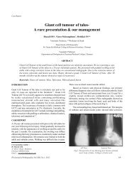
Case ReportA <strong>rare</strong> <strong>tumor</strong> <strong>of</strong> <strong>interosseous</strong> <strong>membrane</strong> <strong>forearm</strong>Kuljit Kumar *, P Prasad **, Ankit Varshney****Assistant Pr<strong>of</strong>essor, **Pr<strong>of</strong>essor, ***Junior ResidentDepartment <strong>of</strong> OrthopaedicsHIMS, DehradunABSTRACT25 year old female presented with swelling on dorsal aspect <strong>of</strong> proximal third <strong>of</strong> <strong>forearm</strong> with difficulty insupination and pronation. Swelling was excised and sent for histopathological examination. Biopsy reportshowed as benign spindle cell <strong>tumor</strong> favouring fibromatosis. Extra adbominal fibromatosis from <strong>interosseous</strong><strong>membrane</strong> <strong>of</strong> <strong>forearm</strong> is <strong>rare</strong> presentation. Although it is <strong>rare</strong> entity differential diagnosis should be kept inmind if there is swelling in proximal third <strong>of</strong> <strong>forearm</strong> presented with restriction <strong>of</strong> supination and pronation.Because these <strong>tumor</strong>s are locally aggressive, early diagnosis and treatment are crucial to minimize morbidity.Keywords: Interosseous <strong>membrane</strong> fibromatosis, Extra abdominal fibromatosisINTRODUCTIONFibromatosis, as proposed by Stout, 13 comprise a broad group<strong>of</strong> benign fibrous tissue proliferations <strong>of</strong> similar microscopicappearance whose biologic behavior is intermediate betweenthat <strong>of</strong> benign fibrous lesions and fibrosarcoma. Likefibrosarcoma, the fibromatoses are characterized by infiltrativegrowth and a tendency toward recurrence, but unlike this <strong>tumor</strong>they never metastasize.The fibromatosis can be divided into two major groupssuperficialand deep fibromatosis. Superficial fibromatosisarise from fascia or aponeurosis. Deep fibromatosis as namesuggests involve deep structure, particularly the musculature<strong>of</strong> the trunk and the extremities. Deep fibromatosis isfurther divided into extraabdominal and intra abdominalfibromatosis. The descriptive term desmoid <strong>tumor</strong>, coined byMueller in 1838, is still widely used in the literature as asynonym for fibromatosis. It is estimated that 3-4 cases <strong>of</strong> extraabdominal fibromatosis per million occur in United Statesannually 2 . Although extra abdominal fibromatosis from<strong>interosseous</strong> <strong>membrane</strong> is <strong>rare</strong> entity, differential diagnosisshould be kept in mind if there is swelling in proximal third <strong>of</strong><strong>forearm</strong> presented with restriction <strong>of</strong> supination andpronation.CASE REPORTA 25 year old female presented with a swelling on the back <strong>of</strong>her right <strong>forearm</strong> since 3 ½ years. Swelling was small at onsetand gradually increased in size. It was associated with nonradiating,mild pricking type <strong>of</strong> on and <strong>of</strong>f pain. There was nohistory <strong>of</strong> significant trauma, weight loss, fever and loss <strong>of</strong>appetite.On physical examination there was a single 8 X 4 cm firm,non-tender swelling over the dorsal aspect <strong>of</strong> the right <strong>forearm</strong>5 cm below the elbow joint. The margins were not welldemarcated and the swelling was attached to the underlyings<strong>of</strong>t tissue. There were no signs <strong>of</strong> inflammation. The distalneurovascular status was normal. Range <strong>of</strong> movement were-Supination (0-10) 0 , Pronation (0-20) 0 . Range <strong>of</strong> movements <strong>of</strong>elbow and wrist were normal. Radiographic evaluation revealedCorresponding Author :Dr. Kuljit KumarDepartment <strong>of</strong> Orthopaedics,HIMS, DehradunEmail: koolguy0062008@yahoo.comFig1. AP X-ray <strong>of</strong> <strong>forearm</strong>Pb Journal <strong>of</strong> Orthopaedics Vol-XIII, No.1, 201279
A <strong>rare</strong> <strong>tumor</strong> <strong>of</strong> <strong>interosseous</strong> <strong>membrane</strong> <strong>forearm</strong>Fig 5. Clinical photo showing <strong>tumor</strong> massFig 6. Clinical photo showing <strong>tumor</strong> massFig 7. Clinical photo showing <strong>tumor</strong> massFig 8. Tumor mass after excisionFig 9. Cross-section after excision <strong>of</strong> <strong>tumor</strong> massarise from <strong>interosseous</strong> <strong>membrane</strong> <strong>of</strong> <strong>forearm</strong> and not from anymuscle. The recurrence rate recorded in literature for extraabdominal fibromatoses is ranging from as low as 19% 4 to asFig 10. Clinical photo showing <strong>forearm</strong> after <strong>tumor</strong> mass excisionhigh as 77%. 8 Numerous studies have found that the extent <strong>of</strong>initial excision is prognostically significant. 5,7,11 Because themicroscopic picture does not reliably reflect the growth potential<strong>of</strong> the <strong>tumor</strong>, therapy is predicated on its extent and anatomicalPb Journal <strong>of</strong> Orthopaedics Vol-XIII, No.1, 201281