Studio delle prestazioni di un sistema a fosfori per mammografia ...
Studio delle prestazioni di un sistema a fosfori per mammografia ...
Studio delle prestazioni di un sistema a fosfori per mammografia ...

Create successful ePaper yourself
Turn your PDF publications into a flip-book with our unique Google optimized e-Paper software.
photons will reach the detector and the ra<strong>di</strong>ation dose to the tissue will be very high in order<br />
to obtain a signal to noise ratio similar to the one obtained with a more energetic beam. The<br />
choice of energy will therefore be a compromise between the requirements of low dose and high<br />
contrast [3].<br />
At the energy values typical of ra<strong>di</strong>ography, the most important photons interactions are the<br />
photoelectric effect and scattering. In soft tissue the photoelectric cross section is larger than<br />
the scatter cross section for energies up to about 25 keV. The photoelectric cross section varies<br />
approximately as the fourth power of the atomic number and inversely as the third power of<br />
the photon energy and it shows <strong>di</strong>scontinuities at the absorption edges. The photon scattering<br />
cross section varies more slowly with energy than does the photoelectric cross section and is<br />
approximately proportional to atomic number [4].<br />
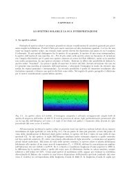
The voltage applied to a X-ray tube de<strong>di</strong>cated to mammography is usually between 20 and<br />
30 kVp (Vmax), depen<strong>di</strong>ng on the clinical <strong>di</strong>agnosis and patient morphology. The X-ray ra-<br />
<strong>di</strong>ation is generated by Bremmstrahl<strong>un</strong>g and by an X-ray emission characteristic of the anode<br />
material. The anode material used in mammography X-ray <strong>un</strong>its is molybdenum, since, with<br />
respect to t<strong>un</strong>gsten targets, it produces lower energy X-rays, which are more appropriate for<br />
imaging thinner body sections at high contrast. Usually the spectrum is attenuated by an added<br />
molybdenum filter (see Chapter 3.1 for the system <strong>un</strong>der investigation) which reduces both the<br />
X-ray component above its K-edge of about 20 keV and the lower energy components. This<br />
is important since it reduces the soft component of the spectrum, which contributes only to the<br />
patient dose and not to image contrast.<br />
1.2 The imaging plate IP<br />
The Imaging Plate is the detecting element of a CR system. It is a multilayer film composed<br />
of a support layer and a sensible layer made of photo-stimulable phosphors. Photo-stimulable<br />
phosphors, also known as storage phosphor, is one of the most successful detectors for <strong>di</strong>gital<br />
ra<strong>di</strong>ography to date.<br />
The photo-stimulable phosphor (PSP) stores the absorbed X-ray energy in crystal structure<br />
traps. The trapped energy can be released in a later moment if stimulated by ad<strong>di</strong>tional light<br />
7