Studio delle prestazioni di un sistema a fosfori per mammografia ...
Studio delle prestazioni di un sistema a fosfori per mammografia ...
Studio delle prestazioni di un sistema a fosfori per mammografia ...

Create successful ePaper yourself
Turn your PDF publications into a flip-book with our unique Google optimized e-Paper software.
ea<strong>di</strong>ng system and of the IP <strong>un</strong>der investigation are describe more in detail in Chapter 3.<br />
1.3 The <strong>di</strong>gitization process<br />
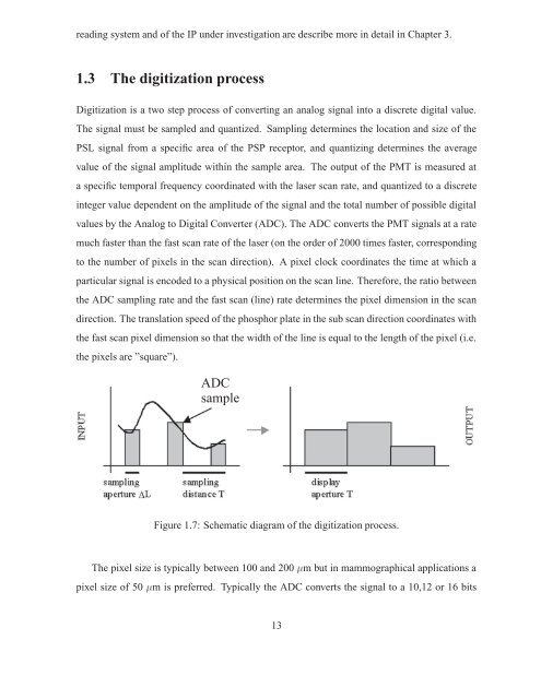
Digitization is a two step process of converting an analog signal into a <strong>di</strong>screte <strong>di</strong>gital value.<br />
The signal must be sampled and quantized. Sampling determines the location and size of the<br />
PSL signal from a specific area of the PSP receptor, and quantizing determines the average<br />
value of the signal amplitude within the sample area. The output of the PMT is measured at<br />
a specific temporal frequency coor<strong>di</strong>nated with the laser scan rate, and quantized to a <strong>di</strong>screte<br />
integer value dependent on the amplitude of the signal and the total number of possible <strong>di</strong>gital<br />
values by the Analog to Digital Converter (ADC). The ADC converts the PMT signals at a rate<br />
much faster than the fast scan rate of the laser (on the order of 2000 times faster, correspon<strong>di</strong>ng<br />
to the number of pixels in the scan <strong>di</strong>rection). A pixel clock coor<strong>di</strong>nates the time at which a<br />
particular signal is encoded to a physical position on the scan line. Therefore, the ratio between<br />
the ADC sampling rate and the fast scan (line) rate determines the pixel <strong>di</strong>mension in the scan<br />
<strong>di</strong>rection. The translation speed of the phosphor plate in the sub scan <strong>di</strong>rection coor<strong>di</strong>nates with<br />
the fast scan pixel <strong>di</strong>mension so that the width of the line is equal to the length of the pixel (i.e.<br />
the pixels are ”square”).<br />
ADC<br />
sample<br />
Figure 1.7: Schematic <strong>di</strong>agram of the <strong>di</strong>gitization process.<br />
The pixel size is typically between 100 and 200 �m but in mammographical applications a<br />
pixel size of 50 �m is preferred. Typically the ADC converts the signal to a 10,12 or 16 bits<br />
13