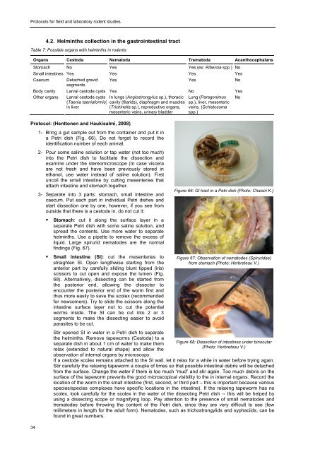
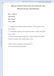
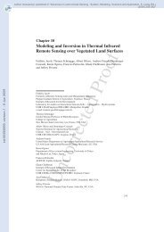
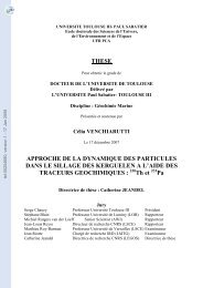
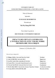
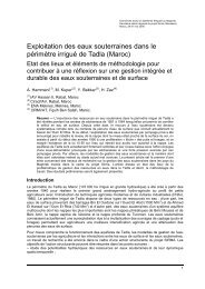
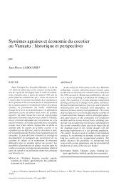
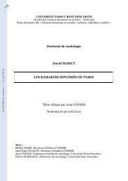
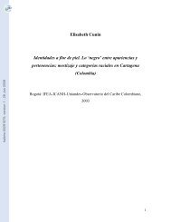
<strong>Protocols</strong> <strong>for</strong> <strong>field</strong> <strong>and</strong> <strong>laboratory</strong> <strong>rodent</strong> <strong>studies</strong>344.2. Helminths collection in the gastrointestinal tractTable 7: Possible organs with helminths in <strong>rodent</strong>sOrgans Cestoda Nematoda Trematoda AcanthocephalansStomach No Yes Yes (ex: Riberoia spp.) NoSmall intestines Yes Yes Yes YesCaecum Detached gravidsegmentsYes Yes NoBody cavity Larval cestode cysts Yes No YesOther organs Larval cestode cysts(Taenia taenia<strong>for</strong>mis)in liverNoProtocol: (Henttonen <strong>and</strong> Haukisalmi, 2008)In lungs (Angiostrongylus sp.), thoraciccavity (filarids), diaphragm <strong>and</strong> muscles(Trichinella sp.), reproductive organs,mesenteric veins, urinary bladder1- Bring a gut sample out from the container <strong>and</strong> put it ina Petri dish (Fig. 66). Do not <strong>for</strong>get to record theidentification number of each animal.2- Pour some saline solution or tap water (not too much)into the Petri dish to facilitate the dissection <strong>and</strong>examine under the stereomicroscope (In case visceraare not fresh <strong>and</strong> have been previously stored inethanol, use water instead of saline solution). Firstuncoil the small intestine by cutting mesenteries thatattach intestine <strong>and</strong> stomach together.3- Separate into 3 parts: stomach, small intestine <strong>and</strong>caecum. Put each part in individual Petri dishes <strong>and</strong>start dissection one by one, however, if you see fromoutside that there is a cestode in, do not cut it:• Stomach: cut it along the surface layer in aseparate Petri dish with some saline solution, <strong>and</strong>spread the contents. Use more water to separatehelminths. Use a pipette to remove the excess ofliquid. Large spirurid nematodes are the normalfindings (Fig. 67).• Small intestine (SI): cut the mesenteries tostraighten SI. Open lengthwise starting from theanterior part by carefully sliding blunt tipped (iris)scissors to cut open <strong>and</strong> expose the lumen (Fig.68). Alternatively, dissecting can be started fromthe posterior end, allowing the dissector toencounter the posterior end of the worm first <strong>and</strong>thus more easily to save the scolex (recommended<strong>for</strong> newcomers). Try to slide the scissors along theintestine surface layer not to cut the potentialworms inside. The SI can be cut into 2 or 3segments to make the dissecting easier to avoidparasites to be cut.Stir opened SI in water in a Petri dish to separatethe helminths. Remove tapeworms (Cestoda) to aseparate dish in about 1 cm of water to make themrelax (extended to natural shape) <strong>and</strong> allow theobservation of internal organs by microscopy.Lung (Paragonimussp.), liver, mesentericveins, (Schistosomaspp.)Figure 66: GI tract in a Petri dish (Photo: Chaisiri K.)Figure 67: Observation of nematodes (Spiruridae)from stomach (Photo: Herbreteau V.)Figure 68: Dissection of intestines under binocular(Photo: Herbreteau V.)If a cestode scolex remains attached to the SI wall, let it relax <strong>for</strong> a while in water be<strong>for</strong>e trying again.Stir carefully the relaxing tapeworm a couple of times so that possible intestinal debris will be detachedfrom the surface. Change the water if there is too much “mud” <strong>and</strong> stir again. Too much debris on thesurface of the tapeworm prevents the good microscopical visibility to the in internal organs. Record thelocation of the worm in the small intestine (first, second, or third part – this is important because variousspecies/species complexes have specific locations in the intestine). If the relaxing tapeworm has noscolex, look carefully <strong>for</strong> the scolex in the water of the dissecting Petri dish -- this will be helped byusing a dissecting scope or magnifying loop. Pay attention to the presence of small nematodes <strong>and</strong>trematodes be<strong>for</strong>e throwing the content of the Petri dish, since they are very difficult to see (fewmillimeters in length <strong>for</strong> the adult <strong>for</strong>m). Nematodes, such as trichostrongylids <strong>and</strong> syphaciids, can befound in great numbers.
4- Isolating helminths in <strong>rodent</strong>s• Caecum: nematodes are the normal findings in the caecum. Use of a stereomicroscope ormagnifying glass is recommended since some nematodes are relatively small <strong>and</strong> hard to observe.The genus Trichuris shows the thin oesophagus strongly attached to the mucosa of the caecum.Gently use tweezers to detach oesophagus from mucosa. Trematodes might be covered with“greenish mud” <strong>and</strong> are difficult to separate from plant materials. It may be useful <strong>for</strong> a newcomer torun material through a small sieve to first discard some fine particulates.4- Collect helminths:• Cestodes should be held <strong>and</strong> washed (stirred) in water at least 2 hours, depending on their size,until totally relaxed, i.e. flat <strong>and</strong> straightened. This is necessary to enhance their observation,especially to examine the internal structure of the proglottids (segments). Following relaxation <strong>and</strong>death in water, all tapeworms will be preserved in 70% ethanol. Fixation should be done flat withoutpressure, <strong>and</strong> let the cestode stay (preferably) overnight on covered Petri dish. Use fresh ethanolwhen preserving cestodes in vials. Ethanol should be changed once after 24 hours, especially <strong>for</strong>the large-sized species. It is recommended that ethanol-preserved cestode materials used later <strong>for</strong>genetic <strong>studies</strong> will primarily be stored in refrigerator.• Trematodes can be relaxed <strong>and</strong> washed in water, which often allows specimens to expel eggs thatmight otherwise obscure some organs. Preserve flukes in 70% ethanol.• Nematodes should be held in saline as osmotic pressure will cause the specimen to burst if kept innormal water. Specimens should be washed in saline <strong>and</strong> then soon preserved in 70% ethanol.Keep parasites from different organ systems separated.5- Separate parasites by group, based on morphology, <strong>for</strong> each individual animal, to help furtheridentification. Count helminths under the stereomicroscope. Label (by pencil) the details of collection ondrawing paper <strong>and</strong> put it into the tube (at least: identifying number of parasite <strong>and</strong> host, group or genus ofparasite, date, location in the GI tract). The absence of helminths should also be recorded <strong>for</strong> furtheranalyses.6- Keep vials in a cool place <strong>and</strong> clean instruments between the individuals to avoid contamination ofsamples.4.3. Method <strong>for</strong> karyological analysis of helminthsFirst of all, a subsample of the helminth to study must be preserved <strong>for</strong> morphological study (see above), as thismethod will destroy the sample.Protocol:1- Treat living worms with 0,025 % colchicine in normal saline <strong>for</strong> 30-60 min at 37°C in order to increase thenumber of observed metaphases.2- Subject the whole worms to hypotonic treatment (0.6 sodium citrate or 75 mM KCl, 20-60 min at 37°C).3- Dissect testes or ovary (or small tissue pieces containing the organs), put them into the hypotonic solution<strong>for</strong> 15 min.4- Put the tissue pieces into the fixative (cold, freshly mixed Carnoy: methanol: acetic acid 3:1 - if methanol isproblem, you can provisionally use ethanol) <strong>for</strong> 30-60 min., then change the fixative one or two times.5- Put the samples within the new fixative to refrigerator <strong>and</strong> let them frozen till next use <strong>for</strong> chromosomepreparations.6- Then, dissociate one small tissue piece in a drop of 60% acetic acid on the slide, put the slide onto aheating plate at 45°C <strong>and</strong> spread the drop of acetic acid with the cells slowly on the slide. Let the slide dry<strong>and</strong> use it <strong>for</strong> staining (Giemsa, C- b<strong>and</strong>ing, fluorescent dyes etc.). Alternatively, slides can be dehydratedin an ethanol series (70%, 80%, <strong>and</strong> 100%, 1 min each). Store slides at -20ºC until use.4.4. Helminths fixation <strong>for</strong> scanning electron microscopyEven if using hot ethanol is correct, a higher quality of images can be obtained by following this protocol:Protocol:1- Different reagents must be mixed the day of use <strong>for</strong> an optimal conservation:1 mL of gluteraldeide + 4 mL of distilled water, then add 5 mL of cacodilate.This fixative must be refrigerated be<strong>for</strong>e <strong>and</strong> after use.2- Once helminths are as clean as possible (in physiologic solution), place them in a microtube with thefixative. Fill microtubes completely with fixative to ensure that specimens remain immersed duringtransportation. Very small individuals can be stored together in the same microtube to be mounted (this willallow a better chance to get them mounted in a perfect position in the support).3- Samples have to be kept in a fridge until complete dehydration process is achieved.35