download PDF version - Dr Harold Hillman
download PDF version - Dr Harold Hillman
download PDF version - Dr Harold Hillman
- No tags were found...

Create successful ePaper yourself
Turn your PDF publications into a flip-book with our unique Google optimized e-Paper software.
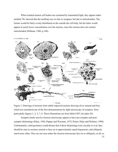
When isolated neuron cell bodies are examined by transmitted light, they appear rathermottled. We showed that the mottling was not due to synapses, but due to mitochondria. Theformer would be fairly evenly distributed on the outside the cell body, but the latter wouldappear in much lower concentration over the nucleus, since the nucleus does not containmitochondria (<strong>Hillman</strong>, 1986, p.148).Figure 3. <strong>Dr</strong>awings of neurons from rabbit trapezoid nucleus showing silver stained end-feet,which are considered one of the first demonstrations by light microscopy of synapses. Noteparticularly figures 1, 2, 3, 7, 8. These illustrations are from Held (1897, his plate XI)Synaptic knobs seen by electron microscopy appear to have pre-synaptic and postsynapticthickenings (Palay, 1956; Pappas and Waxman, 1972; Peters, Palay and Webster, 1998).Unfortunately, solid geometry would dictate that if these thickenings were circular or oval, theyshould be seen in sections normal or face-on in approximately equal frequencies, and obliquelymuch more often. They are not seen under the electron microscope face-on or obliquely, at all, so17