download PDF version - Dr Harold Hillman
download PDF version - Dr Harold Hillman
download PDF version - Dr Harold Hillman
- No tags were found...
Create successful ePaper yourself
Turn your PDF publications into a flip-book with our unique Google optimized e-Paper software.
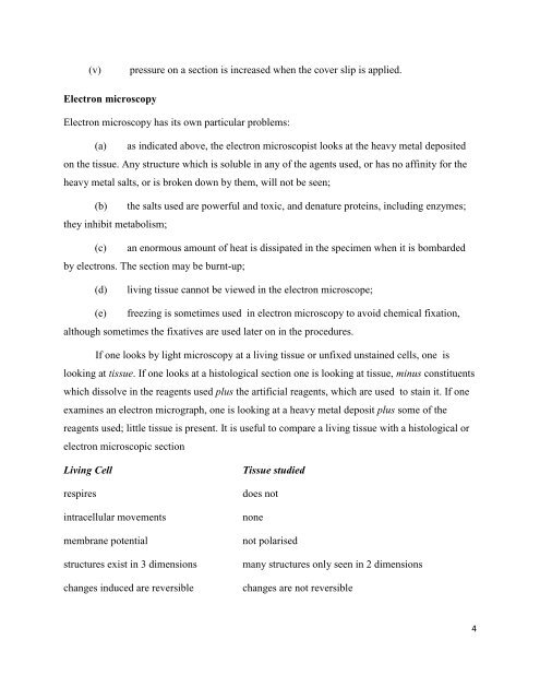
(v)pressure on a section is increased when the cover slip is applied.Electron microscopyElectron microscopy has its own particular problems:(a) as indicated above, the electron microscopist looks at the heavy metal depositedon the tissue. Any structure which is soluble in any of the agents used, or has no affinity for theheavy metal salts, or is broken down by them, will not be seen;(b) the salts used are powerful and toxic, and denature proteins, including enzymes;they inhibit metabolism;(c) an enormous amount of heat is dissipated in the specimen when it is bombardedby electrons. The section may be burnt-up;(d)living tissue cannot be viewed in the electron microscope;(e) freezing is sometimes used in electron microscopy to avoid chemical fixation,although sometimes the fixatives are used later on in the procedures.If one looks by light microscopy at a living tissue or unfixed unstained cells, one islooking at tissue. If one looks at a histological section one is looking at tissue, minus constituentswhich dissolve in the reagents used plus the artificial reagents, which are used to stain it. If oneexamines an electron micrograph, one is looking at a heavy metal deposit plus some of thereagents used; little tissue is present. It is useful to compare a living tissue with a histological orelectron microscopic sectionLiving Cellrespiresintracellular movementsmembrane potentialstructures exist in 3 dimensionschanges induced are reversibleTissue studieddoes notnonenot polarisedmany structures only seen in 2 dimensionschanges are not reversible4


