CMA-Microdialysis.pdf - somapharm.ch
CMA-Microdialysis.pdf - somapharm.ch
CMA-Microdialysis.pdf - somapharm.ch
- No tags were found...

Create successful ePaper yourself
Turn your PDF publications into a flip-book with our unique Google optimized e-Paper software.
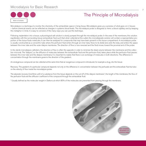
<strong>Microdialysis</strong> for Basic Resear<strong>ch</strong>The Principle of <strong>Microdialysis</strong>7<strong>Microdialysis</strong> is a te<strong>ch</strong>nique to monitor the <strong>ch</strong>emistry of the extracellular space in living tissue. <strong>Microdialysis</strong> gives you a preview of what goes on in tissues– before <strong>ch</strong>emical events can be reflected as <strong>ch</strong>anges in systemic blood levels. The microdialysis probe is designed to mimic a blood capillary and by keepingthis metaphor in mind, it is easy to conceive of the many ways you can use this te<strong>ch</strong>nique.Following implantation into a tissue, a physiological salt solution is slowly pumped through the microdialysis probe. In the area of the membrane, this solutionequilibrates with the surrounding tissue extracellular fluid su<strong>ch</strong> that when collected at the outlet, this microdialysate solution will contain a representative proportionof the tissue fluids molecules. It can then be analyzed for compounds that may have been present in this tissue compartment. A microdialysis probeis usually constructed as a concentric tube where the perfusion fluid enters through an inner tube, flows to its distal end, exits the tube, and enters the spacebetween the inner tube and the outer dialysis membrane. The direction of flow is now reversed and the fluid moves toward the proximal end of the probe.In the sterile microdialysis catheters, the direction of flow is often the opposite in order to minimize the dead volume between the membrane and the collectionmicrovial. The “dialysis”, i.e. the diffusion of molecules between the extracellular fluid and the perfusion fluid, takes place while the perfusion fluid passesbetween the inner tube and the dialysis membrane. It is important to realize that there is an ex<strong>ch</strong>ange of molecules in both directions. The difference inconcentration through the membrane governs the direction of the gradient.An endogenous compound can be collected at the same time that an exogenous compound is introduced, for example a drug, into the tissue.Recovery: The gradient of a particular compound depends not only on the difference in concentration between the perfusate and the extracellular fluid but alsoon the velocity of flow inside the microdialysis probe.The absolute recovery (mol/time unit) of a substance from the tissue depends on the cut-off of the dialysis membrane*, the length of the membrane, the flow ofthe perfusion fluid and the diffusion coefficient of the compound through the extracellular fluid.* Usually defined as the molecular weight in Daltons at whi<strong>ch</strong> 80% of the molecules are prevented from passing through the membrane.