CASE OF THE MONTHAhmed Ismail Manjra, MB ChB, FC Paed (SA), DipAllergology (SA), M Clin Pharm, FAAAAIWestville, Durban, South AfricaC Motala, MB ChB, DCH (SA), FC Paed (SA), FACAAI,FAAAAI<strong>Allergy</strong> Clinic, Red Cross Children’s Hospital, CapeTown, South AfricaMASTOCYTOMA IN A 3-MONTH-OLDINFANTMastocytosis is a heterogeneous group of diseasescharacterised by the abnormal infiltration of mast cells(MC) in the skin <strong>and</strong>, sometimes, other organs.Symptoms in mastocytosis are caused by biologicalmediators released from MC <strong>and</strong>/or the infiltration ofneoplastic MC in various organs, the skin <strong>and</strong> the bonemarrow being predominantly involved. A WHO consensusclassification for mastocytosis 1 exists, which iswidely accepted <strong>and</strong> includes three major categories:1. Cutaneous mastocytosis (CM), a benign disease inwhich MC infiltration is confined to the skin, is preferentiallyseen in young children <strong>and</strong> exhibits amarked tendency to regress spontaneously.2. Systemic mastocytosis (SM), a rather rare disorder,it is commonly diagnosed in adults <strong>and</strong> includesfour major subtypes:(i) indolent SM (ISM, the most common forminvolving mainly skin <strong>and</strong> bone marrow);(ii) a unique subcategory termed SM with an associatednon-mast-cell clonal haematological disease(SM-AHNMD);(iii) aggressive SM, usually presenting without skinlesions, <strong>and</strong>(iv) MC leukaemia (MCL), probably representingthe rarest variant of human leukaemias.The diagnosis of SM, SM-AHNMD, <strong>and</strong> MCL might beconfused with a variety of endocrinological, vascular, orimmunological disorders. It is therefore of particularimportance to be aware of the possibility of an underlying(malignant) MC disease in patients with unexplainedvascular instability, unexplained (anaphylactoid)shock, idiopathic flushing, diarrhoea, headache, <strong>and</strong>other symptoms that might be mediator related. Animportant diagnostic clue in such cases is an increasedserum tryptase level.Systemic manifestations dependent on whether thereis multiorgan/specific organ involvement, e.g. recurrentflushing with multiorgan involvement; gastrointestinaltract involvement: increase in gastric <strong>and</strong> colonicmucosal mast cells, gastroduodenitis, bonemarrow:anaemia, thrombocytopenia, hepatosplenomegaly. 23. The extremely rare localised extracutaneous MCneoplasms, either presenting as malignancy (MCsarcoma) or as benign tumour termed extracutaneousmastocytoma.Diagnostic criteria for mastocytosis 1 are available <strong>and</strong>are widely accepted. SM criteria include one major criterion(multifocal compact tissue infiltration by MC) <strong>and</strong>four minor criteria:(i) prominent spindling of MC;(ii) atypical immunophenotype of MC with coexpressionof CD2 <strong>and</strong>/or CD25 (antigens whichhave not been found to be expressed on normal/reactiveMC);(iii) activating (somatic) point mutations of the c-kitproto-oncogene usually involving exon 17, withthe imatinib-resistant type D816V being mostfrequent, <strong>and</strong>(iv) persistently elevated serum tryptase level(>20 ng/ml).To establish the diagnosis of SM, at least one major<strong>and</strong> one minor criterion, or at least three minor criteria,have to be fulfilled.Case reportA 3-month-old child presented with a solitary, progressivelyenlarging lesion on the abdomen. There were noassociated systemic manifestations. There was nofamily history of mastocytosis. On examination thechild was well-looking <strong>and</strong> had a brownish-red lesion, 5cm in diameter on the abdominal wall. It was firm,shiny, nodular, non-tender with a typical peau d’ orangeappearance. (Fig 1). Stroking of the lesion with a tongueblade elicited a typical urticarial lesion (Darier’s sign).Abdominal examination was otherwise normal. Therewere no other cutaneous lesions. A punch biopsy wasperformed after application of an emla patch. Histologyrevealed dense infiltration of mast cells, typical featuresof a mastocytoma (Figs 2-4). The child was notgiven any treatment. The follow-up visit a month laterThis is a new feature aimed at highlighting instructivecase studies that clinicians encounter. If youhave an interesting case that you would like to sharewith our readiers, please submit your case report tothe Section Editor, Prof Cas Motala, e-mailcassim.motala@uct.ac.zaCorrespondence: Dr AI Manjra, PO Box 1085, Westville 3630, tel 031-265-1024, fax 031-265-1029, e-mail manjra@mweb.co.zaFig. 1. Mastocytoma on abdomen<strong>Current</strong> <strong>Allergy</strong> & <strong>Clinical</strong> <strong>Immunology</strong>, <strong>March</strong> <strong>2008</strong> Vol 21, No. 1 33
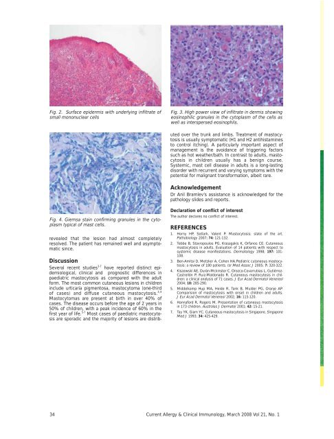
Fig. 2. Surface epidermis with underlying infiltrate ofsmall mononuclear cellsFig. 3. High power view of infiltrate in dermis showingeosinophilic granules in the cytoplasm of the cells aswell as interspersed eosinophils.DiscussionSeveral recent studies 3-7 have reported distinct epidemiological,clinical <strong>and</strong> prognostic differences inpaediatric mastocytosis as compared with the adultform. The most common cutaneous lesions in childreninclude urticaria pigmentosa, mastocytoma (one-thirdof cases) <strong>and</strong> diffuse cutaneous mastocytosis. 3,4Mastocytomas are present at birth in over 40% ofcases. The disease occurs before the age of 2 years in50% of children, with a peak incidence of 60% in thefirst year of life. 5-7 Most cases of paediatric mastocytosisare sporadic <strong>and</strong> the majority of lesions are distributedover the trunk <strong>and</strong> limbs. Treatment of mastocytosisis usually symptomatic (H1 <strong>and</strong> H2 antihistaminesto control itching). A particularly important aspect ofmanagement is the avoidance of triggering factorssuch as hot weather/bath. In contrast to adults, mastocytosisin children usually has a benign course.Systemic, mast cell disease in adults is a long-lastingdisorder with recurrent <strong>and</strong> varying symptoms with thepotential for malignant transformation, albeit rare.AcknowledgementDr Anil Bramlev’s assistance is acknowledged for thepathology slides <strong>and</strong> reports.Fig. 4. Giemsa stain confirming granules in the cytoplasmtypical of mast cells.revealed that the lesion had almost completelyresolved. The patient has remained well <strong>and</strong> asymptomaticsince.Declaration of conflict of interestThe author declares no conflict of interest.REFERENCES1. Horny HP, Sotlark, Valent P. Mastocytosis: state of the art.Pathobiology 2007; 74: 121-132.2. Tebbe B, Stavropoulos PG, Krasagakis K, Orfanos CE. Cutaneousmastocytosis in adults. Evaluation of 14 patients with respect tosystemic disease manifestations. Dermatology 1998; 197: 101-108.3. Ben-Amitai D, Metzker A, Cohen HA.Pediatric cutaneous mastocytosis:a review of 180 patients. Isr Med Assoc J 2005; 7: 320-322.4. Kiszewski AE, Durán-Mckinster C, Orozco-Covarrubias L, Gutiérrez-Castrellón P, Ruiz-Maldonado R. Cutaneous mastocytosis in children:a clinical analysis of 71 cases. J Eur Acad Dermatol Venereol2004; 18: 285-290.5. Middelkamp Hup MA, Heide R, Tank B, Mulder PG, Oranje AP.Comparision of mastocytosis with onset in children <strong>and</strong> adults.J Eur Acad Dermatol Venereol 2002; 16: 115-120.6. Hannaford R, Rogers M. Presentation of cutaneous mastocytosisin 173 children. Australas J Dermatol 2001; 42: 15-21.7. Tay YK, Giam YC. Cutaneous mastocytosis in Singapore. SingaporeMed J 1993; 34: 425-429.34 <strong>Current</strong> <strong>Allergy</strong> & <strong>Clinical</strong> <strong>Immunology</strong>, <strong>March</strong> <strong>2008</strong> Vol 21, No. 1