Methods: <strong>27</strong>2 T cell CDC and FLXM and 269 B cell CDC and FLXM’s were reviewed and the resultscompared to Mean Fluorescent Intensity (MFI) of LSAB Donor Specific Antibody (DSA).Results: DSA MFI’s were grouped into seven levels and are presented in the table below:[table1]Conclusions: From this data there was a very low frequency of positive crossmatches between 0 and 5<strong>00</strong>MFI. At an MFI of 1<strong>00</strong>0-2<strong>00</strong>0, T cell FLXM and B cell FLXM were positive but CDC was still negative.FLXM did not achieve a 90% rate until the 5<strong>00</strong>0-1<strong>00</strong><strong>00</strong> MFI group. It is interesting that even at >15<strong>00</strong>0MFI’s T and B cell CDC crossmatches were positive 62.5% and 77% respectively and B FLXM werepositive 92.3%. It is evident that LSAB can be used to predict FLXM and CDC within specific MFI rangesbut not with 1<strong>00</strong>% accuracy. Comparing LSAB MFI’s to FLXM and CDC can aid in establishing effectivelimits <strong>for</strong> Unacceptable Antigens. LSAB MFI is useful as a guide <strong>for</strong> estimating the risk <strong>for</strong> a positiveFLXM and CDC.45-PDISCREPANT LUMINEX SINGLE ANTIGEN BEAD AND FLOW PRA RESULT IN RECIPIENTAWAITING HEART TRANSPLANT.Cecil L. Rhodes, Ijeoma Okere, Bryna L. Cook, Vijaya Hegde, Alpa Patel, Monica Ramirez, Prakash Rao.NJ Sharing Network Transplant Lab, New Jersey Organ and Tissue Sharing Network, New Providence, NJ,USA.Aim: To address unexpected results between Luminex Single Antigen Beads (LSAB) and Flow PRABeads (FLPRA).Methods: Patient serum with unexpected results between Luminex Single Antigen Beads (LSAB) andFLOW PRA (FLPRA) was tested by Flow Cytometer Crossmatch (FLXM) to confirm result.Results: Potential Heart Recipient MP was negative <strong>for</strong> both Class I and Class II antibodies by FLPRA butpositive <strong>for</strong> Class II LSAB. Subsequent testing by LSAB revealed many positive Class II beads with MFI’sranging from 2021 to 9041. Flow Single Antigen Class II Beads confirmed the LSAB result. Most LSABreactions appear as allele specificities with a lack of groups of positive alleles. Reactions that present asallele specificities are not confirmed by additional beads bearing the same antigen,i.e., theDQA1*0201/DQB1*0401 bead was strongly positive with an MFI of 9041 however the DQA1*0201 andDQB1*0401 are found independently on other beads and were both negative (0 MFI). Two flowcrossmatches (FLXM) were per<strong>for</strong>med with donors matched <strong>for</strong> a positive allele specific DSA. One donorpositive <strong>for</strong> DRB1*0102 had a corresponding DSA of 4609 MFI. A second donor positive <strong>for</strong> DRB3*0202had a corresponding DSA of 6969 MFI. Expected crossmatch MCS <strong>for</strong> the DRB1*0102 and theDRB3*0202 were 1<strong>00</strong> and 150 MCS respectively, however, B cell FLXM’s were negative in both caseswith a MCS of 0.Conclusions: Reactions between DQA and DQB beads indicate that this antibody is not directed to HLAepitopes or depends on con<strong>for</strong>mational characteristics of the DQ alpha and beta chains. Lack of reactivityby FLXM with normal lymphocytes may be due to cryptic epitopes that are only assessable in denaturedproteins on the beads.Important questions are raised by this data. 1. Are these antibodies relevant to graftsurvival. 2. What other method can confirm Flow Negative Luminex Positive specificities.46-PDETECTION OF DONOR SPECIFIC ANTIBODIES WITH DynaChip TECHNOLOGY: A PILOTSTUDY.Dave L. Roelen, Simone Brand-Schaaf, Marissa van der Linden, Marian Witvliet, Ilias I.N. Doxiadis, FransH.J. Claas. Dept. of Immunohematology and Blood Transfusion, Leiden University Medical Center, Leiden,Netherlands.Aim: In the recent years many new technologies <strong>for</strong> HLA specific antibody screening have beendeveloped, among which the DynaChip assay (Invitrogen, UK). This Solid Phase Assay (SPA) makes useof a chip coated with purified HLA antigens of single donors. We have evaluated whether this assay ismore sensitive compared to the complement dependent cytotoxicity assay (CDC) and whether the definedantibodies are clinically relevant.Methods: In total 142 immunized kidney patients, transplanted between 1995 and 2<strong>00</strong>4, with a negativeCDC cross-match (with current and historical sera) were tested. Sera be<strong>for</strong>e transplantation were selected
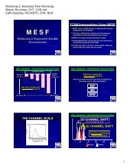
and tested in the DynaChip assay <strong>for</strong> Donor-Specific-HLA-Antibodies (DSA), which could not be detectedin the CDC.Results: In 51 patients the actual presence of DSA could not be determined because of their high PRA.These patients were excluded from the study. In 8 of the 91 in<strong>for</strong>mative patients DSA against HLA werefound. One patient lost the graft within 2 weeks with an anti HLA-A2. Three patients lost their graft within3 to 4 years, (two anti HLA-A9, one anti HLA-DR4). One patient, which died after 2 1/2 years with afunctioning graft, had DSA anti HLA-B18. In three cases DSA were found in patients with wellfunctioning organs (anti HLA-A2, HLA-DR11 and HLA-DR4).Conclusions: In conclusion, testing sera of sensitized patients on the kidney waiting list with the SPAbased DynaChip assay led to the identification of additional specificities not detected by CDC. The clinicalstudy confirms that the presence of DSA only detectable in the more sensitive SPA’s is rather a risk factorthan a contra-indication <strong>for</strong> transplantation. In some patients these antibodies may be associated withcomplications (one early rejection) whereas other patients have a functioning graft, one even more than 15years, in the presence of DSA. Future studies, including the titer and the antibody (sub)class of the DSA,should lead to a clearer identification of the patients at risk <strong>for</strong> early rejection.47-PTITER OF ANTIBODIES CAN BE ESTIMATED FROM MEDIAN CHANNEL SHIFTS OFPOSITIVE FLOW CROSSMATCHES.Jerry C. Rosenberg, Chak-Sum Ho, Mary Jackowski, Dorothy Levis, Christine Pieter, Kyle Putnam, A.Bradley Eisenbrey. Histocompatibilty Laboratory, Gift of Life Michigan, Ann Arbor, MI, USA.Aim: Transplant clinicians increasingly want to know the median channel shifts (MCS) when thecrossmatch is positive so as to gauge the strength of the antibody. Conventionally, the latter has beenassessed by the antibody’s titer. The following experiments were carried out to determine if the MCS canestimate the antibody’s titer and there<strong>for</strong>e the strength of the antibody or antibodies.Methods: Seven different serum samples that were highly positive on antibody testing were titered usingthe AHG cytotoxicity assay and then crossmatched to the same cell using flow cytometry. The resultingMCS and MESF were then correlated with the titer.Results:[figure1]A statistically significant correlation exists between the titer of an antibody and the MCSof a flow crossmatch. The correlation is best at the extremes of the titer, (when the titer is very high or verylow) and less reliable when the titer is between 1:4 and 1:8. The correlation with MESF was also significantat a p of 0.05. However, MESF is a linear function and MCS is a log function, resulting in a semi-log plotthat is not linear. Using the above <strong>for</strong>mula <strong>for</strong> the linear relationship between titer and MCS, andcalculating the titer from the MCS, the average deviation of from the true titer was 17%. The differencewas greatest at a titer of 1:2 which was overestimated as a titer of 1:6.5. At titers of 1:128 and 1:1024 thedifferences varied between 0% and -7%.Conclusions: The titer of a highly sensitized serum sample against a lymphocyte can be reasonablyestimated in a linear fashion from the MCS obtained by flow cytometry.48-PCAN WE PREVENT SENSITIZATION AFTER GRAFT LOSS?Juan Scornik, Herwig-Ulf Meyer-Kriesche. Pathology and Medicine, University of Florida College ofMedicine, Gainesville, FL, USA.Aim: Transplant (Tx) loss, a major cause of sensitization, is generally considered a natural consequence of<strong>for</strong>eign antigen (Ag) exposure, but few if any studies have looked systematically to determine if that is thecase.Methods: In the present work HLA antibodies (Abs) were measured by Luminex single Ag and other solidphase tests in patients, originally unsensitized, who lost a kidney Tx (Tx duration: 1 day to several years).Results: Overall, 73/104 (70%) patients became sensitized (90% donor specific). Abs be<strong>for</strong>e Tx loss werepresent in 16/85 patients (19%), that is, sensitization is potentially preventable in 81% of the patients.Patients already sensitized were excluded from subsequent analysis. Tx nephrectomy was per<strong>for</strong>med in 39patients and 31 (79%) became Ab positive, but 72% were also transfused. Conversely, 26/38 (68%)transfused patients made Abs, but 92% were also nephrectomized and most, in either group, were also offimmunosuppression (IS). To circumvent these confounding variables we identified 25 patients who had
- Page 1 and 2: Monday, September 27, 20102:00 PM -
- Page 3 and 4: 5-ORTWO CASES OF DONOR SPECIFIC ANT
- Page 5 and 6: Monday, September 27, 20102:00 PM -
- Page 7 and 8: Results: PHA-induced mRNA expressio
- Page 9 and 10: conjugated IgM (BD Biosciences), Ig
- Page 11 and 12: Methods: Peripheral blood and endom
- Page 13 and 14: Aim: NK cells have an established r
- Page 15 and 16: Methods: Sera (n=22) were tested fo
- Page 17 and 18: expression on lymphocytes, we hypot
- Page 19 and 20: Conclusions: Clean SA beads can eli
- Page 21 and 22: goal is to re-examine our previous
- Page 23 and 24: Aim: There is increasing evidence t
- Page 25 and 26: esult in poor graft survival. The g
- Page 27 and 28: Aim: Pronase treatment eliminates i
- Page 29 and 30: 34-PCLINICAL AND PATHOLOGICAL SIGNI
- Page 31 and 32: Nabil Mohsin, Isabelle G. Wood, Ind
- Page 33: donor/recipient pairs for transplan
- Page 37 and 38: average difference between Ave and
- Page 39 and 40: Conclusions: The XM-One assay can b
- Page 41 and 42: 59-PCYTOTOXIC AND NON-CYTOTOXIC ANT
- Page 43 and 44: data suggests that DTT pretreatment
- Page 45 and 46: Bhavna Lavingia 1 , Chantale Lacell
- Page 47 and 48: ATG INTERFERNCE IN SINGLE ANTIGEN L
- Page 49 and 50: 75-PPREFORMED LOW REACTIVE ANTI-HLA
- Page 51 and 52: Sabarinathan Ramachandran 1 , Naohi
- Page 53 and 54: 83-PALLELIC DIVERSITY OF KIR2DL4 AN
- Page 55 and 56: São Paulo, Brazil; 3 Commissariat
- Page 57 and 58: 91-PCHARACTERIZATION OF COMMON AND
- Page 59 and 60: Conclusions: The extensive diversit
- Page 61 and 62: 99-PPOTENTIAL COMMON NOVEL ALLELES
- Page 63 and 64: 103-PTOWARDS A FUNCTIONAL HLA-DPB1
- Page 65 and 66: 107-PWHOLE GENOME AMPLIFICATION (WG
- Page 67 and 68: 111-PLOSS OF MISMATCHED HLA IN FAMI
- Page 69 and 70: 115-PDONOR EPITHELIAL CELL CHIMERIS
- Page 71 and 72: Caucasian patients. MRs vary signif
- Page 73 and 74: Results: We obtained the total No.
- Page 75 and 76: cord blood and platelet transfusion
- Page 77 and 78: 131-PASSOCIATION OF IL-2-330(G/T) W
- Page 79 and 80: Institute, Children’s Hospital Oa
- Page 81 and 82: Methods: To prove the robustness of
- Page 83 and 84: populations.We studied the involvem
- Page 85 and 86:
amplicons generated from different
- Page 87 and 88:
Results: The sequence of the unexpe
- Page 89 and 90:
concentration of the Taq Polymerase
- Page 91 and 92:
160-PASSOCIATION OF HAPLOTYPES WITH
- Page 93 and 94:
164-PDETECTION OF A DE NOVO DONOR S
- Page 95 and 96:
168-PA NOVEL HLA-DQB1 ALLELE CONFIR
- Page 97 and 98:
172-PSCREENING FOR HLA-SPECIFIC ALL
- Page 99 and 100:
176-PIMPLEMENTATION OF THE WORLD HE
- Page 101 and 102:
epertoire of displayed ligands and
- Page 103 and 104:
mucosa. No other samples were avail
- Page 105 and 106:
Conclusions: In previously reported
- Page 107 and 108:
Conclusions: Our findings can demon
- Page 109 and 110:
Conclusions: In analyzing this data
- Page 111 and 112:
examined on microarrays from a huma
- Page 113 and 114:
Methods: Serial analysis of sera ob
- Page 115 and 116:
Conclusions: These new technologies
- Page 117 and 118:
allograft injury. Four of these 18
- Page 119 and 120:
A NOVEL HLA CLASS I SINGLE ANTIGEN
- Page 121 and 122:
Results: Approximately twice as man
- Page 123:
Methods: Molecular HLA typing was p