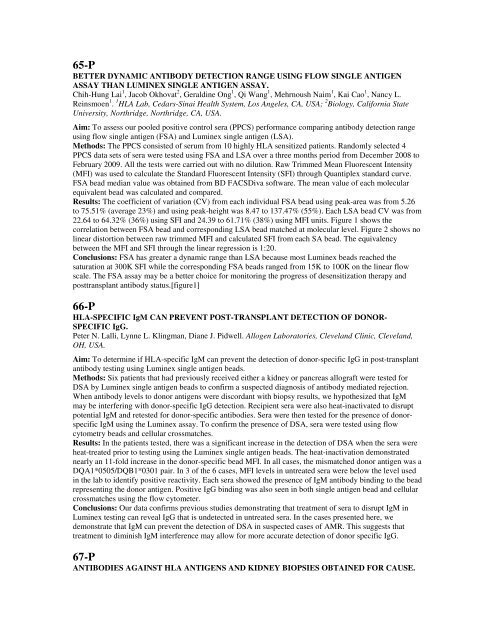
65-PBETTER DYNAMIC ANTIBODY DETECTION RANGE USING FLOW SINGLE ANTIGENASSAY THAN LUMINEX SINGLE ANTIGEN ASSAY.Chih-Hung Lai 1 , Jacob Okhovat 2 , Geraldine Ong 1 , Qi Wang 1 , Mehrnoush Naim 1 , Kai Cao 1 , Nancy L.Reinsmoen 1 . 1 HLA Lab, Cedars-Sinai Health System, Los Angeles, CA, USA; 2 Biology, Cali<strong>for</strong>nia StateUniversity, Northridge, Northridge, CA, USA.Aim: To assess our pooled positive control sera (PPCS) per<strong>for</strong>mance comparing antibody detection rangeusing flow single antigen (FSA) and Luminex single antigen (LSA).Methods: The PPCS consisted of serum from 10 highly HLA sensitized patients. Randomly selected 4PPCS data sets of sera were tested using FSA and LSA over a three months period from December 2<strong>00</strong>8 toFebruary 2<strong>00</strong>9. All the tests were carried out with no dilution. Raw Trimmed Mean Fluorescent Intensity(MFI) was used to calculate the Standard Fluorescent Intensity (SFI) through Quantiplex standard curve.FSA bead median value was obtained from BD FACSDiva software. The mean value of each molecularequivalent bead was calculated and compared.Results: The coefficient of variation (CV) from each individual FSA bead using peak-area was from 5.26to 75.51% (average 23%) and using peak-height was 8.47 to 137.47% (55%). Each LSA bead CV was from22.64 to 64.32% (36%) using SFI and 24.39 to 61.71% (38%) using MFI units. Figure 1 shows thecorrelation between FSA bead and corresponding LSA bead matched at molecular level. Figure 2 shows nolinear distortion between raw trimmed MFI and calculated SFI from each SA bead. The equivalencybetween the MFI and SFI through the linear regression is 1:20.Conclusions: FSA has greater a dynamic range than LSA because most Luminex beads reached thesaturation at 3<strong>00</strong>K SFI while the corresponding FSA beads ranged from 15K to 1<strong>00</strong>K on the linear flowscale. The FSA assay may be a better choice <strong>for</strong> monitoring the progress of desensitization therapy andposttransplant antibody status.[figure1]66-PHLA-SPECIFIC IgM CAN PREVENT POST-TRANSPLANT DETECTION OF DONOR-SPECIFIC IgG.Peter N. Lalli, Lynne L. Klingman, Diane J. Pidwell. Allogen Laboratories, Cleveland Clinic, Cleveland,OH, USA.Aim: To determine if HLA-specific IgM can prevent the detection of donor-specific IgG in post-transplantantibody testing using Luminex single antigen beads.Methods: Six patients that had previously received either a kidney or pancreas allograft were tested <strong>for</strong>DSA by Luminex single antigen beads to confirm a suspected diagnosis of antibody mediated rejection.When antibody levels to donor antigens were discordant with biopsy results, we hypothesized that IgMmay be interfering with donor-specific IgG detection. Recipient sera were also heat-inactivated to disruptpotential IgM and retested <strong>for</strong> donor-specific antibodies. Sera were then tested <strong>for</strong> the presence of donorspecificIgM using the Luminex assay. To confirm the presence of DSA, sera were tested using flowcytometry beads and cellular crossmatches.Results: In the patients tested, there was a significant increase in the detection of DSA when the sera wereheat-treated prior to testing using the Luminex single antigen beads. The heat-inactivation demonstratednearly an 11-fold increase in the donor-specific bead MFI. In all cases, the mismatched donor antigen was aDQA1*0505/DQB1*0301 pair. In 3 of the 6 cases, MFI levels in untreated sera were below the level usedin the lab to identify positive reactivity. Each sera showed the presence of IgM antibody binding to the beadrepresenting the donor antigen. Positive IgG binding was also seen in both single antigen bead and cellularcrossmatches using the flow cytometer.Conclusions: Our data confirms previous studies demonstrating that treatment of sera to disrupt IgM inLuminex testing can reveal IgG that is undetected in untreated sera. In the cases presented here, wedemonstrate that IgM can prevent the detection of DSA in suspected cases of AMR. This suggests thattreatment to diminish IgM interference may allow <strong>for</strong> more accurate detection of donor specific IgG.67-PANTIBODIES AGAINST HLA ANTIGENS AND KIDNEY BIOPSIES OBTAINED FOR CAUSE.
Bhavna Lavingia 1 , Chantale Lacelle 1 , Christopher Lu 2 , Miguel Vazquez 2 , Juan Arenas 3 , Mouin Seikaly 4 ,Peter Stastny 1,5 . 1 Pathology, UT Southwestern Medical Center, Dallas, TX, USA; 2 Internal Medicine -Nephrology, UT Southwestern Medical Center, Dallas, TX, USA; 3 Surgery - Surgical Transplantation, UTSouthwestern Medical Center, Dallas, TX, USA; 4 Pediatrics, UT Southwestern Medical Center, Dallas, TX,USA; 5 Internal Medicine - Transplant Immunology, UT Southwestern Medical Center, Dallas, TX, USA.Aim: Kidney transplant recipients with acute dysfunction can be analyzed <strong>for</strong> events in tissue and blood.Tissue histology, complement deposition and donor-specific HLA antibodies (DSA) were studied tocorrelate between DSA and biopsy findings.Methods: Sera from 41 kidney transplant recipients with acute dysfunction, suspected to be rejecting, werestudied between 2<strong>00</strong>4 and 2<strong>00</strong>9 <strong>for</strong> de novo production of HLA antibodies, biopsy readings and C4ddeposition. All were transplanted with negative T cell AHG, and negative T cell and B cell flow cytometrycross-matches.Results: New antibodies against donor HLA developed in 24/41 (59%) recipients. Among the DSAnegativerecipients, 11 (<strong>27</strong>%) had no antibodies to HLA, 4 (10%) developed HLA antibodies not donorspecific and 2 (5%) had antibodies cross-reactive with donor HLA. In 6 of the 24 (25%), DSA were HLAclass I, in 9 (38%) HLA class II and in 9 (38%) HLA class I and class II. Interestingly, among the 18recipients with DSA HLA class II, 15 (83%) had only anti-HLA-DQ and not DR. Biopsies were availablein 37 recipients, 22 had DSA and 15 without DSA. Among the 22 with DSA, 12 (55%) recipients had C4ddeposition in peritubular capillaries. In the 15 without DSA, only one (7%) had deposition of C4d(p=0.<strong>00</strong>2). All 13 recipients with C4d deposition had histological evidence of rejection. In the groupwithout C4d deposition, 5/25 (20%) recipients had histological rejection: 3 cellular and 2 humoral.Presence of DSA was significantly associated with rejection by biopsy (p
- Page 1 and 2: Monday, September 27, 20102:00 PM -
- Page 3 and 4: 5-ORTWO CASES OF DONOR SPECIFIC ANT
- Page 5 and 6: Monday, September 27, 20102:00 PM -
- Page 7 and 8: Results: PHA-induced mRNA expressio
- Page 9 and 10: conjugated IgM (BD Biosciences), Ig
- Page 11 and 12: Methods: Peripheral blood and endom
- Page 13 and 14: Aim: NK cells have an established r
- Page 15 and 16: Methods: Sera (n=22) were tested fo
- Page 17 and 18: expression on lymphocytes, we hypot
- Page 19 and 20: Conclusions: Clean SA beads can eli
- Page 21 and 22: goal is to re-examine our previous
- Page 23 and 24: Aim: There is increasing evidence t
- Page 25 and 26: esult in poor graft survival. The g
- Page 27 and 28: Aim: Pronase treatment eliminates i
- Page 29 and 30: 34-PCLINICAL AND PATHOLOGICAL SIGNI
- Page 31 and 32: Nabil Mohsin, Isabelle G. Wood, Ind
- Page 33 and 34: donor/recipient pairs for transplan
- Page 35 and 36: and tested in the DynaChip assay fo
- Page 37 and 38: average difference between Ave and
- Page 39 and 40: Conclusions: The XM-One assay can b
- Page 41 and 42: 59-PCYTOTOXIC AND NON-CYTOTOXIC ANT
- Page 43: data suggests that DTT pretreatment
- Page 47 and 48: ATG INTERFERNCE IN SINGLE ANTIGEN L
- Page 49 and 50: 75-PPREFORMED LOW REACTIVE ANTI-HLA
- Page 51 and 52: Sabarinathan Ramachandran 1 , Naohi
- Page 53 and 54: 83-PALLELIC DIVERSITY OF KIR2DL4 AN
- Page 55 and 56: São Paulo, Brazil; 3 Commissariat
- Page 57 and 58: 91-PCHARACTERIZATION OF COMMON AND
- Page 59 and 60: Conclusions: The extensive diversit
- Page 61 and 62: 99-PPOTENTIAL COMMON NOVEL ALLELES
- Page 63 and 64: 103-PTOWARDS A FUNCTIONAL HLA-DPB1
- Page 65 and 66: 107-PWHOLE GENOME AMPLIFICATION (WG
- Page 67 and 68: 111-PLOSS OF MISMATCHED HLA IN FAMI
- Page 69 and 70: 115-PDONOR EPITHELIAL CELL CHIMERIS
- Page 71 and 72: Caucasian patients. MRs vary signif
- Page 73 and 74: Results: We obtained the total No.
- Page 75 and 76: cord blood and platelet transfusion
- Page 77 and 78: 131-PASSOCIATION OF IL-2-330(G/T) W
- Page 79 and 80: Institute, Children’s Hospital Oa
- Page 81 and 82: Methods: To prove the robustness of
- Page 83 and 84: populations.We studied the involvem
- Page 85 and 86: amplicons generated from different
- Page 87 and 88: Results: The sequence of the unexpe
- Page 89 and 90: concentration of the Taq Polymerase
- Page 91 and 92: 160-PASSOCIATION OF HAPLOTYPES WITH
- Page 93 and 94: 164-PDETECTION OF A DE NOVO DONOR S
- Page 95 and 96:
168-PA NOVEL HLA-DQB1 ALLELE CONFIR
- Page 97 and 98:
172-PSCREENING FOR HLA-SPECIFIC ALL
- Page 99 and 100:
176-PIMPLEMENTATION OF THE WORLD HE
- Page 101 and 102:
epertoire of displayed ligands and
- Page 103 and 104:
mucosa. No other samples were avail
- Page 105 and 106:
Conclusions: In previously reported
- Page 107 and 108:
Conclusions: Our findings can demon
- Page 109 and 110:
Conclusions: In analyzing this data
- Page 111 and 112:
examined on microarrays from a huma
- Page 113 and 114:
Methods: Serial analysis of sera ob
- Page 115 and 116:
Conclusions: These new technologies
- Page 117 and 118:
allograft injury. Four of these 18
- Page 119 and 120:
A NOVEL HLA CLASS I SINGLE ANTIGEN
- Page 121 and 122:
Results: Approximately twice as man
- Page 123:
Methods: Molecular HLA typing was p


