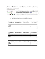
Download the Workshop Document - comPADRE
Download the Workshop Document - comPADRE
Download the Workshop Document - comPADRE
- No tags were found...

Create successful ePaper yourself
Turn your PDF publications into a flip-book with our unique Google optimized e-Paper software.
BEGINNER LAB EXERCISES± Lab 0: Introduction to DeskCATIn this lab, students will scan a simple 3D object (i.e. a mouse phantom) tobecome familiar with <strong>the</strong> DeskCAT scanner procedures and software interface.This will help students become oriented with <strong>the</strong> operations of a CT scannerand associated software for data capture, image reconstruction, and 3D imagedisplay.This exercise will introduce <strong>the</strong> students to <strong>the</strong> user interface of <strong>the</strong> DeskCATscanner, explain <strong>the</strong> concept of a reference versus data scan, and allow <strong>the</strong>m toexplore <strong>the</strong> different navigational and visualization tools that <strong>the</strong> software offersfor both 2D projections and 3D reconstructions.± Lab 1: Introduction to Medical Imaging – 3D LocalizationIn this lab, students will make geometric measurements of <strong>the</strong> 3D positions offiducial markers embedded in a cylindrical phantom. Measurements are madefrom 2 orthogonal radiographic (camera) projections, as well as from a CTreconstructed 3D image. Students can compare and contrast <strong>the</strong> two imagingmodalities with calculations of coordinates by each approach. By <strong>the</strong> end of <strong>the</strong>lab, students should find that <strong>the</strong> marker positions are in close agreement usingan orthogonal pair of 2D radiographic images and <strong>the</strong> full 3D CT image.This exercise will fur<strong>the</strong>r introduce <strong>the</strong> students to <strong>the</strong> user interface of <strong>the</strong>DeskCAT scanner. Students will determine <strong>the</strong> geometric positions of fiducialmarkers in 3D space, and will also learn how to adjust window and level toimprove visibility of targets in an image.± Lab 2: System LinearityIn this lab, students will perform an experiment to assess <strong>the</strong> linearity of <strong>the</strong>DeskCAT scanner performance. Students are given a water-filled jar andconcentrated black dye solution. They must acquire a data-set of scans withincreasing optical attenuation coefficients. By <strong>the</strong> end of <strong>the</strong> lab, students shouldobserve a linear trend and accurate reconstructions to within 10%, with resultsdepending on <strong>the</strong> concentration of <strong>the</strong> dye. Students must identify sources oferror and offer suggestions to reduce <strong>the</strong> experimental error.This exercise will illustrate <strong>the</strong> importance of an imaging system’s linearity on itssuitability as a quantitative tool.± Lab 3: Spatial Resolution and Modulation Transfer Function (MTF)In this lab, <strong>the</strong> student will assess <strong>the</strong> spatial resolution of <strong>the</strong> DeskCATscanner at 2 levels: The spatial resolution of a single 2D projection radiographicimage, and <strong>the</strong> spatial resolution of a 3D CT reconstructed image. Studentswill acquire images of a test bar pattern with a series of “picket fence” linethicknesses. By measuring <strong>the</strong> contrast of line pairs, students plot pixel intensity(or modulation) vs. spatial frequency to estimate <strong>the</strong> shape of <strong>the</strong> modulationtransfer function (MTF).This exercise will introduce students to <strong>the</strong> concept of MTF, its practicalimplications and demonstrate one method of measuring it.© 2012 Modus Medical Devices Inc. Version 1.1 Modus Medical Devices Inc. reserves <strong>the</strong> right to make changes without notice and shall not be liable for any errors or omissions contained in this document.