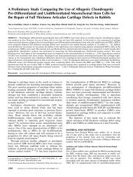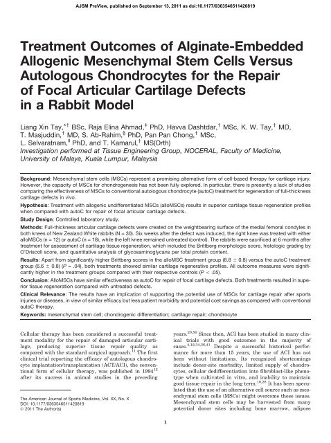
Liang Xin Tay, Raja Elina Ahmad, Havva Dashtdar - High Impact ...
Liang Xin Tay, Raja Elina Ahmad, Havva Dashtdar - High Impact ...
Liang Xin Tay, Raja Elina Ahmad, Havva Dashtdar - High Impact ...

Create successful ePaper yourself
Turn your PDF publications into a flip-book with our unique Google optimized e-Paper software.
Treatment Outcomes of Alginate-Embedded<br />
Allogenic Mesenchymal Stem Cells Versus<br />
Autologous Chondrocytes for the Repair<br />
of Focal Articular Cartilage Defects<br />
in a Rabbit Model<br />
<strong>Liang</strong> <strong>Xin</strong> <strong>Tay</strong>,* y BSc, <strong>Raja</strong> <strong>Elina</strong> <strong>Ahmad</strong>, z PhD, <strong>Havva</strong> <strong>Dashtdar</strong>, y MSc, K. W. <strong>Tay</strong>, y MD,<br />
T. Masjuddin, y MD, S. Ab-Rahim, § PhD, Pan Pan Chong, y MSc,<br />
L. Selvaratnam, ll PhD, and T. Kamarul, y MS(Orth)<br />
Investigation performed at Tissue Engineering Group, NOCERAL, Faculty of Medicine,<br />
University of Malaya, Kuala Lumpur, Malaysia<br />
Background: Mesenchymal stem cells (MSCs) represent a promising alternative form of cell-based therapy for cartilage injury.<br />
However, the capacity of MSCs for chondrogenesis has not been fully explored. In particular, there is presently a lack of studies<br />
comparing the effectiveness of MSCs to conventional autologous chondrocyte (autoC) treatment for regeneration of full-thickness<br />
cartilage defects in vivo.<br />
Hypothesis: Treatment with allogenic undifferentiated MSCs (alloMSCs) results in superior cartilage tissue regeneration profiles<br />
when compared with autoC for repair of focal articular cartilage defects.<br />
Study Design: Controlled laboratory study.<br />
Methods: Full-thickness articular cartilage defects were created on the weightbearing surface of the medial femoral condyles in<br />
both knees of New Zealand White rabbits (N = 30). Six weeks after the defect was induced, the right knee was treated with either<br />
alloMSCs (n = 12) or autoC (n = 18), while the left knee remained untreated (control). The rabbits were sacrificed at 6 months after<br />
treatment for assessment of cartilage tissue regeneration, which included the Brittberg morphologic score, histologic grading by<br />
O’Driscoll score, and quantitative analysis of glycosaminoglycans per total protein content.<br />
Results: Apart from significantly higher Brittberg scores in the alloMSC treatment group (8.8 6 0.8) versus the autoC treatment<br />
group (6.6 6 0.8) (P = .04), both treatments showed similar cartilage regenerative profiles. All outcome measures were significantly<br />
higher in the treatment groups compared with their respective controls (P \ .05).<br />
Conclusion: AlloMSCs have similar effectiveness as autoC for repair of focal cartilage defects. Both treatments resulted in superior<br />
tissue regeneration compared with untreated defects.<br />
Clinical Relevance: The results have an implication of supporting the potential use of MSCs for cartilage repair after sports<br />
injuries or diseases, in view of similar efficacy but less patient morbidity and potential cost savings as compared with conventional<br />
autoC therapy.<br />
Keywords: mesenchymal stem cell; chondrogenic differentiation; cartilage repair; chondrocyte<br />
Cellular therapy has been considered a successful treatment<br />
modality for the repair of damaged articular cartilage,<br />
producing superior tissue repair quality as<br />
compared with the standard surgical approach. 11 The first<br />
clinical trial reporting the efficacy of autologous chondrocyte<br />
implantation/transplantation (ACT/ACI), the conventional<br />
form of cellular therapy, was published in 1994 12<br />
after its success in animal studies in the preceding<br />
The American Journal of Sports Medicine, Vol. XX, No. X<br />
DOI: 10.1177/0363546511420819<br />
Ó 2011 The Author(s)<br />
AJSM PreView, published on September 13, 2011 as doi:10.1177/0363546511420819<br />
1<br />
years. 20,50 Since then, ACI has been studied in many clinical<br />
trials with good outcomes in the majority of<br />
cases. 4,12,34,36,41 Despite a successful historical performance<br />
for more than 15 years, the use of ACI has not<br />
been without limitations. Its recognized shortcomings<br />
include donor-site morbidity, limited supply of chondrocytes,<br />
cellular dedifferentiation into fibroblast-like phenotype<br />
when cultivated in vitro, and inability to maintain<br />
good tissue repair in the long term. 10,28 It has been speculated<br />
that the use of an alternative cell source such as mesenchymal<br />
stem cells (MSCs) might overcome these issues.<br />
Mesenchymal stem cells may be harvested from many<br />
potential donor sites including bone marrow, adipose
2 <strong>Tay</strong> et al The American Journal of Sports Medicine<br />
tissue, trabecular bone, and other tissues without causing<br />
damage to the unaffected cartilage. 40 In addition, they<br />
have a high proliferative capacity, and owing to their multipotency,<br />
these cells can conveniently be manipulated in<br />
vitro to differentiate into chondrocytes for subsequent<br />
use in cartilage regeneration. 14,18,31,43<br />
There have been many previous reports involving in vivo<br />
experiments describing good repair outcomes after transplantation<br />
of MSCs in cartilage defects. Wakitani et al 49<br />
in 1994 were among the first research groups to report<br />
the successful transplantation of bone marrow–derived<br />
MSCs in osteochondral defects in rabbit models. Other<br />
researchers have also studied the application of allogenic<br />
or autologous bone marrow–derived MSCs using different<br />
scaffolds with or without the addition of growth factors to<br />
treat cartilage defects in various animal models. 19,22,37<br />
While the use of autologous chondrocytes (autoC) and allogenic<br />
undifferentiated MSCs (alloMSCs) to repair damaged<br />
cartilage have been forthcoming in many studies, it has<br />
been demonstrated that additional use of scaffolds enhances<br />
the tissue repair. 2,13,22,48,55 These scaffolds not only provide<br />
a convenient method for delivering cells into focal defect<br />
sites, but also provide structural support to the construct<br />
and induce cartilage matrix formation within the defective<br />
sites. 47,48,55 Alginate is a well-established biomaterial for<br />
use in these conditions. 8,23,30,33,48,52 Notonlyisthismaterial<br />
biocompatible, the embedded chondrocytes maintain their<br />
phenotype and produce more depositions of extracellular<br />
cartilaginous matrix in alginate. 16<br />
Despite a multitude of available experimental reports on<br />
a cell-based approach for articular cartilage repair, there is<br />
presently limited exploratory research comparing alloMSCs<br />
with other sources of chondrocytes, particularly those<br />
obtained from the autologous transplantation procedure<br />
(autoC). To date, there is only 1 prospective clinical study<br />
comparing the effectiveness of MSCs against autoC for<br />
repairing damaged cartilage in humans. 38 The study<br />
revealed similar clinical outcomes between MSCs and autoC<br />
assessed postoperatively by a multifaceted questionnairebased<br />
clinical evaluation carried out throughout a temporal<br />
course of 2 years. While it may be appropriate to use the validated<br />
knee cartilage outcome clinical instruments to assess<br />
the overall function of the repaired cartilage tissue, it is<br />
equally important to ascertain the changes in tissue repair<br />
quality in response to both treatments. However, these forms<br />
of investigation are usually confined to animal models since<br />
the invasiveness of the tissue biopsy procedure may not necessarily<br />
be tolerated by human subjects in the clinical setting.<br />
A study incorporating more objective assessments of<br />
tissue repair is therefore required to determine the cellular<br />
changes that may exist during the transplantation process.<br />
This may demonstrate a clear distinction in the physical features<br />
of the repaired cartilage tissue resulting from the individual<br />
treatment. No parallel comparative analysis of<br />
tissue-healing outcome has been previously performed to<br />
evaluate the effectiveness of alloMSCs compared with autoC<br />
in animal studies. Therefore, the aim of the current study is<br />
to compare in a more objective manner the effectiveness of<br />
the 2 different forms of cell-based therapy (alloMSCs vs<br />
autoC) in repairing focal cartilage defects in an in vivo rabbit<br />
model, by analyzing the gross morphology, histology, and<br />
extracellular matrix protein content such as glycosaminoglycan<br />
in response to transplantation of chondrocytes from<br />
these distinct sources into the defective areas in the joint.<br />
MATERIALS AND METHODS<br />
Rabbit Focal Articular Cartilage Defect Model<br />
Thirty male New Zealand White (NZW) rabbits aged between<br />
5 and 6 months and weighing approximately 2.5 kg were randomly<br />
assigned into 2 treatment groups: ACI group (autoC; n<br />
= 18) and allogenic MSC group (alloMSC; n = 12). The sample<br />
size of each treatment group was calculated based on 2 independent<br />
studies previously conducted in our laboratory comparing<br />
MSCs and autoC treatments with their respective<br />
control groups, 15,29,30 in view of the lack of reference data<br />
involving direct parallel comparison between the 2 treatment<br />
groups using similar objective measurements. The stated<br />
number of subjects in each group was required to detect 2fold<br />
difference in Brittberg scores between the treated and<br />
untreated groups with 80% power at a .05% significance<br />
level. In addition, 3 rabbits were sacrificed separately to harvest<br />
bone-marrow–derived MSCs for use in the alloMSCtreated<br />
group. This study was approved by University of<br />
Malaya animal ethics committee and the experiments were<br />
carried out in accordance to the regulations imposed by the<br />
university and the appropriate Malaysian government regulatory<br />
body (OS/05/07/2006/TKZ/A[R] and PM/24/06/2008/<br />
TKZ[c][R]).<br />
The rabbits were anesthetized before both femorotibial<br />
joints were surgically exposed. A 5-mm–diameter fullthickness<br />
articular cartilage defect was created in both knees<br />
using a custom-made chondrotome as previously described<br />
by Kamarul et al in 2008. 30 All defects were created on the<br />
weightbearing portion of the medial femoral condyle. Cartilage<br />
from the operated sites were removed from both knees<br />
and collected aseptically in 13 phosphate-buffered saline<br />
solution (PBS; pH 7.4) (Gibco/Invitrogen, Carlsbad,<br />
*Address correspondence to <strong>Liang</strong> <strong>Xin</strong> <strong>Tay</strong>, BSc, Tissue Engineering Group, National Orthopaedic Surgery Centre of Excellence for Research and<br />
Learning (NOCERAL), Department of Orthopaedic Surgery, Faculty of Medicine, University of Malaya, 50603 Kuala Lumpur, Malaysia (e-mail:<br />
liangxin28@gmail.com).<br />
y Tissue Engineering Group, NOCERAL, Faculty of Medicine, University of Malaya, Kuala Lumpur, Malaysia.<br />
z Department of Physiology, Faculty of Medicine, University of Malaya, Kuala Lumpur, Malaysia.<br />
§ Faculty of Medicine, UiTM, Shah Alam, Selangor, Malaysia.<br />
ll School of Medicine and Health Sciences, Monash University, Sunway Campus, Selangor, Malaysia.<br />
One or more of the authors has declared the following potential conflict of interest or source of funding: This study was supported by the University of<br />
Malaya’s HIR-MOHE research grant initiative and 2 grants from the Ministry of Science, Technology and Innovation of Malaysia (IRPA PR-06-03-01 and<br />
TF0106B071).
Vol. XX, No. X, XXXX Cartilage Treatment Using Alginate-Embedded Allogenic MSCs vs Autologous Chondrocytes 3<br />
California) supplemented with 4% penicillin-streptomycin<br />
(Invitrogen). The cartilage specimens were kept at 4°Cbefore<br />
further processing of chondrocytes. The wounds were surgically<br />
closed in layers with 5-0 Vicryl absorbable sutures<br />
(Ethicon, Johnson & Johnson, Somerville, New Jersey) and<br />
bandages. All surgeries were done in accordance with the<br />
standard procedures under sterile surgical conditions. Postoperatively,<br />
the animals were transferred into the cages<br />
without immobilization. All animals were closely monitored<br />
for infections and other complications.<br />
Isolation of AutoC and Allogenic MSCs<br />
Autologous chondrocytes were isolated under sterile conditions<br />
from cartilage tissues in accordance with the techniques<br />
previously described by Ab-Rahim et al in 2008. 1 The<br />
specimens were first digested using 0.6% type II collagenase<br />
(Sigma-Aldrich, St Louis, Missouri) at 37°C and<br />
rotated overnight followed by centrifugation at 500g for<br />
10 minutes. The supernatant was discarded and the cell<br />
pellet was resuspended in 1 mL of Dulbecco’s Modified<br />
Eagle Medium (DMEM; Invitrogen). The resuspended cells<br />
were then seeded into a T75 flask (Corning, Lowell, Massachusetts)<br />
containing DMEM/Ham’s F-12 Nutrient Mixture<br />
(1:1) (Invitrogen) supplemented with 10% fetal bovine<br />
serum (FBS; HyClone, Logan, Utah), 0.0025% ascorbic<br />
acid (Sigma), and 1% penicillin-streptomycin. The autoCs<br />
were maintained in monolayer culture at 37°C, 95%<br />
humidity, and 5% CO 2 for 3 weeks while the growth<br />
medium was exchanged every 2 to 3 days.<br />
To obtain alloMSCs for transplantation, 3 rabbits were<br />
sacrificed using an intravenous overdose of pentobarbital.<br />
Femurs and tibias of both lower limbs were removed and<br />
any adherent tissues were discarded. All harvested bones<br />
were kept on ice in 13 PBS solution supplemented with<br />
4% penicillin-streptomycin until they were processed. Within<br />
3 hours, bone marrow was harvested from femurs and tibias.<br />
AlloMSCs were isolated according to an established protocol<br />
described by Pittenger et al in 1999. 42 Briefly, 2 mL of bone<br />
marrow was diluted with 13 PBS solution (1:2) and loaded<br />
over 3 mL Ficoll-paque (GE Healthcare–Amersham Biosciences,<br />
Piscataway, New Jersey) in a 15-mL Falcon tube (Corning).<br />
Mononuclear cells were harvested from the interface of<br />
plasma and Ficoll-paque after centrifugation at 2200 rpm for<br />
25 minutes. The cells were then washed with prewarmed<br />
DMEM and centrifuged for another 10 minutes. The cell pellet<br />
was resuspended in DMEM containing 10% FBS and 1%<br />
penicillin-streptomycin and seeded into a T75 flask. All cells<br />
were maintained and incubated under the same conditions as<br />
described above. After 8 days, the nonadherent cells were<br />
removed while the adherent cells (alloMSCs) were replenished<br />
with fresh medium. The alloMSC culture was also<br />
maintained in monolayer for 3 weeks in a similar environment<br />
to that of the autoC culture.<br />
Autologous Chondrocytes (AutoC)<br />
and AlloMSC Culture in Alginate<br />
Autologous chondrocytes and alloMSC alginate beads<br />
were prepared in 1.2% low-viscosity alginate solutions<br />
(Sigma-Aldrich) in 0.15 M sodium chloride as previously<br />
described by Kamarul et al 30 after 21 days of monolayer culture.<br />
Autologous chondrocytes and alloMSCs were first<br />
detached from the T75 flask surface using TypLE express<br />
(Invitrogen) and pelletized in a separate 15-mL Falcon<br />
tube. Cell count was performed, followed by the addition of<br />
the appropriate volume of alginate solution to form alginate<br />
beads, each containing approximately 5.0 3 10 5 cells. The<br />
cell suspension was mixed thoroughly and expressed slowly<br />
through a micropipette tip into a 102-mM calcium chloride<br />
solution. After 10 minutes of polymerization in the solution,<br />
the alginate beads were washed with 0.15 M sodium chloride<br />
to remove the excess calcium chloride. Five autoC-alginate<br />
beads were transferred to each of the 24-well culture plates<br />
(Becton Dickinson, Durham, North Carolina) and cultured<br />
in DMEM/F12 containing 10% FBS, 25 mg/mL ascorbic<br />
acid, 50 mg/mL gentamicin (Invitrogen), and 360 mg/mL<br />
L-glutamine (Invitrogen). The alloMSC-alginate beads were<br />
cultured in DMEM low glucose containing 10% FBS and<br />
1% penicillin-streptomycin. Both cultures in the alginate<br />
scaffold were maintained at 37°C in a humidified environment<br />
of 5% CO 2 for another 3 weeks.<br />
Implantation of AutoC-Alginate and AlloMSC-Alginate<br />
Constructs Into Defective Areas<br />
To avoid dedifferentiation of autoC, implantation of autoC<br />
and alloMSC embedded in alginate beads into the rabbits’<br />
cartilage defects was carried out after 3 weeks of cell culture<br />
in the alginate constructs, culminating in an overall culture<br />
period of 6 weeks (ie, 3 weeks in monolayer and 3 weeks in<br />
alginate construct) after the creation of the defect. All transplantations<br />
were performed only in the right knee of each rabbit.<br />
The rabbits were anesthetized and the chondral defects<br />
were exposed and identified. A periosteum flap of 7-mm diameter<br />
was harvested from the medial aspect of the proximal<br />
tibia of the same limb. The periosteum flap was then placed<br />
on top of the defect and anchored with 1 suture in each corner<br />
using 8-0 Vicryl absorbable sutures. Two beads of either<br />
autoC-alginate or alloMSC-alginate (total of 1 3 10 6 cells)<br />
were inserted underneath the flap according to the respective<br />
treatment group. The remaining opening of the periosteum<br />
flap was then sealed using Tisseel fibrin glue (Baxter AG<br />
Industries,Vienna,Austria)and suture (11-0 Vicryl absorbable<br />
suture). The wound was closed in layers using absorbable<br />
suture and bandages, followed by application of<br />
antiseptic solution to prevent infection. The left knees were<br />
left untreated (control group). All transplanted rabbits were<br />
treated with analgesic and antipyretic, Metacam (Boehringer<br />
Ingelheim, Berks, United Kingdom) and Kombitrim (KELA<br />
Laboratoria NV, Hoogstraten, Belgium) for 3 days after the<br />
operation, both of which additionally act as broad-spectrum<br />
antibiotics for both gram-positive and gram-negative bacteria.<br />
Harvesting and Gross Examination of the<br />
Regenerated Tissues From Defective Areas<br />
At 6 months after implantation, the rabbits’ knee joints<br />
were surgically removed and examined. For gross
4 <strong>Tay</strong> et al The American Journal of Sports Medicine<br />
evaluation, all specimens were examined under direct light<br />
microscopy by 2 independent observers, both experienced<br />
orthopaedic surgeons, who were blinded to the sample<br />
groups. The observers were asked to examine and grade<br />
samples according to the Brittberg scoring system. 13 Upon<br />
completion of the scoring, the specimens were halved using<br />
a mechanical bone saw (Fein MultiMaster Accu, C & E Fein<br />
Gmbh, Stuttgart, Germany). This was done carefully to<br />
avoid any alteration or destruction of the tissue specimen<br />
according to the technique previously established by<br />
Kamarul et al. 29,30 Half of each specimen was fixed in<br />
10% phosphate-buffered formalin (4% formaldehyde) for<br />
histology and immunostaining, while the other half was utilized<br />
for analysis of the glycosaminoglycan (GAG) content.<br />
Histologic Examination and<br />
Immunohistochemical Staining<br />
Specimens that were fixed in 4% buffered formalin were<br />
decalcified. Fixed and decalcified tissues were subsequently<br />
dehydrated in ethanol in a stepwise manner from 70% up to<br />
100%, transferred to xylene, and embedded in paraffin. At<br />
the center of each sample, 5-mm paraffin sections were prepared<br />
and placed on glass slides, dried overnight, and stored<br />
at 4°C. The samples were stained with hematoxylin and<br />
eosin for general morphologic evaluation and safranin<br />
O–fast green to detect proteoglycan. Immunohistochemical<br />
staining was performed using DAKO EnVision 1 System<br />
peroxidase kit (DAKO, Glostrup, Denmark). The specimen<br />
section slides were incubated in primary antibody solution<br />
(anti–collagen type II, Santa Cruz Biotechnology, Santa<br />
Cruz, California) followed by secondary antibody and<br />
substrate-chromogen solution in accordance with the manufacturer’s<br />
instructions. The O’Driscoll cartilage scoring was<br />
used for histologic and histochemical assessment of the<br />
repaired cartilage specimens. The scoring was performed<br />
by 2 histologists blinded to the group status.<br />
Biochemical Assay for GAG<br />
The Blyscan GAG assay kit (Biocolor Ltd, Antrim, United<br />
Kingdom) was used to evaluate GAG content of the regenerated<br />
cartilage tissues in both treatment groups. Specimens<br />
were dissected into small pieces using a scalpel and<br />
digested using RIPA buffer (Merck & Co, Whitehouse Station,<br />
New Jersey) supplemented with protease inhibitors<br />
(Sigma) for 1 hour. Aliquots of each sample were mixed<br />
with DMMB (dimethylmethylene blue) dye and reagents<br />
(Biocolor) according to the manufacturer’s instructions.<br />
The absorbance at 656 nm was measured using the spectrophotometer<br />
and compared with a standard plot of chondroitin<br />
sulphate provided by the manufacturer for<br />
quantitative determination of the GAG content.<br />
Statistical Analysis<br />
Statistical analysis was performed using SPSS statistical<br />
software (version 17.0, SPSS Inc, an IBM company, Chicago,<br />
Illinois). The values of Brittberg and O’Driscoll scores,<br />
as well as GAG concentrations for all tissue samples, were<br />
presented as mean 6 standard deviation. Comparisons of<br />
variables between the 2 treatment groups were analyzed<br />
using the parametric 2-sided independent t test. Differences<br />
were considered statistically significant at P \ .05.<br />
RESULTS<br />
Gross Appearance of Defect<br />
Figure 1 illustrates an example of the gross macroscopic<br />
analysis of the representative tissues from the alloMSC,<br />
autoC, and control groups. Macroscopic examinations at<br />
6 months after implantation revealed that defects treated<br />
with both alloMSCs and autoC showed good filling, with<br />
the surface appearing flush and smooth (Figures 1D and<br />
1E). In contrast, none of the untreated defects in the left<br />
knees (control) (Figure 1F) showed complete filling as compared<br />
with the treated knees.<br />
Figure 2 shows the results of Brittberg scores reflecting<br />
quantitative macroscopic evaluation of the tissue regenerate<br />
quality between the alloMSC and autoC treatment groups<br />
together with their respective controls. Both the alloMSC<br />
and autoC implantations showed superior cartilage regeneration<br />
quality as compared with the untreated left knees, as<br />
evidenced by the significantly higher Brittberg scores in the<br />
treatment groups (8.8 6 0.8 vs 3.0 6 0.8 for the alloMSCcontrol<br />
pair, and 6.6 6 0.8 vs 2.3 6 0.6 for the autoC-control<br />
pair; both pairs were associated with a P value of .001). The<br />
difference in Brittberg scores between alloMSC and autoC<br />
was significant (P = .04) from the 2-sided independent<br />
t test. No significant differences in the Brittberg scores<br />
were found between the untreated left knees (control) in<br />
both alloMSC and autoC groups (P =.31).<br />
Histologic and Immunohistochemical Appearance<br />
of the Regenerated Cartilage Tissues<br />
Figure 3 shows the results of the microscopic examination<br />
of the regenerate tissues in 4 groups (ie, the alloMSC-treated<br />
group, the autoC-treated group, the untreated group<br />
[negative control] and a representative specimen of a normal<br />
NZW rabbit’s knee articular cartilage tissue [positive<br />
control]). The microscopic appearance of the tissue using<br />
various staining methods revealed cartilage regeneration<br />
within the treated sites although some areas appeared to<br />
be infiltrated by a small amount of fibrous tissues. In contrast,<br />
the untreated defects did not appear to undergo complete<br />
healing, but were merely filled with soft tissue<br />
instead (Figures 3C, 3G, and 3K). In the hematoxylin<br />
and eosin–stained sections, cartilage from the alloMSCtreated<br />
group (Figure 3A) showed substantial thickening<br />
of the cartilage tissue compared with the untreated defect<br />
(Figure 3C), with the chondrocytes arranged in clusters.<br />
Similar cartilage tissue thickening was also observed in<br />
the autoC-treated group (Figure 3B); however, the chondrocytes<br />
were mostly found in columnar formations. The<br />
regenerated tissues from both treatment groups showed<br />
a continuous surface with a mixture of hyaline and fibrocartilage.<br />
Immunohistochemical staining for type II
Vol. XX, No. X, XXXX Cartilage Treatment Using Alginate-Embedded Allogenic MSCs vs Autologous Chondrocytes 5<br />
Figure 1. Macroscopic evaluation of the defective areas on<br />
articular cartilage from the alloMSC, autoC, and control<br />
groups before treatment (A, B, and C, respectively) and 6<br />
months after transplantation (D, alloMSC; E, autoC; and F,<br />
control). There was incomplete filling of the control knee, while<br />
the defect appeared completely filled by both alloMSC and<br />
autoC transplantation. alloMSC, allogenic undifferentiated<br />
mesenchymal stem cells; autoC, autologous chondrocytes.<br />
Figure 2. Brittberg scores of articular cartilage regeneration<br />
in alloMSC and autoC treatment groups were compared with<br />
their respective control on the contralateral side 6 months<br />
after transplantation. The Brittberg score was significantly<br />
higher in alloMSC compared with autoC. Both treatments<br />
were associated with significantly higher scores compared<br />
with their respective control groups. alloMSC, allogenic<br />
undifferentiated mesenchymal stem cells; autoC, autologous<br />
chondrocytes. *Significant difference from untreated, negative<br />
control (P \ .01). # Significant difference between<br />
alloMSC and autoC group (P \ .05).<br />
collagen further confirmed that both the alloMSC- and<br />
autoC-treated tissue sections showed a relatively higher<br />
expression of cartilaginous collagen (Figures 3E and 3F)<br />
compared with that of the untreated defects (Figure 3G).<br />
The alloMSC- and autoC-treated tissue sections also<br />
showed relatively heavier staining with safranin O<br />
(Figures 3I and 3J) compared with the untreated defect<br />
(Figure 3K), suggesting a higher concentration of proteoglycans,<br />
one of the major components of cartilage tissue.<br />
Figure 3. Immunohistochemical staining of rabbit femoral<br />
condyles from alloMSC-treated (A, E, I), autoC-treated (B,<br />
F, J), and untreated (C, G, K) tissues at 6 months after transplantation.<br />
A representative specimen of a normal cartilage<br />
tissue is shown (D, H, L) as a reference for evaluation of<br />
the normal cartilage healing. The type II collagen is stained<br />
in brown, and the safranin O (ie, a measure of proteoglycans<br />
characterizing cartilage tissue) is stained in red. alloMSC,<br />
allogenic undifferentiated mesenchymal stem cells; autoC,<br />
autologous chondrocytes. Scale bars = 100 mm.<br />
Figure 4. Quantitative histologic evaluation of the regenerated<br />
cartilage using O’Driscoll scores at 6 months after treatment<br />
with alloMSC and autoC together with their respective<br />
controls. Results showed significantly higher scores in the<br />
treatment groups compared with their controls. However,<br />
there are no significant differences in the scores between<br />
the 2 treatment groups. alloMSC, allogenic undifferentiated<br />
mesenchymal stem cells; autoC, autologous chondrocytes.<br />
*Significant difference from untreated, negative control<br />
(P \ .01).<br />
The average difference in the O’Driscoll score (ie, quantitative<br />
histologic grading of the tissues) between the treatment<br />
groups appeared to be very small and nonsignificant<br />
(Figure 4), with mean values of 11.2 6 0.8 and 13.5 6 2.4<br />
for alloMSC and autoC groups, respectively (P . .05). However,<br />
both treatment groups had significantly higher<br />
O’Driscoll scores compared with their respective controls.<br />
The mean O’Driscoll score for the alloMSC-control was<br />
8.2 6 0.7, and that for the autoC-control was 6.7 6 2.1.
6 <strong>Tay</strong> et al The American Journal of Sports Medicine<br />
Figure 5. Comparison of the glycosaminoglycan (GAG) concentrations<br />
in the alloMSC and autoC treatment groups with<br />
their respective control groups, showing significant differences<br />
between each treatment with its respective control but<br />
no significant differences between the 2 treatment groups.<br />
alloMSC, allogenic undifferentiated mesenchymal stem cells;<br />
autoC, autologous chondrocytes. *Significant difference<br />
from untreated, negative control (P \ .05).<br />
Glycosaminoglycan Concentrations<br />
in the Regenerated Tissues<br />
Quantitative determination of GAG levels in the tissue samples<br />
revealed a dramatic and significant increase in GAG<br />
concentrations in both the alloMSC- and autoC-treated knees<br />
as compared with the untreated knees (P = .001). However,<br />
there was no significant difference in GAG levels between<br />
the 2 treatment groups (P = .74). The mean GAG/protein concentrations<br />
in the treated knees were 1.7 6 0.3 and 1.8 6<br />
0.5 mg GAGs/mg proteins for the alloMSC and autoC groups,<br />
respectively. For the control knees, the mean GAG/protein<br />
concentrations for the alloMSC and autoC groups were<br />
1.0 6 0.2 and 1.1 6 0.3 mg GAGs/mg proteins, respectively<br />
(Figure 5). No significant differences for both knees were<br />
observed between these groups (P . .05).<br />
DISCUSSION<br />
The use of both autoC and alloMSCs produced comparable<br />
cartilage repair outcome, and was significantly better than<br />
those left untreated. Although the superior outcome of<br />
using cell-based therapy is not unexpected, it is anticipated<br />
that differences between the use of both cell sources should<br />
have been evident in the study. In particular, alloMSC is<br />
postulated to result in superior tissue repair as compared<br />
with autoC in view of the numerous advantages potentially<br />
conferred by MSCs in cartilage regeneration, as further<br />
discussed in this section. In the current study, the morphologic,<br />
histologic, and biochemical profiles of the regenerated<br />
cartilage tissue using the two cell sources were<br />
compared. This was envisaged to provide more objective<br />
evidence and further insight into the effectiveness of the<br />
repair process following either alloMSC or autoC<br />
treatment. To the best of our knowledge, this study is the<br />
first to report the results of direct parallel comparison<br />
between autoC and alloMSC for repair of focal cartilage<br />
defects in an animal model in vivo.<br />
The complementing results observed from the various<br />
parameters examined in this study support the findings<br />
that cell therapies induce cartilage regeneration after<br />
injury. 13,15,20,22,30,50 Histologic observations revealed several<br />
notable features that may be of interest. In the<br />
alloMSC-treated group, chondrocytes in the histologic sections<br />
appeared to be clustered in groups, while in the<br />
autoC-treated group, cells were in columnar arrangements.<br />
However, the difference in the histologic appearance<br />
between the 2 treatment groups did not appear to affect<br />
the O’Driscoll histologic score. It is unclear whether the different<br />
structural arrangements and orientation of cells<br />
within the repair site result in any significant alterations<br />
in the cartilage biomechanical function. Further studies<br />
investigating potential correlation between the different<br />
histologic structures of the regenerated cartilage and its biomechanical<br />
integrity would be of substantial value.<br />
The outcomes of the present study appear to support the<br />
clinical findings published by Nejadnik et al, 38 who concluded<br />
that transplanted autologous bone marrow–derived MSCs in<br />
patients is as effective as ACT. An apparent limitation noted<br />
in the referred study is that there was an absence of standardization<br />
of variables such as the size and the depth of cartilagedefectsaswellaspatientage.Thesefactorsmight<br />
affect the healing process and therefore could potentially confound<br />
the results of the study. It was previously demonstrated<br />
that increased passages and donor age contributed<br />
toward reducing the rate of MSC differentiation, which is critical<br />
to cartilage healing. 7,9,45,46 Thus, we attempted to overcome<br />
this issue by conducting an animal study in a more<br />
controlled environment, which included ensuring the uniformity<br />
of defect size, homogeneous cartilage tissue, similar animal<br />
age, and using an animal from a smaller genetic pool.<br />
Apart from using animals within a narrow margin of age<br />
group, parallel comparison between the treated and<br />
untreated groups was conducted within the same animal in<br />
the present study. The use of cells embedded in alginates<br />
for transplantation was necessary to ensure that transplantation<br />
could be conducted more conveniently in a standardized<br />
manner. In addition, MSCs maintained in a 3-dimensional<br />
environment such as that provided by the alginate scaffold<br />
promotes chondrogenic differentiation and produces selective<br />
upregulation of cartilage-specific genes. 17,26,52 Previous studies<br />
have also shown that chondrocytes in alginate beads<br />
remain viable up to 8 months in culture and produce cellassociated<br />
matrix similar to hyaline articular cartilage. 21,23-25<br />
The use of MSCs as a treatment approach for articular<br />
cartilage injury should be considered since it is associated<br />
with a greater number of advantages and has comparable<br />
efficacy to that of ACI. One of the established limitations of<br />
ACI is the insufficiency in cell supply. Autologous chondrocyte<br />
implantation is commonly associated with limited<br />
ability of the harvested chondrocytes to proliferate and<br />
undergo differentiation. 35 In contrast, MSCs have a higher<br />
capacity of self-replication and differentiation both in vitro<br />
and in vivo. 44 Mesenchymal stem cells are also versatile
Vol. XX, No. X, XXXX Cartilage Treatment Using Alginate-Embedded Allogenic MSCs vs Autologous Chondrocytes 7<br />
and can be expanded for a prolonged period of time without<br />
transforming into other cell phenotypes. 32,54 On the contrary,<br />
chondrocytes often lose their cartilaginous phenotype<br />
during expansion in monolayer culture. 5,6<br />
Mesenchymal stem cells can facilitate homing and engraftment<br />
of other stem cells, which may facilitate tissue healing.<br />
They can potentially be mobilized into the circulation<br />
in response to injury signals, and exert their reparative<br />
effects at the site of injury. 31 However, despite all of these<br />
advantages, it should be appreciated that the entire process<br />
of MSC harvest and implantation is rather complex<br />
and should be well understood before its application in<br />
the clinical field. The use of allogenic MSCs (or alloMSCs)<br />
in clinical applications has yet to be ascertained, although<br />
several studies using animal models have shown promising<br />
results. 3,15,37,49,51,53 While there may be concerns of graft<br />
rejection and poor tissue healing because of immuneincompatibility<br />
between alloMSCs and the host tissues,<br />
in vitro studies suggest that human leukocyte antigen<br />
(HLA)–mismatched MSCs do not provoke an immune<br />
response in the host and are even able to suppress allogenic<br />
lymphocyte proliferation. 39 However, alloMSC transplantation<br />
in humans needs to be further investigated in<br />
a rigorous manner to ensure that this potential therapeutic<br />
strategy remains free from any considerable problems.<br />
Our study presents several limitations that are worth<br />
noting. Because of constrained financial resources, the tissue<br />
analyses could not be further extended into biochemical<br />
evaluation and/or gene expression analysis. Financial<br />
restriction also prohibited functional assessment of the<br />
repaired site (eg, biomechanical testing). Thus, it was difficult<br />
to ascertain whether the observed difference in histologic<br />
appearance of the regenerated cartilage between<br />
alloMSCs and autoC affects the biomechanical function of<br />
the tissues. Nevertheless, we consider that this study still<br />
provides valuable findings since the conclusions were<br />
drawn using well-accepted experimental techniques utilized<br />
in several other previous studies. 27,38,53 Introduction<br />
of another treatment arm that utilizes a standard treatment<br />
approach for cartilage repair, such as subchondral<br />
drilling or microfracture, would have led to a more comprehensive<br />
conclusion. However, since the study was not<br />
designed to validate alloMSC or autoC against a standard<br />
treatment procedure, as well as the limitation in resources,<br />
this action was not pursued. Improvement could also be<br />
made to the study by using autologous MSCs instead of<br />
allogenic sources. This, however, can only be performed<br />
in larger animal models, given that extracting MSCs<br />
from rabbit marrow may prove to be too challenging. The<br />
issue of cell viability following cell culture in alginate<br />
between the 2 treatment groups was not specifically<br />
addressed in the current study, which could be further<br />
incorporated in future studies.<br />
In conclusion, chondral defects treated with allogenic<br />
MSCs or autoC produced hyaline-like repair tissue that<br />
had significantly better quality than that of the control<br />
untreated knees. However, both alloMSC and autoC demonstrated<br />
comparable tissue regenerative profiles. In view of<br />
the additional advantages conferred by MSCs, such as<br />
self-renewal ability ensuring sufficient cell supply and<br />
lesser donor-site morbidity, the use of MSCs to treat full<br />
thickness articular cartilage defects remains a viable and<br />
attractive therapeutic option. Nevertheless, caution should<br />
be exercised when translating these findings into clinical<br />
applications owing to the intrinsically rapid healing potential<br />
of rabbits compared with human cartilage. A better<br />
understanding of the complex molecular events that are<br />
involved in chondrogenesis induced by MSCs and the<br />
in vivo integration of cell constructs with the native articular<br />
cartilage is required before widespread use of MSCs<br />
could be strongly advocated in the clinical field.<br />
ACKNOWLEDGMENT<br />
The authors thank Dr Rekha Govindan for critically reading<br />
the manuscript, as well as Ms Azeera Abu Bakar and<br />
Mrs Sahrinanah binti Mappiare for experimental assistance<br />
with animal handling and operation theater preparation<br />
as well as their assistance in preparing the histologic<br />
samples.<br />
REFERENCES<br />
1. Ab-Rahim S, Selvaratnam L, Kamarul T. The effect of TGF-beta1 and<br />
beta-estradiol on glycosaminoglycan and type II collagen distribution<br />
in articular chondrocyte cultures. Cell Biol Int. 2008;32(7):841-847.<br />
2. Adachi N, Ochi M, Deie M, Ito Y. Transplant of mesenchymal stem<br />
cells and hydroxyapatite ceramics to treat severe osteochondral<br />
damage after septic arthritis of the knee. J Rheumatol.<br />
2005;32(8):1615-1618.<br />
3. Anraku Y, Mizuta H, Sei A, et al. Analyses of early events during<br />
chondrogenic repair in rat full-thickness articular cartilage defects.<br />
J Bone Miner Metab. 2009;27(3):272-286.<br />
4. Bartlett W, Skinner JA, Gooding CR, et al. Autologous chondrocyte<br />
implantation versus matrix-induced autologous chondrocyte implantation<br />
for osteochondral defects of the knee: a prospective, randomised<br />
study. J Bone Joint Surg Br. 2005;87(5):640-645.<br />
5. Benya PD, Padilla SR, Nimni ME. Independent regulation of collagen<br />
types by chondrocytes during the loss of differentiated function in<br />
culture. Cell. 1978;15(4):1313-1321.<br />
6. Benya PD, Padilla SR, Nimni ME. The progeny of rabbit articular<br />
chondrocytes synthesize collagen types I and III and type I trimer,<br />
but not type II: verifications by cyanogen bromide peptide analysis.<br />
Biochemistry. 1977;16(5):865-872.<br />
7. Bos PK, Verhaar JA, van Osch GJ. Age-related differences in articular<br />
cartilage wound healing: a potential role for transforming growth factor<br />
beta1 in adult cartilage repair. Adv Exp Med Biol. 2006;585:297-309.<br />
8. Bosnakovski D, Mizuno M, Kim G, Takagi S, Okumura M, Fujinaga T.<br />
Chondrogenic differentiation of bovine bone marrow mesenchymal<br />
stem cells (MSCs) in different hydrogels: influence of collagen type<br />
II extracellular matrix on MSC chondrogenesis. Biotechnol Bioeng.<br />
2006;93(6):1152-1163.<br />
9. Braun J, Hack A, Weis-Klemm M, et al. Evaluation of the osteogenic and<br />
chondrogenic differentiation capacities of equine adipose tissue-derived<br />
mesenchymal stem cells. Am J Vet Res. 2010;71(10):1228-1236.<br />
10. Brittberg M. Autologous chondrocyte transplantation. Clin Orthop<br />
Relat Res. 1999;367(Suppl):S147-S155.<br />
11. Brittberg M. Cell carriers as the next generation of cell therapy for cartilage<br />
repair: a review of the matrix-induced autologous chondrocyte<br />
implantation procedure. Am J Sports Med. 2010;38(6):1259-1271.<br />
12. Brittberg M, Lindahl A, Nilsson A, Ohlsson C, Isaksson O, Peterson L.<br />
Treatment of deep cartilage defects in the knee with autologous<br />
chondrocyte transplantation. N Engl J Med. 1994;331(14):889-895.
8 <strong>Tay</strong> et al The American Journal of Sports Medicine<br />
13. Brittberg M, Nilsson A, Lindahl A, Ohlsson C, Peterson L. Rabbit<br />
articular cartilage defects treated with autologous cultured chondrocytes.<br />
Clin Orthop Relat Res. 1996;326:270-283.<br />
14. Caplan AI. Adult mesenchymal stem cells for tissue engineering versus<br />
regenerative medicine. J Cell Physiol. 2007;213(2):341-347.<br />
15. <strong>Dashtdar</strong> H, Rothan HA, <strong>Tay</strong> T, et al. A preliminary study comparing<br />
the use of allogenic chondrogenic pre-differentiated and undifferentiated<br />
mesenchymal stem cells for the repair of full thickness articular<br />
cartilage defects in rabbits. J Orthop Res. 2011;29(9):1336-1342.<br />
16. Diduch DR, Jordan LC, Mierisch CM, Balian G. Marrow stromal cells<br />
embedded in alginate for repair of osteochondral defects. Arthroscopy.<br />
2000;16(6):571-577.<br />
17. Duggal S, Fronsdal KB, Szoke K, Shahdadfar A, Melvik JE, Brinchmann<br />
JE. Phenotype and gene expression of human mesenchymal stem cells<br />
in alginate scaffolds. Tissue Eng Part A. 2009;15(7):1763-1773.<br />
18. Friedenstein AJ. Precursor cells of mechanocytes. Int Rev Cytol.<br />
1976;47:327-359.<br />
19. Gao J, Knaack D, Goldberg VM, Caplan AI. Osteochondral defect<br />
repair by demineralized cortical bone matrix. Clin Orthop Relat Res.<br />
2004;427(Suppl):S62-S66.<br />
20. Grande DA, Pitman MI, Peterson L, Menche D, Klein M. The repair of<br />
experimentally produced defects in rabbit articular cartilage by autologous<br />
chondrocyte transplantation. J Orthop Res. 1989;7(2):208-218.<br />
21. Guo JF, Jourdian GW, MacCallum DK. Culture and growth characteristics<br />
of chondrocytes encapsulated in alginate beads. Connect Tissue<br />
Res. 1989;19(2-4):277-297.<br />
22. Han SH, Kim YH, Park MS, et al. Histological and biomechanical<br />
properties of regenerated articular cartilage using chondrogenic<br />
bone marrow stromal cells with a PLGA scaffold in vivo. J Biomed<br />
Mater Res A. 2008;87(4):850-861.<br />
23. Hauselmann HJ, Aydelotte MB, Schumacher BL, Kuettner KE, Gitelis<br />
SH, Thonar EJ. Synthesis and turnover of proteoglycans by human<br />
and bovine adult articular chondrocytes cultured in alginate beads.<br />
Matrix. 1992;12(2):116-129.<br />
24. Hauselmann HJ, Fernandes RJ, Mok SS, et al. Phenotypic stability of<br />
bovine articular chondrocytes after long-term culture in alginate<br />
beads. J Cell Sci. 1994;107(Pt 1):17-27.<br />
25. Hauselmann HJ, Masuda K, Hunziker EB, et al. Adult human chondrocytes<br />
cultured in alginate form a matrix similar to native human<br />
articular cartilage. Am J Physiol. 1996;271(3 Pt 1):C742-C752.<br />
26. Herlofsen SR, Kuchler AM, Melvik JE, Brinchmann JE. Chondrogenic<br />
differentiation of human bone marrow-derived mesenchymal stem<br />
cells in self-gelling alginate discs reveals novel chondrogenic signature<br />
gene clusters. Tissue Eng Part A. 2011;17(7-8):1003-1013.<br />
27. Hui JH, Chen F, Thambyah A, Lee EH. Treatment of chondral lesions<br />
in advanced osteochondritis dissecans: a comparative study of the<br />
efficacy of chondrocytes, mesenchymal stem cells, periosteal graft,<br />
and mosaicplasty (osteochondral autograft) in animal models.<br />
J Pediatr Orthop. 2004;24(4):427-433.<br />
28. Hunziker EB. Articular cartilage repair: basic science and clinical<br />
progress—a review of the current status and prospects. Osteoarthritis<br />
Cartilage. 2002;10(6):432-463.<br />
29. Kamarul T, Ab-Rahim S, Tumin M, Selvaratnam L, <strong>Ahmad</strong> TS. A preliminary<br />
study of the effects of glucosamine sulphate and chondroitin<br />
sulphate on surgically treated and untreated focal cartilage damage.<br />
Eur Cell Mater. 2011;21:259-271; discussion 270-271.<br />
30. Kamarul T, Selvaratnam L, Masjuddin T, et al. Autologous chondrocyte<br />
transplantation in the repair of full-thickness focal cartilage damage<br />
in rabbits. J Orthop Surg (Hong Kong). 2008;16(2):230-236.<br />
31. Karp JM, Leng Teo GS. Mesenchymal stem cell homing: the devil is<br />
in the details. Cell Stem Cell. 2009;4(3):206-216.<br />
32. Kim J, Kang JW, Park JH, et al. Biological characterization of longterm<br />
cultured human mesenchymal stem cells. Arch Pharm Res.<br />
2009;32(1):117-126.<br />
33. Ma HL, Hung SC, Lin SY, Chen YL, Lo WH. Chondrogenesis of<br />
human mesenchymal stem cells encapsulated in alginate beads.<br />
J Biomed Mater Res A. 2003;64(2):273-281.<br />
34. Manfredini M, Zerbinati F, Gildone A, Faccini R. Autologous chondrocyte<br />
implantation: a comparison between an open periostealcovered<br />
and an arthroscopic matrix-guided technique. Acta Orthop<br />
Belg. 2007;73(2):207-218.<br />
35. Marlovits S, Zeller P, Singer P, Resinger C, Vecsei V. Cartilage repair:<br />
generations of autologous chondrocyte transplantation. Eur J Radiol.<br />
2006;57(1):24-31.<br />
36. Minas T. Autologous chondrocyte implantation for focal chondral<br />
defects of the knee. Clin Orthop Relat Res. 2001;391(Suppl):S349-S361.<br />
37. Murphy JM, Fink DJ, Hunziker EB, Barry FP. Stem cell therapy in a caprine<br />
model of osteoarthritis. Arthritis Rheum. 2003;48(12):3464-3474.<br />
38. Nejadnik H, Hui JH, Feng Choong EP, Tai BC, Lee EH. Autologous<br />
bone marrow-derived mesenchymal stem cells versus autologous<br />
chondrocyte implantation: an observational cohort study. Am J<br />
Sports Med. 2010;38(6):1110-1116.<br />
39. Niemeyer P, Krause U, Kasten P, et al. Mesenchymal stem cellbased<br />
HLA-independent cell therapy for tissue engineering of bone<br />
and cartilage. Curr Stem Cell Res Ther. 2006;1(1):21-27.<br />
40. Pelttari K, Steck E, Richter W. The use of mesenchymal stem cells for<br />
chondrogenesis. Injury. 2008;39(Suppl 1):S58-S65.<br />
41. Peterson L, Minas T, Brittberg M, Nilsson A, Sjogren-Jansson E, Lindahl<br />
A. Two- to 9-year outcome after autologous chondrocyte transplantation<br />
of the knee. Clin Orthop Relat Res. 2000;374:212-234.<br />
42. Pittenger MF, Mackay AM, Beck SC, et al. Multilineage potential of<br />
adult human mesenchymal stem cells. Science. 1999;284(5411):<br />
143-147.<br />
43. Prockop DJ, Gregory CA, Spees JL. One strategy for cell and gene<br />
therapy: harnessing the power of adult stem cells to repair tissues.<br />
Proc Natl Acad Sci U S A. 2003;100(Suppl 1):11917-11923.<br />
44. Sekiya I, Larson BL, Smith JR, Pochampally R, Cui JG, Prockop DJ.<br />
Expansion of human adult stem cells from bone marrow stroma: conditions<br />
that maximize the yields of early progenitors and evaluate<br />
their quality. Stem Cells. 2002;20(6):530-541.<br />
45. Szeparowicz P, Popko J, Sawicki B, Wolczynski S. Is the repair of<br />
articular cartilage lesion by costal chondrocyte transplantation donor<br />
age-dependent? An experimental study in rabbits. Folia Histochem<br />
Cytobiol. 2006;44(3):201-206.<br />
46. Vasara AI, Hyttinen MM, Pulliainen O, et al. Immature porcine knee<br />
cartilage lesions show good healing with or without autologous<br />
chondrocyte transplantation. Osteoarthritis Cartilage. 2006;14(10):<br />
1066-1074.<br />
47. Vinatier C, Bouffi C, Merceron C, et al. Cartilage tissue engineering:<br />
towards a biomaterial-assisted mesenchymal stem cell therapy.<br />
Curr Stem Cell Res Ther. 2009;4(4):318-329.<br />
48. Vinatier C, Mrugala D, Jorgensen C, Guicheux J, Noel D. Cartilage<br />
engineering: a crucial combination of cells, biomaterials and biofactors.<br />
Trends Biotechnol. 2009;27(5):307-314.<br />
49. Wakitani S, Goto T, Pineda SJ, et al. Mesenchymal cell-based repair<br />
of large, full-thickness defects of articular cartilage. J Bone Joint<br />
Surg Am. 1994;76(4):579-592.<br />
50. Wakitani S, Kimura T, Hirooka A, et al. Repair of rabbit articular surfaces<br />
with allografts of chondrocytes embedded in collagen gels [in<br />
Japanese]. Nippon Seikeigeka Gakkai Zasshi. 1989;63(5):529-538.<br />
51. Wilke MM, Nydam DV, Nixon AJ. Enhanced early chondrogenesis in<br />
articular defects following arthroscopic mesenchymal stem cell<br />
implantation in an equine model. J Orthop Res. 2007;25(7):913-925.<br />
52. Xu J, Wang W, Ludeman M, et al. Chondrogenic differentiation of<br />
human mesenchymal stem cells in three-dimensional alginate gels.<br />
Tissue Eng Part A. 2008;14(5):667-680.<br />
53. Yan H, Yu C. Repair of full-thickness cartilage defects with cells of<br />
different origin in a rabbit model. Arthroscopy. 2007;23(2):178-187.<br />
54. Yoo JU, Barthel TS, Nishimura K, et al. The chondrogenic potential of<br />
human bone-marrow-derived mesenchymal progenitor cells. J Bone<br />
Joint Surg Am. 1998;80(12):1745-1757.<br />
55. Zippel N, Schulze M, Tobiasch E. Biomaterials and mesenchymal<br />
stem cells for regenerative medicine. Recent Pat Biotechnol.<br />
2010;4(1):1-22.<br />
For reprints and permission queries, please visit SAGE’s Web site at http://www.sagepub.com/journalsPermissions.nav