

002 Sinus Rhythm, atrial rate 90 Mobitz II - Blaufuss Multimedia
002 Sinus Rhythm, atrial rate 90 Mobitz II - Blaufuss Multimedia
002 Sinus Rhythm, atrial rate 90 Mobitz II - Blaufuss Multimedia

You also want an ePaper? Increase the reach of your titles
YUMPU automatically turns print PDFs into web optimized ePapers that Google loves.
Copyright © 2006 <strong>Blaufuss</strong> <strong>Multimedia</strong>. All rights reserved. Page 16<br />
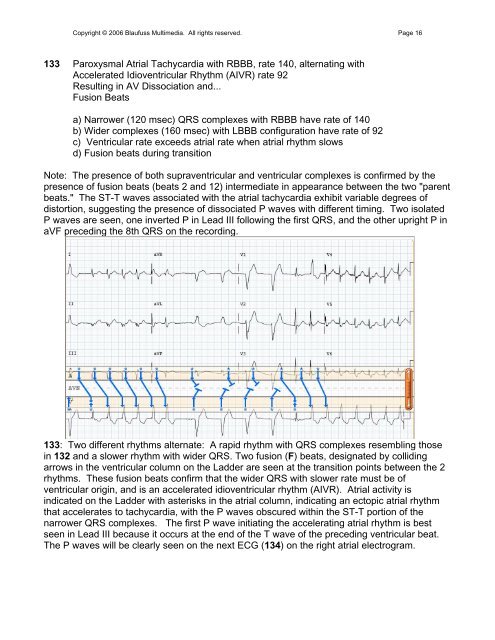
133 Paroxysmal Atrial Tachycardia with RBBB, <strong>rate</strong> 140, alternating with<br />
Accele<strong>rate</strong>d Idioventricular <strong>Rhythm</strong> (AIVR) <strong>rate</strong> 92<br />
Resulting in AV Dissociation and...<br />
Fusion Beats<br />
a) Narrower (120 msec) QRS complexes with RBBB have <strong>rate</strong> of 140<br />
b) Wider complexes (160 msec) with LBBB configuration have <strong>rate</strong> of 92<br />
c) Ventricular <strong>rate</strong> exceeds <strong>atrial</strong> <strong>rate</strong> when <strong>atrial</strong> rhythm slows<br />
d) Fusion beats during transition<br />
Note: The presence of both supraventricular and ventricular complexes is confirmed by the<br />
presence of fusion beats (beats 2 and 12) intermediate in appearance between the two "parent<br />
beats." The ST-T waves associated with the <strong>atrial</strong> tachycardia exhibit variable degrees of<br />
distortion, suggesting the presence of dissociated P waves with different timing. Two isolated<br />
P waves are seen, one inverted P in Lead <strong>II</strong>I following the first QRS, and the other upright P in<br />
aVF preceding the 8th QRS on the recording.<br />
133: Two different rhythms alternate: A rapid rhythm with QRS complexes resembling those<br />
in 132 and a slower rhythm with wider QRS. Two fusion (F) beats, designated by colliding<br />
arrows in the ventricular column on the Ladder are seen at the transition points between the 2<br />
rhythms. These fusion beats confirm that the wider QRS with slower <strong>rate</strong> must be of<br />
ventricular origin, and is an accele<strong>rate</strong>d idioventricular rhythm (AIVR). Atrial activity is<br />
indicated on the Ladder with asterisks in the <strong>atrial</strong> column, indicating an ectopic <strong>atrial</strong> rhythm<br />
that accele<strong>rate</strong>s to tachycardia, with the P waves obscured within the ST-T portion of the<br />
narrower QRS complexes. The first P wave initiating the accelerating <strong>atrial</strong> rhythm is best<br />
seen in Lead <strong>II</strong>I because it occurs at the end of the T wave of the preceding ventricular beat.<br />
The P waves will be clearly seen on the next ECG (134) on the right <strong>atrial</strong> electrogram.