

002 Sinus Rhythm, atrial rate 90 Mobitz II - Blaufuss Multimedia
002 Sinus Rhythm, atrial rate 90 Mobitz II - Blaufuss Multimedia
002 Sinus Rhythm, atrial rate 90 Mobitz II - Blaufuss Multimedia

You also want an ePaper? Increase the reach of your titles
YUMPU automatically turns print PDFs into web optimized ePapers that Google loves.
Copyright © 2006 <strong>Blaufuss</strong> <strong>Multimedia</strong>. All rights reserved. Page 36<br />
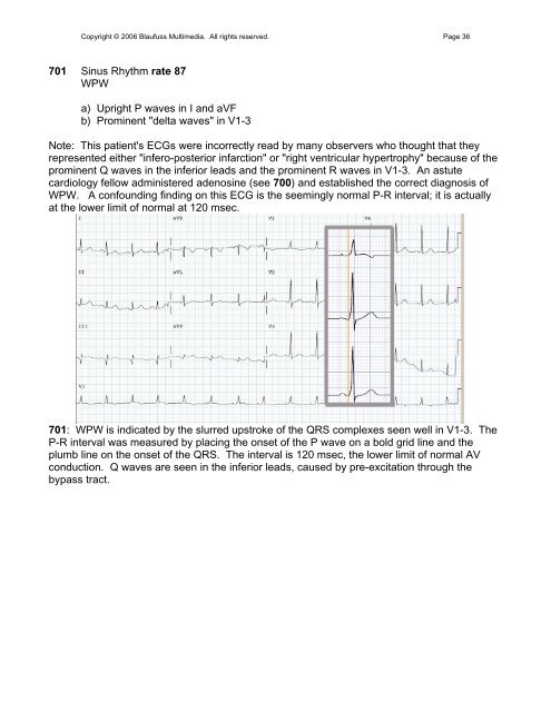
701 <strong>Sinus</strong> <strong>Rhythm</strong> <strong>rate</strong> 87<br />
WPW<br />
a) Upright P waves in I and aVF<br />
b) Prominent "delta waves" in V1-3<br />
Note: This patient's ECGs were incorrectly read by many observers who thought that they<br />
represented either "infero-posterior infarction" or "right ventricular hypertrophy" because of the<br />
prominent Q waves in the inferior leads and the prominent R waves in V1-3. An astute<br />
cardiology fellow administered adenosine (see 700) and established the correct diagnosis of<br />
WPW. A confounding finding on this ECG is the seemingly normal P-R interval; it is actually<br />
at the lower limit of normal at 120 msec.<br />
701: WPW is indicated by the slurred upstroke of the QRS complexes seen well in V1-3. The<br />
P-R interval was measured by placing the onset of the P wave on a bold grid line and the<br />
plumb line on the onset of the QRS. The interval is 120 msec, the lower limit of normal AV<br />
conduction. Q waves are seen in the inferior leads, caused by pre-excitation through the<br />
bypass tract.