

002 Sinus Rhythm, atrial rate 90 Mobitz II - Blaufuss Multimedia
002 Sinus Rhythm, atrial rate 90 Mobitz II - Blaufuss Multimedia
002 Sinus Rhythm, atrial rate 90 Mobitz II - Blaufuss Multimedia

Create successful ePaper yourself
Turn your PDF publications into a flip-book with our unique Google optimized e-Paper software.
Copyright © 2006 <strong>Blaufuss</strong> <strong>Multimedia</strong>. All rights reserved. Page 22<br />
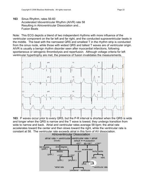
163 <strong>Sinus</strong> <strong>Rhythm</strong>, <strong>rate</strong>s 56-60<br />
Accele<strong>rate</strong>d Idioventricular <strong>Rhythm</strong> (AIVR) <strong>rate</strong> 58<br />
Resulting in Atrioventricular Dissociation and...<br />
Fusion Beats<br />
Note: This ECG depicts a blend of two independent rhythms with more influence of the<br />
ventricular component on the far left and far right, and the conducted supraventricular beats in<br />
the middle. The beat with the narrowest QRS and smallest T in the rhythm strip is conducted<br />
from the sinus node, while those with widest QRS and tallest T waves are of ventricular origin.<br />
AIVR is usually a benign rhythm disorder seen after myocardial infarctions, following<br />
spontaneous or iatrogenic thrombolysis and reperfusion. Although voltage criteria for left<br />
ventricular hypertrophy are met, the presence of fusion invalidates the measurements.<br />
163: P waves occur prior to every QRS, but the P-R interval is shortest when the QRS is wide<br />
and longer when the QRS is narrow and the T wave is lowest; they undergo transition from<br />
wide to narrow and back. Atrial and ventricular <strong>rate</strong>s average 58 bpm; the <strong>atrial</strong> <strong>rate</strong><br />
accele<strong>rate</strong>s toward the center and then slows toward the right, while the ventricular <strong>rate</strong> is<br />
constant at 58. The ventricular <strong>rate</strong> exceeds <strong>atrial</strong> in this form of AV dissociation.