

002 Sinus Rhythm, atrial rate 90 Mobitz II - Blaufuss Multimedia
002 Sinus Rhythm, atrial rate 90 Mobitz II - Blaufuss Multimedia
002 Sinus Rhythm, atrial rate 90 Mobitz II - Blaufuss Multimedia

You also want an ePaper? Increase the reach of your titles
YUMPU automatically turns print PDFs into web optimized ePapers that Google loves.
Copyright © 2006 <strong>Blaufuss</strong> <strong>Multimedia</strong>. All rights reserved. Page 6<br />
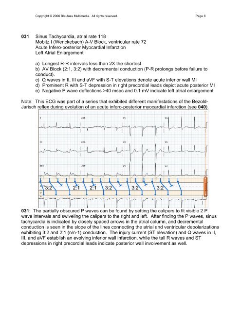
031 <strong>Sinus</strong> Tachycardia, <strong>atrial</strong> <strong>rate</strong> 118<br />
<strong>Mobitz</strong> I (Wenckebach) A-V Block, ventricular <strong>rate</strong> 72<br />
Acute Infero-posterior Myocardial Infarction<br />
Left Atrial Enlargement<br />
a) Longest R-R intervals less than 2X the shortest<br />
b) AV Block (2:1, 3:2) with decremental conduction (P-R prolongs before failure to<br />
conduct).<br />
c) Q waves in <strong>II</strong>, <strong>II</strong>I and aVF with S-T elevations denote acute inferior wall MI<br />
d) Prominent R with S-T depression in right precordial leads depict acute posterior MI<br />
e) Negative P wave deflections >40 msec and 0.1 mV indicate left <strong>atrial</strong> enlargement<br />
Note: This ECG was part of a series that exhibited different manifestations of the Bezold-<br />
Jarisch reflex during evolution of an acute infero-posterior myocardial infarction (see 040).<br />
031: The partially obscured P waves can be found by setting the calipers to fit visible 2 P<br />
wave intervals and swiveling the calipers to the right and left. After finding the P waves, sinus<br />
tachycardia is indicated by closely spaced arrows in the <strong>atrial</strong> column, and decremental<br />
conduction is seen in the slope of the lines connecting the <strong>atrial</strong> and ventricular depolarizations<br />
exhibiting 3:2 and 2:1 (n/n-1) conduction. The injury current (ST elevation) and Q waves in <strong>II</strong>,<br />
<strong>II</strong>I, and aVF establish an evolving inferior wall infarction, while the tall R waves and ST<br />
depressions in right precordial leads indicate posterior wall involvement as well.