

002 Sinus Rhythm, atrial rate 90 Mobitz II - Blaufuss Multimedia
002 Sinus Rhythm, atrial rate 90 Mobitz II - Blaufuss Multimedia
002 Sinus Rhythm, atrial rate 90 Mobitz II - Blaufuss Multimedia

You also want an ePaper? Increase the reach of your titles
YUMPU automatically turns print PDFs into web optimized ePapers that Google loves.
Copyright © 2006 <strong>Blaufuss</strong> <strong>Multimedia</strong>. All rights reserved. Page 20<br />
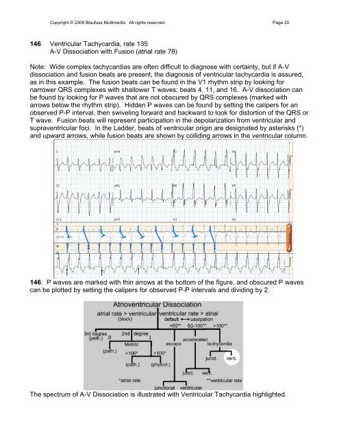
146 Ventricular Tachycardia, <strong>rate</strong> 135<br />
A-V Dissociation with Fusion (<strong>atrial</strong> <strong>rate</strong> 78)<br />
Note: Wide complex tachycardias are often difficult to diagnose with certainty, but if A-V<br />
dissociation and fusion beats are present, the diagnosis of ventricular tachycardia is assured,<br />
as in this example. The fusion beats can be found in the V1 rhythm strip by looking for<br />
narrower QRS complexes with shallower T waves: beats 4, 11, and 16. A-V dissociation can<br />
be found by looking for P waves that are not obscured by QRS complexes (marked with<br />
arrows below the rhythm strip). Hidden P waves can be found by setting the calipers for an<br />
observed P-P interval, then swiveling forward and backward to look for distortion of the QRS or<br />
T wave. Fusion beats will represent participation in the depolarization from ventricular and<br />
supraventricular foci. In the Ladder, beats of ventricular origin are designated by asterisks (*)<br />
and upward arrows, while fusion beats are shown by colliding arrows in the ventricular column.<br />
146: P waves are marked with thin arrows at the bottom of the figure, and obscured P waves<br />
can be plotted by setting the calipers for observed P-P intervals and dividing by 2.<br />
The spectrum of A-V Dissociation is illust<strong>rate</strong>d with Ventricular Tachycardia highlighted.