Use of two-dimensional matched filters for estimating a length of ...
Use of two-dimensional matched filters for estimating a length of ...
Use of two-dimensional matched filters for estimating a length of ...

You also want an ePaper? Increase the reach of your titles
YUMPU automatically turns print PDFs into web optimized ePapers that Google loves.
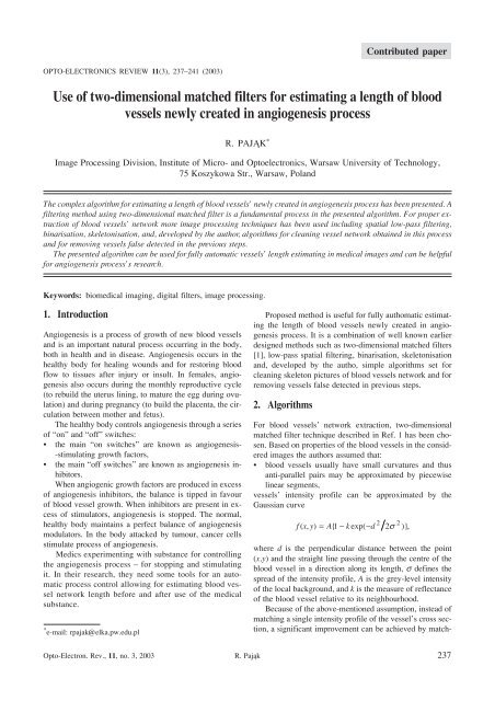
Contributed paperOPTO-ELECTRONICS REVIEW 11(3), 237–241 (2003)<strong>Use</strong> <strong>of</strong> <strong>two</strong>-<strong>dimensional</strong> <strong>matched</strong> <strong>filters</strong> <strong>for</strong> <strong>estimating</strong> a <strong>length</strong> <strong>of</strong> bloodvessels newly created in angiogenesis processR. PAJ¥K *Image Processing Division, Institute <strong>of</strong> Micro- and Optoelectronics, Warsaw University <strong>of</strong> Technology,75 Koszykowa Str., Warsaw, PolandThe complex algorithm <strong>for</strong> <strong>estimating</strong> a <strong>length</strong> <strong>of</strong> blood vessels’ newly created in angiogenesis process has been presented. Afiltering method using <strong>two</strong>-<strong>dimensional</strong> <strong>matched</strong> filter is a fundamental process in the presented algorithm. For proper extraction<strong>of</strong> blood vessels’ ne<strong>two</strong>rk more image processing techniques has been used including spatial low-pass filtering,binarisation, skeletonisation, and, developed by the author, algorithms <strong>for</strong> cleaning vessel ne<strong>two</strong>rk obtained in this processand <strong>for</strong> removing vessels false detected in the previous steps.The presented algorithm can be used <strong>for</strong> fully automatic vessels’ <strong>length</strong> <strong>estimating</strong> in medical images and can be helpful<strong>for</strong> angiogenesis process’s research.Keywords: biomedical imaging, digital <strong>filters</strong>, image processing.1. IntroductionAngiogenesis is a process <strong>of</strong> growth <strong>of</strong> new blood vesselsand is an important natural process occurring in the body,both in health and in disease. Angiogenesis occurs in thehealthy body <strong>for</strong> healing wounds and <strong>for</strong> restoring bloodflow to tissues after injury or insult. In females, angiogenesisalso occurs during the monthly reproductive cycle(to rebuild the uterus lining, to mature the egg during ovulation)and during pregnancy (to build the placenta, the circulationbetween mother and fetus).The healthy body controls angiogenesis through a series<strong>of</strong> “on” and “<strong>of</strong>f” switches:• the main “on switches” are known as angiogenesis--stimulating growth factors,• the main “<strong>of</strong>f switches” are known as angiogenesis inhibitors.When angiogenic growth factors are produced in excess<strong>of</strong> angiogenesis inhibitors, the balance is tipped in favour<strong>of</strong> blood vessel growth. When inhibitors are present in excess<strong>of</strong> stimulators, angiogenesis is stopped. The normal,healthy body maintains a perfect balance <strong>of</strong> angiogenesismodulators. In the body attacked by tumour, cancer cellsstimulate process <strong>of</strong> angiogenesis.Medics experimenting with substance <strong>for</strong> controllingthe angiogenesis process – <strong>for</strong> stopping and stimulatingit. In their research, they need some tools <strong>for</strong> an automaticprocess control allowing <strong>for</strong> <strong>estimating</strong> blood vesselne<strong>two</strong>rk <strong>length</strong> be<strong>for</strong>e and after use <strong>of</strong> the medicalsubstance.* e-mail: rpajak@elka.pw.edu.plProposed method is useful <strong>for</strong> fully authomatic <strong>estimating</strong>the <strong>length</strong> <strong>of</strong> blood vessels newly created in angiogenesisprocess. It is a combination <strong>of</strong> well known earlierdesigned methods such as <strong>two</strong>-<strong>dimensional</strong> <strong>matched</strong> <strong>filters</strong>[1], low-pass spatial filtering, binarisation, skeletonisationand, developed by the autho, simple algorithms set <strong>for</strong>cleaning skeleton pictures <strong>of</strong> blood vessels ne<strong>two</strong>rk and <strong>for</strong>removing vessels false detected in previous steps.2. AlgorithmsFor blood vessels’ ne<strong>two</strong>rk extraction, <strong>two</strong>-<strong>dimensional</strong><strong>matched</strong> filter technique described in Ref. 1 has been chosen.Based on properties <strong>of</strong> the blood vessels in the consideredimages the authors assumed that:• blood vessels usually have small curvatures and thusanti-parallel pairs may be approximated by piecewiselinear segments,vessels’ intensity pr<strong>of</strong>ile can be approximated by theGaussian curve2 2fxy ( , ) = A{ 1-kexp( -d2s )},where d is the perpendicular distance between the point(x,y) and the straight line passing through the centre <strong>of</strong> theblood vessel in a direction along its <strong>length</strong>, s defines thespread <strong>of</strong> the intensity pr<strong>of</strong>ile, A is the grey-level intensity<strong>of</strong> the local background, and k is the measure <strong>of</strong> reflectance<strong>of</strong> the blood vessel relative to its neighbourhood.Because <strong>of</strong> the above-mentioned assumption, instead <strong>of</strong>matching a single intensity pr<strong>of</strong>ile <strong>of</strong> the vessel’s cross section,a significant improvement can be achieved by match-Opto-Electron. Rev., 11, no. 3, 2003 R. Paj¹k 237
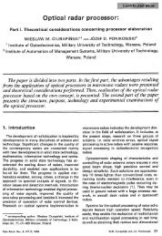
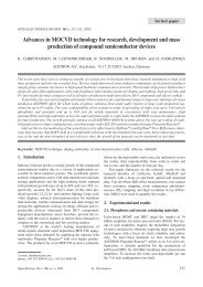
<strong>Use</strong> <strong>of</strong> <strong>two</strong>-<strong>dimensional</strong> <strong>matched</strong> <strong>filters</strong> <strong>for</strong> <strong>estimating</strong> a <strong>length</strong> <strong>of</strong> blood vessels newly created in angiogenesis processNext, it is necessary to make a skeleton picture from abinary image. An algorithm <strong>for</strong> this operation has beentaken from Ref. 3. Then, it is necessary to use some cleaningalgorithms <strong>for</strong> removing false detected, unconnectedblood vessels, <strong>for</strong> removing standalone pixels, and <strong>for</strong> removingfalse detected <strong>for</strong>k points. The applied algorithmsbased on neighbourhood properties <strong>of</strong> local pixels’. Schematicdiagram <strong>of</strong> the whole process is presented in Fig. 1.3. Application and resultsPictures <strong>of</strong> chicken egg surface with a few weeks old embryoshave been used <strong>for</strong> experiments. A photo <strong>of</strong> thechicken egg surface is presented in Fig. 2.Fig. 1. Schematic diagram <strong>of</strong> the image processing algorithm.ing a number <strong>of</strong> cross sections <strong>of</strong> identical pr<strong>of</strong>ile simultaneously.Thus, a kernel can be used which mathematicalexpression isK(x,y) = –exp(-x 2 /2s 2 )<strong>for</strong> |y| £ L/2,where L is the <strong>length</strong> <strong>of</strong> the segment <strong>for</strong> which fixed orientationhas been assumed. The direction <strong>of</strong> the blood vesselhere is assumed to be aligned along the y-axis and <strong>for</strong> differentorientations the kernel has to be rotated accordingly.Convolution <strong>of</strong> the generated kernel with a vessel intensitypr<strong>of</strong>ile function allows deciding if the pixel belongs to thevessel or not.Be<strong>for</strong>e using a <strong>matched</strong> filter <strong>for</strong> vessel detection it isrecommended to use a low-pass filter <strong>for</strong> noise removing.Simple mean filter with a 5´5 mask has been used.Next step after filtering stage is binarisation. A differentbinarisation methods has been tested and Otsu’s methoddescribed in Ref. 2 has been chosen as the one giving bestresults. In short, this method consists in finding optimalthreshold <strong>for</strong> image based on selecting the lowest point between<strong>two</strong> pixels’ classes. First, variance <strong>of</strong> the <strong>two</strong> classesis calculated separately (within-class variance) and a totalvariance <strong>for</strong> a whole image. The criterion function involvesminimising the ratio <strong>of</strong> the between classes variance to thetotal variance. Because the criterion function depends onthe threshold value we can find the optimal threshold calculatingcriterion function’s value iterative and findingminimum function’s value in this way.Fig. 2. A photo <strong>of</strong> chicken egg’s. An analysed part <strong>of</strong> the image ismarked with a black line.Fig. 3. Zoomed photo <strong>of</strong> the blood vessels’ structure.238 Opto-Electron. Rev., 11, no. 3, 2003 © 2003 COSiW SEP, Warsaw
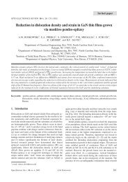
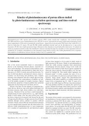
Contributed paperFig. 4. RGB components <strong>of</strong> the image (green component in the middle).Fig. 5. Matched filter output <strong>for</strong> the smallest (left) and biggest (right) vessel’s diameter.The pictures have been obtained using digital cameraconnected to a microscope set. Size <strong>of</strong> pictures taken in theacquisition process was 3200´2400 pixels and the pixel bitdepth was 24 bits. Colour picture has been split into threeRGB components and <strong>for</strong> further analysis only the greencomponent has been taken because <strong>of</strong> the highest contrast.From this picture a smaller one, including analysed part <strong>of</strong>blood vessels’ ne<strong>two</strong>rk has been cut out and passed <strong>for</strong> furtheranalysis.Zoomed picture <strong>of</strong> the part <strong>of</strong> blood vessels’ ne<strong>two</strong>rkand three RGB components are presented in Figs. 3 and 4,respectively. After smoothing the greyscale picture withthe spatial mean filter (5´5 mask has been used) a set <strong>of</strong><strong>matched</strong> <strong>filters</strong> have been used. A set <strong>of</strong> <strong>filters</strong> has beenconstructed with different kernel sizes because <strong>of</strong> differentblood vessel diameters detected. For every supposed vesseldiameter a new kernel has been built with different widthsand <strong>length</strong>s. A width <strong>of</strong> the kernel depends on the detectedvessel’s diameter and the <strong>length</strong> depends on the assumedfixed orientation <strong>length</strong>. Because <strong>of</strong> the need to detect vessels<strong>of</strong> different spatial orientation, the kernel has to be rotatedaccordingly. Assuming an angular resolution <strong>of</strong> 15°,12 different kernels are needed to span all possible orientations.Corresponding responses from every rotated filter arecompared and <strong>for</strong> each pixel only the maximum responsevalue is to be retained.A filtering procedure is repeated as many times as thedifferent values <strong>of</strong> vessels’ diameter is assumed. At the end<strong>of</strong> every filterbank, new output image is generated. The result<strong>of</strong> application <strong>of</strong> the <strong>matched</strong> filter to the image <strong>for</strong><strong>two</strong> assumed vessels’ diameters is given in Fig. 5.For every kernel size its output image is binarised usingthe Otsu’s method. A result <strong>of</strong> the binarisation processis given in Fig. 6. After that, all binary pictures areadded up (binary sum) and new image is generated as abinary representation <strong>of</strong> the blood vessels ne<strong>two</strong>rk(Fig. 7). Such a solution has been proposed because <strong>of</strong>the effects <strong>of</strong> the applied <strong>matched</strong> filter. It can be seen inFig. 5 that <strong>for</strong> small kernel width blood vessels with thebigger diameters are improperly represented in the sameway as the thin vessels are (only edges <strong>of</strong> the vessels aredetected). On the contrary, <strong>for</strong> the bigger kernels, the in<strong>for</strong>mationabout smaller vessels could be lost. For thisreason a set <strong>of</strong> the <strong>matched</strong> <strong>filters</strong> followed by a separatedbinarisation and the final sum image has been proposed.Opto-Electron. Rev., 11, no. 3, 2003 R. Paj¹k 239
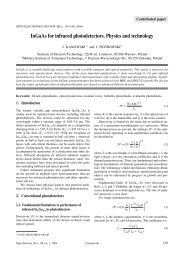
<strong>Use</strong> <strong>of</strong> <strong>two</strong>-<strong>dimensional</strong> <strong>matched</strong> <strong>filters</strong> <strong>for</strong> <strong>estimating</strong> a <strong>length</strong> <strong>of</strong> blood vessels newly created in angiogenesis processFig. 6. Binarised filter outputs <strong>for</strong> the pictures <strong>of</strong> Fig. 5.Next step in presented algorithm is picture skeletonising.The goal <strong>of</strong> the operation is to bring the in<strong>for</strong>mationabout vessels’ ne<strong>two</strong>rk topology, ending points’ location,<strong>for</strong>k points, passing over the in<strong>for</strong>mation about bloodvessels’ thickness. The result <strong>of</strong> this operation is presentedin Fig. 8. As it can be seen, the marked connectionne<strong>two</strong>rk differs from the expected one. It is caused by thealgorithm properties. It marks <strong>for</strong>ks and small vessels thatin fact do not exist. Because <strong>of</strong> that additional operationsare needed.First, all isolated pixels are removed from the picture.Next, all short vessels not connected with the rest <strong>of</strong> thevesssels’ ne<strong>two</strong>rk are removed from the picture either. Every<strong>for</strong>k point marked in the previous step is tested now if itis a real <strong>for</strong>k point. For every <strong>for</strong>k, the point blood vesselwidth is determined. The algorithm uses in<strong>for</strong>mation fromthe binary image presented in Fig. 7 <strong>for</strong> approximatingblood vessel’s width at the given point characterised by theproperties <strong>of</strong> the skeleton image. False detected <strong>for</strong>k pointsare unchecked and marked blood vessels connected to themare removed.Two images are given in Fig. 9. One <strong>of</strong> them is a separatedblood vessels structure taken from the image and theother is a corresponding skeleton image after initial “cleaning”procedure.The final, cleaned skeleton image is used <strong>for</strong> markingvessels’ ending points and <strong>for</strong>k points. Coordinates <strong>of</strong>these points are used <strong>for</strong> <strong>estimating</strong> the blood vessels<strong>length</strong>. A vessels geometry is approximated by apiecewise linear segments and a sum <strong>of</strong> their <strong>length</strong>s istaken as a final vessels <strong>length</strong>. Except <strong>of</strong> that it is possibleto estimate the <strong>length</strong> <strong>of</strong> the newly created vessels or theFig. 7. Sum <strong>of</strong> all binary pictures.Fig. 8. Binary image after skeletonising.240 Opto-Electron. Rev., 11, no. 3, 2003 © 2003 COSiW SEP, Warsaw
Contributed paperFig. 9. Separated blood vessels’ ne<strong>two</strong>rk (left) and its skeleton representation.total area <strong>of</strong> the blood vessels’ ne<strong>two</strong>rk in the analysedpart <strong>of</strong> the image.4. ConclusionsPresented set <strong>of</strong> algortihms can be used as a powerful tool<strong>for</strong> fully automatic blood veesels’ ne<strong>two</strong>rk or newly createdvessels’ <strong>length</strong> <strong>estimating</strong>. It can be helpful in monitoring<strong>of</strong> the angiogenesis process stimulated or stopped bythe stimulators or the inhibitors accordingly making ananalysis <strong>of</strong> the results easier and faster.References1. S. Chaudhuri, S. Chatterjee, N. Katz, M. Nelson, and M.Goldbaum, “Detection <strong>of</strong> blood vessels in retinal images using<strong>two</strong>-<strong>dimensional</strong> <strong>matched</strong> <strong>filters</strong>”, IEEE Trans. on MedicalImaging 8, 263–269 (1989).2. N. Otsu, “A threshold selection method from grey-level histograms”,IEEE Trans. Syst., Man., Cybern. SMC-9, 62–66(1979).3. C. Quek, G.S. Ng, and R.W. Zhou, “A novel single-passthinning algorithm and an effective set <strong>of</strong> per<strong>for</strong>mance criteria”,Pattern Recognition Letters 16, 1267–1275 (1995).Opto-Electron. Rev., 11, no. 3, 2003 R. Paj¹k 241
SpringerMarian A. Herman, Polish Academy <strong>of</strong> Sciences, Warsaw, Poland;Helmut Sitter, University <strong>of</strong> Linz, AustriaMolecular Beam EpitaxyFundamentals and Current Status1996. 2nd rev. and updated ed.. XIV, 453 pp. 260 figs., 25 in color,(Springer Series in Materials Science, Volume 7)Hardcover. EUR 54.95; £ 38.50; sFr 107ISBN 3-540-60594-0Molecular Beam Epitaxy describes a technique in wide-spread use <strong>for</strong> the production <strong>of</strong> high-quality semiconductor devices. It discusses the mostimportant aspects <strong>of</strong> the MBE apparatus, the physics and chemistry <strong>of</strong> the crystallization <strong>of</strong> various materials and device structures, and the characterizationmethods that relate the structural parameters <strong>of</strong> the grown (or growing) film or structure to the technologically relevant procedure. In thissecond edition <strong>two</strong> new fields have been added: crystallization <strong>of</strong> as-grown low-<strong>dimensional</strong> heterostructures, mainly quantum wires and quantumdots, and in-growth control <strong>of</strong> the MBE crystallization process <strong>of</strong> strained-layer structures. Out-<strong>of</strong>-date material has been removed.Jacob Greenberg, The Hebrew University, Jerusalem, IsraelThermodynamic Basis <strong>of</strong> Crystal GrowthP-T-X Phase Equilibrium and Non-Stoichiometry2002. VIII, 249 pp. 126 figs., 28 tabs.(Springer Series in Materials Science, Volume 44)Hardcover. EUR 79.95; £ 56.00; sFr 133.ISBN 3-540-41246-8This book covers the thermodynamic foundations <strong>of</strong> inorganic materials science and the controlled synthesis <strong>of</strong> inorganic materials. A new thermodynamicapproach to the non-stoichiometry <strong>of</strong> crystalline solids, known as vapour pressure scanning, has been developed by the author andis described in detail in this book. It is based on the high-precision experimental determination <strong>of</strong> the boundaries <strong>of</strong> the single-phase volume <strong>of</strong>the solid in the pressure-temperature-composition P-T-X phase space. This approach has been tested on a number <strong>of</strong> inorganic materials and hasbeen shown to have an unparalleled precision (up to 10-4 at.%) in the determination <strong>of</strong> non-stoichiometry directly at high temperatures (up to1200°C). Along with the results obtained by the author and his colleagues, the P-T-X diagrams <strong>of</strong> other important materials (e.g., III-V, IV-VIsemiconductors) are also discussed.Vitaly Shchukin, Nikolai N. Ledentsov, Dieter Bimberg, Technische Universität Berlin, GermanySelf-organized Formation <strong>of</strong> Semiconductor Nanostructures2003. Approx. 250 pp. 70 figs., 10 in color.(NanoScience and Technology)Hardcover. Approx. EUR 64.50ISBN 3-540-67817-4The main focus <strong>of</strong> the book are physical mechanisms <strong>of</strong> spontaneous <strong>for</strong>mation <strong>of</strong> ordered nanostructures at semiconductor surfaces. Thesemechanisms lie behind recent breakthroughs in advanced nanotechnology <strong>of</strong> quantum wire-and quantum dot fabrication. Generic theoreticalmodels are presented addressing <strong>for</strong>mation <strong>of</strong> all basic types <strong>of</strong> nanostructures, including periodically faceted surfaces, arrays <strong>of</strong> step-bunches <strong>of</strong>equal heights and single- and multi-sheet arrays <strong>of</strong> both 2- and 3-D islands. Decisive experiments on both structural and optical characterization<strong>of</strong> nanostructures are discussed to verify theoretical models and link them to practical examples. Experimental tools are described that enableone to intentionally control parameters <strong>of</strong> self-organized nanostructures, like chemical composition, shape, size, density and relative arrangement<strong>of</strong> quantum dots and wires.Boyan Mutaftschiev, Universites de Paris VI et VII, Paris, FranceThe Atomistic Nature <strong>of</strong> Crystal Growth2001. XIII, 368 pp. 98 figs.(Springer Series in Materials Science, Volume 43)Hardcover. EUR 94.95; £ 66.50; sFr 157.50ISBN 3-540-66496-3Crystal growth and nucleation are treated in the specialized literature in different ways depending on the discipline in question (physics, physicalchemistry, chemical engineering) and on the theoretical approaches (atomistic vs continuum approach as regards crystal growth, phase vs chemicalconcept as regards nucleation). This book relates the different approaches to one another, giving preference to atomistic treatments by themethods <strong>of</strong> statistical thermodynamics and chemical kinetics. This unified approach also facilitates an understanding <strong>of</strong> some related phenomena<strong>of</strong> surface physics, such as adsorption, wetting etc. The book allows research novices and graduate students to get an insight into the physics<strong>of</strong> the phenomena and to interpret some <strong>of</strong> the experimental results.All EUR and GBP prices are net prices subject to local VAT; e.g. in Germany 7% VAT <strong>for</strong> books. Prices and other details aresubject to change without notice. Create your own interest pr<strong>of</strong>ile at: http://www.springer.de/alert242 Opto-Electron. Rev., 11, no. 3, 2003 © 2003 COSiW SEP, Warsaw