

World Journal of Radiology - World Journal of Gastroenterology
World Journal of Radiology - World Journal of Gastroenterology
World Journal of Radiology - World Journal of Gastroenterology

Create successful ePaper yourself
Turn your PDF publications into a flip-book with our unique Google optimized e-Paper software.
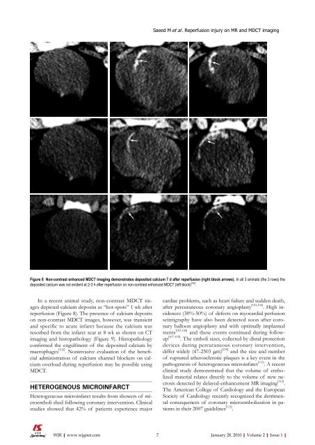
In a recent animal study, non-contrast MDCT images<br />
depicted calcium deposits as “hot-spots” 1 wk after<br />
reperfusion (Figure 8). The presence <strong>of</strong> calcium deposits<br />
on non-contrast MDCT images, however, was transient<br />
and specific to acute infarct because the calcium was<br />
resorbed from the infarct scar at 8 wk as shown on CT<br />
imaging and histopathology (Figure 9). Histopathology<br />
confirmed the engulfment <strong>of</strong> the deposited calcium by<br />
macrophages [102] . Noninvasive evaluation <strong>of</strong> the beneficial<br />
administration <strong>of</strong> calcium channel blockers on calcium<br />
overload during reperfusion may be possible using<br />
MDCT.<br />
HETEROGENOUS MICROINFARCT<br />
Heterogeneous microinfarct results from showers <strong>of</strong> microemboli<br />
shed following coronary intervention. Clinical<br />
studies showed that 42% <strong>of</strong> patients experience major<br />
WJR|www.wjgnet.com<br />
Saeed M et al . Reperfusion injury on MR and MDCT imaging<br />
Figure 8 Non-contrast enhanced MDCT imaging demonstrates deposited calcium 7 d after reperfusion (right block arrows). In all 3 animals (the 3 rows) the<br />
deposited calcium was not evident at 2-3 h after reperfusion on non-contrast enhanced MDCT (left block) [102] .<br />
cardiac problems, such as heart failure and sudden death,<br />
after percutaneous coronary angioplasty [103,104] . High incidences<br />
(30%-50%) <strong>of</strong> defects on myocardial perfusion<br />
scintigraphy have also been detected soon after coronary<br />
balloon angioplasty and with optimally implanted<br />
stents [105,106] and these events continued during followup<br />
[107-109] . The emboli sizes, collected by distal protection<br />
devices during percutaneous coronary intervention,<br />
differ widely (47-2503 μm) [110] and the size and number<br />
<strong>of</strong> ruptured atherosclerotic plaques is a key event in the<br />
pathogenesis <strong>of</strong> heterogeneous microinfarct [111] . A recent<br />
clinical study demonstrated that the volume <strong>of</strong> embolized<br />
material relates directly to the volume <strong>of</strong> new necrosis<br />
detected by delayed-enhancement MR imaging [112] .<br />
The American College <strong>of</strong> Cardiology and the European<br />
Society <strong>of</strong> Cardiology recently recognized the detrimental<br />
consequences <strong>of</strong> coronary microembolization in patients<br />
in their 2007 guidelines [113] .<br />
7 January 28, 2010|Volume 2|Issue 1|


