

World Journal of Radiology - World Journal of Gastroenterology
World Journal of Radiology - World Journal of Gastroenterology
World Journal of Radiology - World Journal of Gastroenterology

Create successful ePaper yourself
Turn your PDF publications into a flip-book with our unique Google optimized e-Paper software.
Ignee A et al . CEUS <strong>of</strong> renal masses<br />
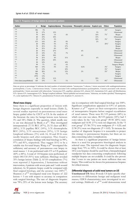
Table 2 Frequency <strong>of</strong> benign lesions in consecutive patients<br />
Author n Benign Angiomyolipomas Oncocytomas Metanephric adenomas Atypical cysts Others Population<br />
[103]<br />
396 5% 1% 7<br />
2% 8<br />
Surgery<br />
[104]<br />
30 17% 13% 3% Ultrasound<br />
[105]<br />
70 26% 1% 4% 14% 7% 3<br />
Biopsy<br />
[106]<br />
40 23% 10% 13% Ultrasound<br />
[107]<br />
35 17% 3% 10% 3% 1<br />
Surgery<br />
[108]<br />
173 14% 4% 8% 2% Surgery<br />
[109]<br />
20 35% 5% 5% 25% 2<br />
Surgery<br />
[110]<br />
78 21% 3% 17% Biopsy<br />
[111]<br />
26 21% 8% 12% Biopsy<br />
[60]<br />
26 31% 27% 4% Ultrasound<br />
[80]<br />
29 10% 3% 7% Ultrasound<br />
[61]<br />
23 34% 30% 4% Ultrasound<br />
[112]<br />
54 21% 4% 11% 6% Biopsy<br />
[113]<br />
97 25% 25% Ultrasound<br />
[4]<br />
30 10% 10% Ultrasound<br />
[114]<br />
99 7% 1% 6% Surgery/Biopsy<br />
[115]<br />
543 15% 5% 6% 0.2% 3% 4<br />
Surgery<br />
[73]<br />
31 13% 10% 3% 5<br />
Surgery<br />
[59]<br />
100 20% 3% 13% 2% 2% 6<br />
Biopsy<br />
[64]<br />
954 7% 7% Surgery<br />
[100]<br />
2770 13% Surgery<br />
[78]<br />
18 33% 11% 22% Ultrasound<br />
Own data (unpublished) 143 15% 3% 1% 1% 1% 8% 9<br />
Ultrasound<br />
Data are given as percentage; N indicates the total number <strong>of</strong> included patients. 1 Leiomyoma; 2 1 abscess, 1 lesion associated with xanthogranulomatous<br />
pyelonephritis, 2 cysts, 1 arteriovenous fistula; 3 1 lesion associated with xanthogranulomatous pyelonephritis, 3 lesions associated with chronic<br />
pyelonephritis, 1 lesion associated with tuberculosis; 4 Leiomyoma 0.9%, papillary adenoma 0.6%, abscess 0.4%, haematoma 0.4%, giant cell fibroblastoma<br />
0.2%, lipoma 0.2%, haemangioma 0.2%; 5 Benign lymphoid infiltrate; 6 Mixed epithelial and stromal tumor; 7 Adenoma, not further specified; 8 Haemangioma<br />
1%, cystic nephroma 0.5%; 9 Abscess 2%, pseudotumour 4%,focal cystic dysplasia 1%, necrosis 1%.<br />
Renal mass biopsy<br />
Since there is a significant proportion <strong>of</strong> lesions with<br />
benign diagnosis especially in small lesions (Table 2),<br />
several studies reported on percutaneous renal mass<br />
biopsy guided either by CECT or US. In the analysis <strong>of</strong><br />
the literature the rates for benign lesions were between<br />
5% and 34% (Table 2). The question, which needle size<br />
to use was discussed by Breda et al [73] . They investigated<br />
intraoperatively 27/31 RCC (87%), 21/31 clear cell RCC<br />
(68%), 3/31 papillary RCC (10%), 3/31 chromophobe<br />
RCC (10%), 3/31 oncocytomas (10%), 1/31 benign<br />
lymphoid infiltrates (3%) with 14, 18 and 20 G core<br />
needle biopsies each after extirpation. They found a<br />
correlation <strong>of</strong> biopsy findings with final histology in 94%,<br />
97% and 81%, respectively. They suggest 18 G to be a<br />
suitable size for renal biopsy. Wang et al [59] investigated the<br />
sufficiency and accuracy <strong>of</strong> percutaneous core biopsy in<br />
renal masses < 4 cm performed with CT or US guidance<br />
(60% vs 40%). A total <strong>of</strong> 110 biopsies were performed, <strong>of</strong><br />
which 100/110 (91%) were sufficient. Histology revealed<br />
35% benign lesions (Table 2). 8/110 complications (7%)<br />
were reported (1 hypotension, 2 hematomas without<br />
intervention, 4 patients with severe pain and 1 with wound<br />
infection). In 34 patients biopsy could be compared with<br />
final surgical histology, and the accuracy was 100% [59] .<br />
Shannon et al [116] investigated renal core biopsies <strong>of</strong> 222<br />
lesions < 5 cm with respect to accuracy in comparison<br />
with surgical histology. The rate <strong>of</strong> diagnostic biopsies<br />
was 78%; 25% <strong>of</strong> the lesions were benign. The accuracy<br />
WJR|www.wjgnet.com<br />
rate in comparison with final surgical histology was 100%.<br />
Significant complications appeared in 0.9% <strong>of</strong> patients.<br />
Kramer et al [117] report on their retrospective analysis<br />
<strong>of</strong> intraoperative biopsies before surgical cryoablation<br />
<strong>of</strong> renal tumors. There were 81/119 patients (68%) in<br />
which one core was taken, 38/119 patients (32%) had 3<br />
cores taken. In the “one core group” 49/81 (60%) were<br />
malignant and 14/81 (17%) were not diagnostic. In the “3<br />
core group” 27/38 (71%) were malignant (P = 0.25) and<br />
2/38 (5%) were not diagnostic (P = 0.03). To increase the<br />
number <strong>of</strong> diagnostic biopsies it is reasonable to project<br />
this strategy to percutaneous biopsies, but there are no<br />
data concerning safety/complications.<br />
As there is a significant proportion <strong>of</strong> benign lesions<br />
there is a need for preoperative histological analysis in<br />
selected cases. The reported rates for diagnostic biopsy<br />
range from 75% to 100%. It could be shown that at least<br />
18 G core biopsies should be used from a histopathological<br />
standpoint but data concerning complications following<br />
multiple biopsies are not available. It could be also proven<br />
that 3 cores in one patient are more sufficient than one<br />
biopsy. This could not be shown for percutaneous biopsies<br />
for ethical reasons.<br />
Differential diagnosis <strong>of</strong> solid renal tumors on US<br />
Unenhanced US: Since B-mode US lacks specific characteristics<br />
to differentiate benign and malignant renal<br />
masses, CDUS characteristics were investigated in several<br />
settings. Habboub et al [118] could demonstrate renal<br />
22 January 28, 2010|Volume 2|Issue 1|


