

World Journal of Radiology - World Journal of Gastroenterology
World Journal of Radiology - World Journal of Gastroenterology
World Journal of Radiology - World Journal of Gastroenterology

You also want an ePaper? Increase the reach of your titles
YUMPU automatically turns print PDFs into web optimized ePapers that Google loves.
nostic value in predicting a future diagnosis <strong>of</strong> ischemia,<br />
infarct, or death. First pass perfusion imaging can be<br />
used to discriminate ischemic myocardium. A recent MRimpact<br />
study in 234 patients reported improved detection<br />
<strong>of</strong> ischemic myocardium distal to coronary stenosis compared<br />
to single photon emission computed tomography<br />
in a multicenter and multivendor randomized trial [27] .<br />
MDCT imaging has also been used in the evaluation<br />
<strong>of</strong> cardiac function, myocardial viability and plaque morphology<br />
[28-30] . A preclinical study demonstrated that this<br />
modality has the potential to detect infarct heterogeneity<br />
in the peri-infarct zone [31] . Recent experimental studies using<br />
modern MDCT technology confirmed the potential<br />
<strong>of</strong> the technique in depicting ischemic myocardium during<br />
the first pass perfusion <strong>of</strong> iodinated contrast media [10,32] .<br />
HOMOGENEOUS MYOCARDIAL INFARCT<br />
Several studies indicated that there is a close correlation<br />
between homogeneous myocardial infarct size, dimensions<br />
(size, circumferential extent and transmurality) and<br />
LV remodeling. Inversion-recovery low-angle-shot MR<br />
imaging and helical MDCT imaging have been recently<br />
introduced and performed following the intravenous administration<br />
<strong>of</strong> contrast media with a delay <strong>of</strong> 5-10 min to<br />
define myocardial infarct dimensions [7,9,10,33-39] (Figure 2).<br />
Investigators found that differentially contrast enhanced<br />
regions on MR and MDCT imaging correlate well with<br />
areas <strong>of</strong> decreased flow [32,40] and dobutamine stress on<br />
echocardiography [41] . Furthermore, the combined use<br />
<strong>of</strong> cine and DE-MR imaging are able to differentiate regional<br />
transitional dysfunction in stunned and hibernating<br />
myocardium from permanent dysfunction on con-<br />
WJR|www.wjgnet.com<br />
Saeed M et al . Reperfusion injury on MR and MDCT imaging<br />
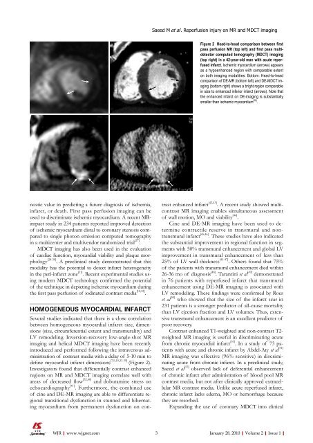
Figure 2 Head-to-head comparison between first<br />
pass perfusion MR (top left) and first pass multidetector<br />
computed tomography (MDCT) imaging<br />
(top right) in a 42-year-old man with acute reperfused<br />
infarct. Ischemic myocardium (arrows) appears<br />
as a hypoenhanced region with comparable extent<br />
on both imaging modalities. Bottom: Head-to-head<br />
comparison <strong>of</strong> DE-MR (bottom left) and DE-MDCT imaging<br />
(bottom right) shows a bright region comparable<br />
in size to enhanced inferior infarct (arrows). Note that<br />
the enhanced infarct on DE-imaging is substantially<br />
smaller than ischemic myocardium [38] .<br />
trast enhanced infarct [42,43] . A recent study showed multicontrast<br />
MR imaging enables simultaneous assessment<br />
<strong>of</strong> wall motion, MO and viability [44] .<br />
Cine and DE-MR imaging have been used to determine<br />
contractile reserve in transmural and nontransmural<br />
infarct [45,46] . These studies have also indicated<br />
the substantial improvement in regional function in segments<br />
with 50% transmural enhancement and global LV<br />
improvement in transmural enhancement <strong>of</strong> less than<br />
25% <strong>of</strong> LV wall thickness [45-47] . Others found that 75%<br />
<strong>of</strong> the patients with transmural enhancement died within<br />
26-36 mo <strong>of</strong> diagnosis [48] . Tarantini et al [49] demonstrated<br />
in 76 patients with reperfused infarct that transmural<br />
enhancement using DE-MR imaging is associated with<br />
LV remodeling. These findings were confirmed by Roes<br />
et al [50] who showed that the size <strong>of</strong> the infarct scar in<br />
231 patients is a stronger predictor <strong>of</strong> all-cause mortality<br />
than LV ejection fraction and LV volumes. Thus, extensive<br />
transmural enhancement is an excellent predictor <strong>of</strong><br />
poor recovery.<br />
Contrast enhanced T1-weighted and non-contrast T2weighted<br />
MR imaging is useful in discriminating acute<br />
from chronic myocardial infarct [51] . In a study <strong>of</strong> 73 patients<br />
with acute and chronic infarct by Abdel-Aty et al [51]<br />
MR imaging was effective (96% sensitive) in discriminating<br />
acute from chronic infarct. In a preclinical study,<br />
Saeed et al [52] observed lack <strong>of</strong> deferential enhancement<br />
<strong>of</strong> chronic infarct after administration <strong>of</strong> blood pool MR<br />
contrast media, but not after clinically approved extracellular<br />
MR contrast media. Unlike acute reperfused infarct,<br />
chronic infarct lacks edema, MO or hemorrhage because<br />
they are resorbed.<br />
Expanding the use <strong>of</strong> coronary MDCT into clinical<br />
3 January 28, 2010|Volume 2|Issue 1|


