Breast Cancer - Arab Medical Association Against Cancer
Breast Cancer - Arab Medical Association Against Cancer
Breast Cancer - Arab Medical Association Against Cancer
- No tags were found...

Create successful ePaper yourself
Turn your PDF publications into a flip-book with our unique Google optimized e-Paper software.
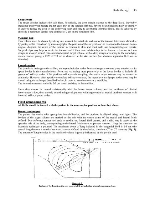
Radiotherapy 145Chest wallThe target volume includes the skin flaps. Posteriorly, the deep margin extends to the deep fascia, inevitablyincluding underlying muscle and rib-cage. Part of the surgical scar may have to be excluded medially or laterallyin order to reduce the dose to the underlying heart and lung to acceptable tolerance limits. This is achieved byallowing a maximum central lung distance of 2 cm on the simulator film.Tumour bedThis volume must be chosen by taking into account the initial site and size of the tumour determined clinically,by photographic record and by mammography, the position of the surgical scar .in relation to the tumour from asurgical diagram, the depth of the tumour in relation to skin and chest wall, and histopathological reports.Surgical clips may help to locate the tumour bed if their exact relationship to the tumour is known. A 2 cmmargin is allowed around this estimated clinical target volume, with a deep margin extending to the underlyingmuscle fascia,. giving a PTV of 7-9 cm in diameter at the skin surface (i.e. electron applicator 8-10 cm indiameter).Lymph nodesThe lymphatic drainage to the axillary and supraclavicular nodes forms an irregular volume lying anteriorly at itsupper border in the supraclavicular fossa, and extending more posteriorly at the lower border to include allgroups of axillary nodes. After positive axillary-node sampling, the entire target volume may be treated incontinuity. However, after a positive complete axillary clearance, the supraclavicular lymph nodes alone may betreated using the technique described below, in order to avoid unnecessary morbidity.The internal mammary nodes lie 2-3 cm lateral and deep to the mid-line.Since they cannot be treated satisfactorily with the breast target volume, and the incidence of clinicalinvolvement is low, they are only treated in high-risk patients with large central or medial quadrant tumours withinvolved axillary lymph nodes.Field arrangementsAll fields should be treated with the patient in the same supine position as described above.<strong>Breast</strong> techniqueThe patient lies supine with appropriate immobilization, and her position is aligned using laser lights. Theborders of the target volume are marked on the skin with the centre points of the medial and lateral fieldsdefined. Two reference tattoos are made at medial and lateral field centres, and a third one is made on theopposite side of the body, corresponding to the lateral field centre, to prevent rotation. Using the simulator, anisocentric technique is planned. The maximum depth of lung included in the tangential field is 2-3 cm (thecentral lung distance is usually less than 2 cm) as defined by simulation, simulator-CT or CT scanning (Fig. 2).The amount of lung included in the irradiated volume is greatly influenced by the portals used.Figure 8.2.Isodose of the breast on the axis tangential field, including internal mammary chain