- Page 1 and 2:
1Chapter 1Epidemiology and risk ass
- Page 3 and 4:
Epidemiology and risk assessment 3c
- Page 5 and 6:
Epidemiology and risk assessment 55
- Page 7 and 8:
Epidemiology and risk assessment 7t
- Page 9: Epidemiology and risk assessment 9O
- Page 12: Epidemiology and risk assessment 12
- Page 16 and 17: Breast Cancer screening 16Breast ca
- Page 18 and 19: Breast Cancer screening 18situation
- Page 20 and 21: Breast Cancer screening 20cause und
- Page 22 and 23: Breast Cancer screening 22The rate
- Page 24 and 25: Breast Cancer screening 24could be
- Page 26: Breast Cancer screening 26These res
- Page 30 and 31: Staging and end results of therapy
- Page 32 and 33: Staging and end results of therapy
- Page 34 and 35: Staging and end results of therapy
- Page 36 and 37: Staging and end results of therapy
- Page 38 and 39: Staging and end results of therapy
- Page 40 and 41: Staging and end results of therapy
- Page 42 and 43: Staging and end results of therapy
- Page 44 and 45: Staging and end results of therapy
- Page 46 and 47: Breast Cancer and Genetic Predispos
- Page 48 and 49: Breast Cancer and Genetic Predispos
- Page 50 and 51: Breast Cancer and Genetic Predispos
- Page 52 and 53: Breast Cancer and Genetic Predispos
- Page 54 and 55: Breast Cancer and Genetic Predispos
- Page 56 and 57: Breast Cancer and Genetic Predispos
- Page 58 and 59: Breast Imaging 56Breast Cancer is t
- Page 62 and 63: Breast Imaging 60Preoperative views
- Page 64 and 65: Breast Imaging 62Breast diagnosisTh
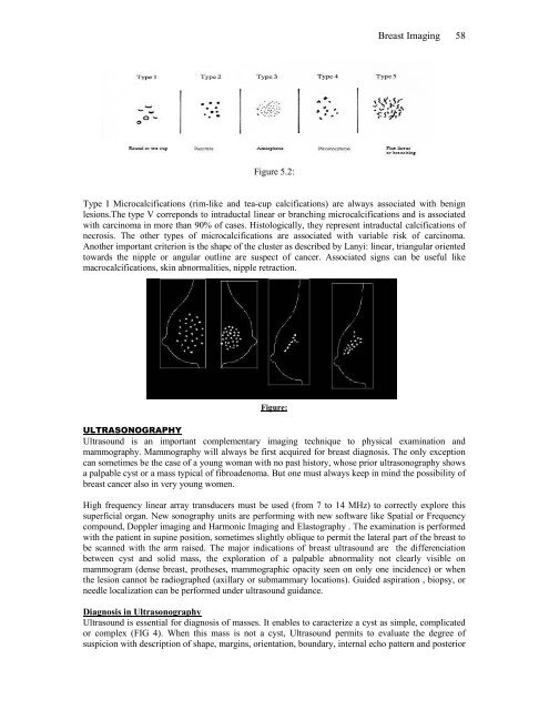
- Page 66 and 67: Breast Imaging 64TYPICAL BENIGN CAL
- Page 68 and 69: Breast Imaging 66Numerousfine linea
- Page 70 and 71: Breast Imaging 68can appear under t
- Page 72 and 73: Pathology and methods of analysiCha
- Page 74 and 75: Pathology and methods of analysis 7
- Page 76 and 77: Pathology and methods of analysis 7
- Page 78 and 79: Pathology and methods of analysis 7
- Page 80 and 81: Pathology and methods of analysis 7
- Page 82 and 83: Pathology and methods of analysis 8
- Page 84 and 85: Pathology and methods of analysis 8
- Page 86 and 87: Pathology and methods of analysis 8
- Page 88 and 89: Pathology and methods of analysis 8
- Page 90 and 91: Pathology and methods of analysis 8
- Page 92 and 93: Pathology and methods of analysis 9
- Page 94 and 95: Pathology and methods of analysis 9
- Page 96 and 97: Chapter 7CPlastic SurgerySurgery 13
- Page 98 and 99: Surgery 135The safety of gel-filled
- Page 100 and 101: Surgery 137Breast Reconstruction Us
- Page 102 and 103: Surgery 139Goal Filled material Sha
- Page 104 and 105: Chapter 8Radiotherapy of BreastRadi
- Page 106 and 107: Radiotherapy 143When a patient pres
- Page 108 and 109: Radiotherapy 145Chest wallThe targe
- Page 110 and 111:
Radiotherapy 147Various techniques
- Page 112 and 113:
Radiotherapy 149CT treatment planni
- Page 114 and 115:
Radiotherapy 151Implementation of p
- Page 116 and 117:
Systemic Adjuvant Therapy 153During
- Page 118 and 119:
Systemic Adjuvant Therapy 155therap
- Page 120 and 121:
Systemic Adjuvant Therapy 157In pra
- Page 122 and 123:
Systemic Adjuvant Therapy 159A. Ova
- Page 124 and 125:
Systemic Adjuvant Therapy 161and pl
- Page 126 and 127:
Chapter 12Management of advanced br
- Page 128 and 129:
Management of advanced breast cance
- Page 130 and 131:
Management of advanced breast cance
- Page 132 and 133:
Management of advanced breast cance
- Page 134 and 135:
Management of advanced breast cance
- Page 136 and 137:
Management of advanced breast cance
- Page 138 and 139:
Hormonal treatment 184Chapter 11Hor
- Page 140 and 141:
Hormonal treatment 186Endocrine Hor
- Page 142 and 143:
Hormonal treatment 188HORMONAL THER
- Page 144 and 145:
Hormonal treatment 190important for
- Page 146 and 147:
Hormonal treatment 192Conference (1
- Page 148 and 149:
Hormonal treatment 194trials have d
- Page 150 and 151:
Hormonal treatment 196combination o
- Page 152 and 153:
Hormonal treatment 198Metastatic br
- Page 154 and 155:
Prognostic factors 200The predictio
- Page 156 and 157:
Prognostic factors 202the term of R
- Page 158 and 159:
Prognostic factors 2043. Histologic
- Page 160 and 161:
Prognostic factors 2066 Other histo
- Page 162 and 163:
Prognostic factors 2087. c-erbB-2c-
- Page 164 and 165:
Prognostic factors 210biology has l
- Page 166 and 167:
Prognostic factors 212changes produ
- Page 168 and 169:
Prognostic factors 214Her2 patients
- Page 170 and 171:
CriteriaP valueN+ 10 -5SBR grade 10
- Page 172 and 173:
Prognostic factors 218Table 14.7. R
- Page 174 and 175:
Prognostic factors 220• Metalloth
- Page 176 and 177:
Persistent Pain 223Although breast
- Page 178 and 179:
Persistent Pain 225• Pain associa
- Page 180 and 181:
Persistent Pain 227• Heavy painfu
- Page 182 and 183:
Persistent Pain 2292. Analgesics ar
- Page 184 and 185:
Persistent Pain 231for long duratio
- Page 186 and 187:
Persistent Pain 233Implantable Syst
- Page 188 and 189:
23518. - M I 11 ygy. I T vg f g
- Page 190 and 191:
23742. Dg .M. . uy f y g.J
- Page 192 and 193:
2373. Hu DJ g D A H . 17 N-y f k
- Page 194 and 195:
241102. Mk gg C A 18 Dv g u
- Page 196 and 197:
243133. J 10 T gy f g- f v uf
- Page 198 and 199:
24510. A w k Pg v vu f 21-g
- Page 200 and 201:
247188. J PP PH F 175 Iu f .
- Page 202 and 203:
2421 C F F P JY 177 Dé I- Cu .
- Page 204 and 205:
251244. F E 175 Hg v f fig u - f .
- Page 206 and 207:
253271. u JM u . 14 N u g yy
- Page 208 and 209:
2552. Hff u T Hzg P M. [Iv MI f
- Page 210 and 211:
257328. u C g u CC Mkk-z N - E F
- Page 212 and 213:
2535. M C C M . I f MI ug y
- Page 214 and 215:
21388. P ED Hk E Yff M . u gg v
- Page 216 and 217:
23417. J g HH Tv FA Iv uy g f u
- Page 218 and 219:
25445. y F Mu T u JP . C D A - g f
- Page 220 and 221:
27475. Tkk Juu H k P 18 T g g-f f
- Page 222 and 223:
2501. A . y V. Pz E. A. T f kg
- Page 224 and 225:
27152. Aww H E-wy A 172 y g f .
- Page 226 and 227:
273557 D T u A C F . 13 Tu u vu fi
- Page 228 and 229:
275584. Hy 1 A ug vw f f . C 24
- Page 230 and 231:
27713 N C Fu P u Fu MC J Cug . y
- Page 232 and 233:
2742 N.. MD E.A. H.Pk .H. I u
- Page 234 and 235:
2817 Ag JC ME T E 178 A A w
- Page 236 and 237:
2832 C D 10 P u v u . I Fw- T C
- Page 238 and 239:
285717 D C F A . 1 Ajuv -y u f w
- Page 240 and 241:
287745 M g g T 182 A wky u f w-
- Page 242 and 243:
28774 M Fuqu A A . 14 Njuv vu ju


