18.1 Studying Viruses and Prokaryotes
18.1 Studying Viruses and Prokaryotes
18.1 Studying Viruses and Prokaryotes

You also want an ePaper? Increase the reach of your titles
YUMPU automatically turns print PDFs into web optimized ePapers that Google loves.
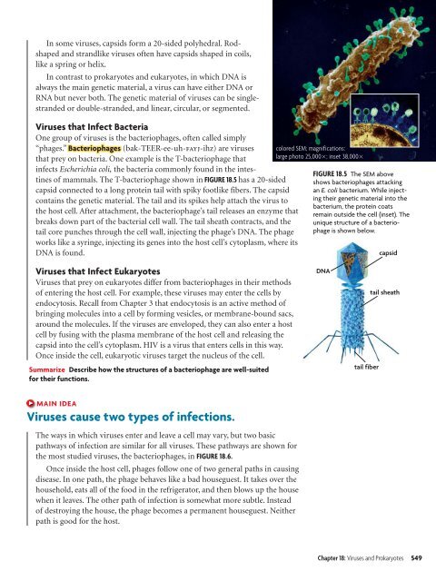
In some viruses, capsids form a 20-sided polyhedral. Rodshaped<strong>and</strong> str<strong>and</strong>like viruses often have capsids shaped in coils,like a spring or helix.In contrast to prokaryotes <strong>and</strong> eukaryotes, in which DNA isalways the main genetic material, a virus can have either DNA orRNA but never both. The genetic material of viruses can be singlestr<strong>and</strong>edor double-str<strong>and</strong>ed, <strong>and</strong> linear, circular, or segmented.<strong>Viruses</strong> that Infect BacteriaOne group of viruses is the bacteriophages, often called simply“phages.” Bacteriophages (bak-TEER-ee-uh-fayj-ihz) are virusesthat prey on bacteria. One example is the T-bacteriophage thatinfects Escherichia coli, the bacteria commonly found in the intestinesof mammals. The T-bacteriophage shown in FIGURE 18.5 has a 20-sidedcapsid connected to a long protein tail with spiky footlike fibers. The capsidcontains the genetic material. The tail <strong>and</strong> its spikes help attach the virus tothe host cell. After attachment, the bacteriophage’s tail releases an enzyme thatbreaks down part of the bacterial cell wall. The tail sheath contracts, <strong>and</strong> thetail core punches through the cell wall, injecting the phage’s DNA. The phageworks like a syringe, injecting its genes into the host cell’s cytoplasm, where itsDNA is found.colored SEM; magnifications:large photo 25,000; inset 38,000FIGURE 18.5 The SEM aboveshows bacteriophages attackingan E. coli bacterium. While injectingtheir genetic material into thebacterium, the protein coatsremain outside the cell (inset). Theunique structure of a bacteriophageis shown below.capsid<strong>Viruses</strong> that Infect Eukaryotes<strong>Viruses</strong> that prey on eukaryotes differ from bacteriophages in their methodsof entering the host cell. For example, these viruses may enter the cells byendocytosis. Recall from Chapter 3 that endocytosis is an active method ofbringing molecules into a cell by forming vesicles, or membrane-bound sacs,around the molecules. If the viruses are enveloped, they can also enter a hostcell by fusing with the plasma membrane of the host cell <strong>and</strong> releasing thecapsid into the cell’s cytoplasm. HIV is a virus that enters cells in this way.Once inside the cell, eukaryotic viruses target the nucleus of the cell.Summarize Describe how the structures of a bacteriophage are well-suitedfor their functions.DNAtail fibertail sheathMAIN IDEA<strong>Viruses</strong> cause two types of infections.The ways in which viruses enter <strong>and</strong> leave a cell may vary, but two basicpathways of infection are similar for all viruses. These pathways are shown forthe most studied viruses, the bacteriophages, in FIGURE 18.6.Once inside the host cell, phages follow one of two general paths in causingdisease. In one path, the phage behaves like a bad houseguest. It takes over thehousehold, eats all of the food in the refrigerator, <strong>and</strong> then blows up the housewhen it leaves. The other path of infection is somewhat more subtle. Insteadof destroying the house, the phage becomes a permanent houseguest. Neitherpath is good for the host.Chapter 18: <strong>Viruses</strong> <strong>and</strong> <strong>Prokaryotes</strong> 549