ruoounu. nnSlunCIS&UINI-rOSIlnS - the Society for Reproductive ...
ruoounu. nnSlunCIS&UINI-rOSIlnS - the Society for Reproductive ...
ruoounu. nnSlunCIS&UINI-rOSIlnS - the Society for Reproductive ...

Create successful ePaper yourself
Turn your PDF publications into a flip-book with our unique Google optimized e-Paper software.
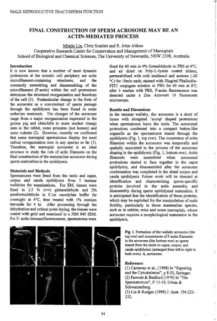
MALE REPROD.UCTIVE TRACT/SPERM FUNCTIONMALE REPRODUCTIVE TRACT/SPERM FUNCTIONFINAL CONSTRUCTION OF SPERM ACROSOME MAY BE ANACTIN-MEDIATED PROCESSMiniie Lin, Chris Scarlett and R. John AitkenCooperative Research Centre <strong>for</strong> Conservation and Management ofMarsupialsSchool ofBiological and Chemical Sciences, The University ofNewcastle, NSW 2308, AustraliaIntroductionIt is now known that a number of most dynamicprotrusions at <strong>the</strong> somatic cell periphery are actinmicrofilament-containing structures, and <strong>the</strong>constantly assembling and disassembling of <strong>the</strong>microfilament (F-actin) within <strong>the</strong> cell protrusionsdetermine <strong>the</strong> structural reorganisation and functionsof <strong>the</strong> cell (1). Posttesticular change in <strong>the</strong> <strong>for</strong>m of<strong>the</strong> acrosome as a concomitant of sperm passagethrough <strong>the</strong> .epididymis has been found in someeu<strong>the</strong>rian mammals. The changes of <strong>the</strong> acrosomerange from a major reorganization expressed in <strong>the</strong>guinea pig and chinchilla to more modest changeseen in <strong>the</strong> rabbit, some primates (not human) andsome rodents (2). However, recently we confirmedthat some marsupial spermatozoa display <strong>the</strong> mostradical reorganization seen in any species so far (3).There<strong>for</strong>e, <strong>the</strong> marsupial acrosome is an idealstructure to study <strong>the</strong> role of actin filaments on <strong>the</strong>final construction of<strong>the</strong> mammalian acrosome duringsperm maturation in <strong>the</strong> epididymis.Materials and MethodsSpermatozoa were freed from <strong>the</strong> testis and caput,corpus and cauda epididymis from 5 tammarwallabies <strong>for</strong> examinations. For EM, tissues werefixed in 2.5 % (v/v) glutaraldehyde and 2%para<strong>for</strong>maldehYde in O.lm cacodylate buffer <strong>for</strong>overnight at 4°C, <strong>the</strong>n treated with 1% osmiumtetroxide <strong>for</strong> 4 hr. After processing through <strong>the</strong>dehydration and critical point drying, <strong>the</strong> tissues werecoated with gold and examined in a JSM 840 SEM.For F- actin immunofluorescences, spermatozoa werefixed <strong>for</strong> 60 min in 4% <strong>for</strong>maldehyde in PBS at 4°C,and air dried on Poly-L-Iysine coated sliders;permeabilised with cold methanol and acetone (-20°C) <strong>for</strong> 10min each; stained with 50J.lg/ml PhalloidinFITC conjugate solution in PBS <strong>for</strong> 60 min at RT;after 3 washes with PBS, F-actin fluorescence wasdetected under a Ziss Axiovert 10 fluorescentmicroscope.Results and DiscussionsIn <strong>the</strong> tammar wallaby, <strong>the</strong> acrosome is a sheet oftissue with elongated 'scoop' shaped protrusionswhen spermatozoa leave <strong>the</strong> testis. The acrosomalprotrusions condensed into a compact button-likeorganelle as <strong>the</strong> spermatozoa transit through <strong>the</strong>epididymis (Fig. 1, top row). The occurrence of actinfilaments within <strong>the</strong> acrosome was temporally andspatially associated to <strong>the</strong> process of <strong>the</strong> acrosomeshaping in <strong>the</strong> epididymis (Fig. 1, bottom row). Actinfilaments were assembled when acrosomalprotrusions started to fuse toge<strong>the</strong>r in <strong>the</strong> caputepididymis, and disassembled after <strong>the</strong> acrosomecondensation was completed in <strong>the</strong> distal corpus andcauda epididymis. Future work will be directed atidentification and characterising sperm-specificproteins involved in <strong>the</strong> actin assembly anddisassembly during sperm epididymal maturation. Itis ~ticipated that <strong>the</strong> identification ofthose proteins,which may be exploited <strong>for</strong> <strong>the</strong> manipulation of malefertility, particularly in those mammalian species,such as in rabbits, mice and some marsupials, whoseacrosome requires a morphological maturation in <strong>the</strong>epididymis.Fig. 1. Fonnation of<strong>the</strong> wallaby acrosome (<strong>the</strong>top row) and occurrences ofF-actin filamentsin <strong>the</strong> acrosome (<strong>the</strong> bottom row) as spermtransit from <strong>the</strong> testis to caput, corpus, andcauda epididymis (arranged from left to right inboth rows). A, acrosome., Reference:(1) Carraway et aI., (1998) In "Signalingand <strong>the</strong> Cytoskeleton", p 8-30, Springer.(2) Fawcett & Bed<strong>for</strong>d (1979) In "TheSpermatozoon", P 11-19, Urban &Schwarzenberg.(3) Lin & Rodger (1999) 1. Anat. 194:223232.DEVELOPMENT OF PSA-IO MONOCLONAL ANTIBODY REACTIVITY WITHBRUSHTAIL POSSUM (TRICHOSURUS VULPECULA) SPERMATOZOA DURINGEPIDIDYMAL MATURATIONM.S. Harris a and J.C. Rodger bCooperative Research Centre <strong>for</strong> <strong>the</strong> Conservation and Management ofMarsupialsaLandcare Research, Lincoln, NZ and bMacquarie University, NSW, AustraliaIntroductionThe PSA-10 monoclonal ant:J.body (mAb) binds to antigen(s) on <strong>the</strong> fibrous sheath and midpiece fibrenetwork (MFN) of mature tammar wallaby (Macropus eugenii) 'spermatozoa and <strong>the</strong> fibrous sheath.,midpiece fibre network and acrosome ofmature bmshtail possum (Trichosurus vulpecula) spermatozoa(1). This study examined <strong>the</strong> development ofPSA-l0 mAb immunoreactivity with <strong>the</strong> acrosome andmidpiece ofpossum spermatozoa during epididymal maturation.MethodsSpermatozoa were collected from <strong>the</strong> testes and proximal and distal regions of<strong>the</strong> head, body and tailof <strong>the</strong> epididymides of three possums after mincing <strong>the</strong> tissue in phosphate buffered saline (PBS)containing protease inlnbitors. Sperm were pooled by region, fixed in 4% para<strong>for</strong>maldehyde andlabelled with PSA-I0 ascites fluid or myeloma ascites as described previously (1). The fluorescencepatterns on 100 randomly selected sperm were counted <strong>for</strong> each ofthree subsamples of sperm. pooledfrom different regions of <strong>the</strong> tract. Data was analyzed using a two-tailed Fisher exact test. Freshlydissected tissue from <strong>the</strong> possum and wallaby male reproductive tract was fixed in 4%para<strong>for</strong>maldehyde <strong>for</strong> 5 h on ice be<strong>for</strong>e being processed <strong>for</strong> post-embedding immunogold labellingwith PSA-l0 ascites and myeloma ascites control (1).ResultsPSA-IO acrosomal immunor~activity was evident on most possum spennatozoa from <strong>the</strong> proximalhead of<strong>the</strong> epididymis but not those from <strong>the</strong> testis (Table 1). PSA-I0 immunoreactivity with restrictedregions of <strong>the</strong> midpiece was detected first on some possum sperm from <strong>the</strong> proximal·head of <strong>the</strong>epididymis (Table 1). The proportion of sperm showing midpiece immunoreactivity increased wi<strong>the</strong>pididymal transit, with <strong>the</strong> entire posterior midpiece ofall spermatozoa from <strong>the</strong> proximal body of<strong>the</strong>epididymis being labelled. Immunogold labelling of <strong>the</strong> possum midpiece was associated first withgranular material surrounding <strong>the</strong> mitochondrial sheath, be<strong>for</strong>e <strong>the</strong> <strong>for</strong>mation of <strong>the</strong> midpiece fibrenetwork. Labelling of <strong>the</strong> :MFN was evident after <strong>for</strong>mation in <strong>the</strong> distal head of <strong>the</strong> epididymis.Immunogold labelling of<strong>the</strong> possum acrosome was associated with <strong>the</strong> outer acrosomaI membrane.Table 1. The percentage of fixed possum spermatozoa (n=3) from different regions of <strong>the</strong> malereproductive tract demonstrating acrosomal, midpiece and principal piece immunoreactivity with PSA10 ascites fluid.Percentage ofpossum sperm demonstrating PSA-I0 mAbOrigin ofsperm inmale reproc Immunoreactivity with given structure (Mean % ±SE)Acrosome Midpiece Principal PieceTestis O±O O±O 100±0Proximal Head Epididymis 92.9±1.6* 11.9±2.0* 100±0Distal Head Epididymis 98.8±1.2* 96.0±2.1* 100±0ProximalBody Epididymis 100±0 100±0* 100±0* WIthin column, values are sIgmficantly different from preVIOUS (P