ruoounu. nnSlunCIS&UINI-rOSIlnS - the Society for Reproductive ...
ruoounu. nnSlunCIS&UINI-rOSIlnS - the Society for Reproductive ...
ruoounu. nnSlunCIS&UINI-rOSIlnS - the Society for Reproductive ...
You also want an ePaper? Increase the reach of your titles
YUMPU automatically turns print PDFs into web optimized ePapers that Google loves.
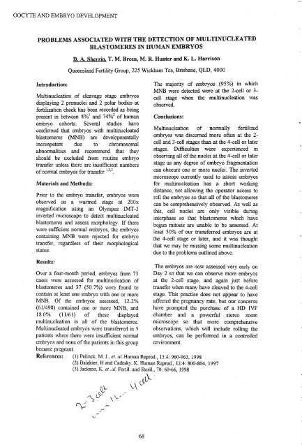
OOCYTE AND EMBRYO DEVELOPMENTOOCYTE AND EMBRYO DEVELOPMENTPROBLEMS ASSOCIATED WITH THE DETECTION OF ~LTINUCLEATEDBLASTOMERES IN HUMAN EMBRYOSIntroduction:D. A. Sherrin, T. M. Breen, M. R. Hunter and K. L. HarrisonQueensland Fertility Group, 225 Wickham Tce, Brisbane, QLD, 4000Multinucleation of cleavage stage embryosdisplaying 2 pronuclei and 2 polar bodies atfertilization check has been recorded as beingpresent in between 8%1 and 74%2 of humanembryo cohorts. Several studies haveconfirmed that embryos with multinucleatedblastomeres (MNB) are developmentallyincompetent due to chromosomalabnormalities and recommend that <strong>the</strong>yshould be excluded from routine embryotransfer unless <strong>the</strong>re are insufficient numbersofnormal embryos <strong>for</strong> transfer I.2,3.Materials and Methods:Prior to <strong>the</strong> embryo transfer, embryos wereobserved on a warmed stage at 200xmagnification using an Olympus IMT-2inverted microscope to detect multinucleatedblastomeres and assess morphology. If <strong>the</strong>rewere sufficient normal embryos, <strong>the</strong> embryoscontaining MNB were rejected <strong>for</strong> embryotransfer, regardless of <strong>the</strong>ir morphologicalstatus.Results:Over a four-month period, embryos from 73cases were assessed <strong>for</strong> multinucleation ofblastomeres and 37 (50.7%) were found tocontain at least one embryo with one or moreMNB. Of <strong>the</strong> embryos assessed, 12.2%(61/498) contained one or more MNB, and18.0% (11161) of <strong>the</strong>se displayedmultinucleation in all of <strong>the</strong> blastomeres.Multinucleated embryos. were transferred in 5patients where <strong>the</strong>re were insufficient normalembryos and none of<strong>the</strong> patients in this groupbecame pregnant.The majority of embryos (95%) in whichMNB were detected were at <strong>the</strong> 2-cell or 3cell stage when <strong>the</strong> multinucleation wasobserved.Conclusions:Multinucleation of normally fertilizedembryos was discerned more often at <strong>the</strong> 2cell and 3-cell stages than at <strong>the</strong> 4-cell or laterstages. Difficulties were experienced inobserving all of<strong>the</strong> nuclei at <strong>the</strong> 4-cell or laterstage as any degree of embryo fragmentationcan obscure one or more nuclei. The invertedmicroscope currently used to assess embryos<strong>for</strong> multinucleation has a short workingdistance, not allowing <strong>the</strong> operator access toroll <strong>the</strong> embryos so that all of<strong>the</strong> blastomerescan be comprehensively observed. As well asthis, cell nuclei are only visible duringinterphase so that blastomeres which havebegun mitosis are unable to be assessed. Atleast 50% of our transferred embryos are at<strong>the</strong> 4-cell stage or later, and it was thoughtthat we may be missing some multinucleationdue to <strong>the</strong> problems outlined above.The embryos are now assessed very early onDay 2 so that we can observe more embryosat <strong>the</strong> 2-cell stage, and again just be<strong>for</strong>etransfer when many have cleaved to <strong>the</strong> 4-cellstage. This practise does not appear to haveaffected <strong>the</strong> pregnancy rate, but our concernshave prompted <strong>the</strong> purchase of a HD IVFchamber and a powerful stereo zoommicroscope so that more comprehensiveobservations, which will include rolling <strong>the</strong>embryos, can be per<strong>for</strong>med in a controlledenvironment.References: (1) Pelinck, M. J., et. al. Human Reprod., 13:4: 960-963, 1998(2) Balakier, Hand Cadesky, K. Human Reprod., 12:4: 800-804, 1997(3) Jackson, K. et .al. Fertil. and Steril., 70: 60-66, 1998~~'v\ L~EXAMINATION OF THE INTERACTION OF EGF, FSH AND ACTIVIN A ONMEIOTIC RESUMPTION IN MOUSE OOCYTESGina Abdelnour, Steven Fleming, Giovanni Coticchio and Peter Illingworth.Department of<strong>Reproductive</strong> Endocrinology and Infertility, University ofSydney, NSW 2145Introduction: A variety of ovarian autocrine and paracrine factors may modulatefolliculogenesis, and hence oocyte maturation, The oocyte developmental programme thatleads to its meiotic resumption is governed by a precise quantitative and temporal pattern ofsignal transduction that triggers germinal vesicle break down (GVBD) proceeding to <strong>the</strong> firstmetaphase (MI) and second metaphase (MIl). FSH plays an essential role in this process. It isknown that a variety of growth factors can modulate FSH action by autocrine and paracrinemechanisms. The purpose ofthis study was to examine <strong>the</strong> effect ofEGF, activin A and FSHon cumulus-enclosed oocytes maintained in <strong>the</strong>ir meiotic arrest in vitro. The presence of adirect physiological role was also investigated by using oocytes denuded from <strong>the</strong>ir cumuluscells.Materials and Methods: Three week old female Quackenbush mice were sacrificed 48 h postPMSG (5 IV/ml) injection. Ovaries were dissected and large antral follicles were punctured torelease cumulus enclosed oocytes (CEO). CEO and denuded oocytes (DO) were cultured in aMEM medium containing 0.3% BSA and 4 mM hypoxanthine, to maintain <strong>the</strong>ir meioticarrest. Medium was supplemented with EGF (0.01 - 1 ng/ml), FSH (0.5 - 75 mIU/ml) andactivin A (50 - 200 ng/ml) ei<strong>the</strong>r alone or in combination. Oocytes with no treatment wereused as controls. Percentages ofGVBD (MI + MIl) were assessed after 17-28 h.Results: The results show that EGF and FSH alone, were able to override <strong>the</strong> inhibitory effectofhypoxanthine and induce GVBD in a dose related manner. A maximal stimulatory effect ofEGF (Ing/ml) and FSH (75 mID/ml) was observed in CEO (73% and 79% GVBDrespectively) but was minimised in DO (11 % and 20% GVBD respectively). Percentages ofGVBD did not increase when <strong>the</strong>y were combined at half-maximal concentrations, however<strong>the</strong> progression from MI to MIl was significantly improved; 39% when combined versus 24%in EGF only, and 26% in FSH only (P< 0.05). Activin A (100 ng/ml) did not affect meioticresumption when used alone (7% GVBD). Fur<strong>the</strong>rmore it had no additive effect on EGF andFSH at half-maximal doses. However when combined with high concentrations of FSH (75mIV/ml), percentages ofGVBD declined significantly by 12% (P


