ruoounu. nnSlunCIS&UINI-rOSIlnS - the Society for Reproductive ...
ruoounu. nnSlunCIS&UINI-rOSIlnS - the Society for Reproductive ...
ruoounu. nnSlunCIS&UINI-rOSIlnS - the Society for Reproductive ...

You also want an ePaper? Increase the reach of your titles
YUMPU automatically turns print PDFs into web optimized ePapers that Google loves.
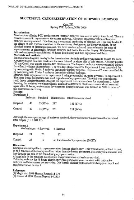
OVARIAN DEVELOPMENT/ASSISTED REPRODUCTION-FEMALEOVARIAN DEVELOPMENT/ASSISTED REPRODUCTION-FEMALEBiopsiedControlSUCCESSFUL CRYOPRESERVATION OF BIOPSIED EMBRYOS404033(82%) 21734(85%) 251Experiment 2# ofembryos # Survived # HatchedBiopsiedControl24252019CattJ.W.Sydney IVF, Sydney, NSW 2000Introduction:Most centres offering PGn produce more 'nonnal' embryos than can be safely transferred. There is<strong>the</strong>re<strong>for</strong>e a need to cryopreserve <strong>the</strong> excess embryos. However, cryopreservation ofbiopsiedembryos is compromised if<strong>the</strong> freezing is conducted on <strong>the</strong> day ofbiopsy(I,2). This may be due to<strong>the</strong> effects ofacid TYrode's solution on <strong>the</strong> remaining blastomeres, <strong>the</strong> biopsy medium, or <strong>the</strong>physical trauma ofblastomere removal. We have used an infra-red laser to breach <strong>the</strong> zona ofsupernumerary or abnonnally fertilised embryos and frozen <strong>the</strong>m after biopsy. We have alsocultured embryos <strong>for</strong> an additional 48 hours post biopsy and <strong>the</strong>n cryopreserved <strong>the</strong>m.Materials and methods:Embryos were biopsied on day3 after insemination. An infra-red laser was used to breach <strong>the</strong> zona.A twenty micron hole was made and <strong>the</strong> zona thinned on ei<strong>the</strong>r side ofthis breach. A biopsy pipette(30 J-lm Cook) was used to aspirate two blastomeres. The biopsied embryos were returned to culture<strong>for</strong> ei<strong>the</strong>r 2- 4 hours (Experiment 1), or two days (Experiment 2). Experiment 1 was controlled <strong>for</strong>by comparing with 40 day 3 embryos thawed <strong>for</strong> clinical purposes. Experiment 2 was controlled <strong>for</strong>by comparing with 25 day 5 embryos thawed <strong>for</strong> clinical purposes.Embryos were cryopreserved in experiment 1 using propanediol or, using glycerol, in experiment 2.The same freeze programme was used regardless ofcryoprotectant. Thawing was conventionalrapid thaws using propanediol/sucrose <strong>for</strong> experiment 1 or sucrose alone <strong>for</strong> experiment 2. Afterthawing, <strong>the</strong> embryos were immediately assessed to determine blastomere survival and assessedagain after 18 hours, to determine development. Embryo survival was defined as 50% or more of<strong>the</strong> blastomeres surviving.Results:Experiment 1Embryos Survived Blastomeres Blastomeres survived17145 (67%)211 (84%)Although <strong>the</strong> same percentage ofembryos survived, <strong>the</strong>re were fewer blastomeres that survivedafter biopsy (p = 0.001 X 2 ).all transferred - 3 pregnancies (10 ET)Discussion:Embryos are susceptible to cryopreservation damage after biopsy. This would seem, at least in part,to be a function of<strong>the</strong> biopsy medium ra<strong>the</strong>r than <strong>the</strong> biopsy procedure. No embryonic material waslost through <strong>the</strong> hole in <strong>the</strong> zona during cryopreservation.A large hole in <strong>the</strong> zona had no effect on cryopreservation and embryo survival.Culturing embryos <strong>for</strong> 48 hours after biopsy gave good embryonic survival with only a fewembryos and blastomeres not surviving. Our current clinical protocol allows biopsy on day 3 andcryopreservation on day 5.FOLLICLE POPULATIONS IN PREPUBERTAL, MATURE AND AGED RED DEER HINDS.Peter R Hurst, Mark W. Fisher* and Bernie J. McLeod*.Department ofAnatomy and Structural Biology, School ofMedical Sciences, U~versity ofOtago,Dunedin, New Zealand and * AgResearch, Invermay Agncultural Centre, Mosglel, New Zealand.Introduction. A previous longevity study in a group ofhinds has shown that <strong>the</strong> success ofweaning calveswas above 60% until aged 16 years and <strong>the</strong>n declined rapidly to only one calfweaned at age 19 and 20 (1).Fur<strong>the</strong>r data on ovaries has been obtained from (n = 7) prepubertal «1 yr), mature (7 yr) and old (20 yr)animals. A stereological analysis was undertaken on 2 ovaries at each age to determine <strong>the</strong> numbers ofsmall, preantral follicles.Methods. Ovaries were weighed and all follicles> 2mm were dissected, counted and measured. For <strong>the</strong>stereological study ovaries were embedded in TechnovitTM: resin and serially sectioned at 40J.lm thickness.Every 20th section was optically sectioned with 100x and lANA objective to estimate.<strong>the</strong> number of .primordial and primary follicles by.<strong>the</strong> dis~~r proced~e. A. follicle ~ sco!ed only if<strong>the</strong> nucleus ofItsconstituent oocyte was detected entirely Wlthin <strong>the</strong> 3-dimenslOnal counting disector frame.Results. The descriptive characteristics (means ±S.D.) of<strong>the</strong> ovaries at 1 yr, 7yrand 20 yr: are shown in<strong>the</strong> Table:arac erlS cvary weI t gm.No. >2mm folliclesDiameter oflargestfollicle, mm 7.25 ± 1.9 8.12 ± 0.77 5.24 ± 1.29o. nmor p :us , ,primary follicles 133,846 6,917Prepubertal and 7 yr. hinds had similar numbers ofantral follicles ofequivalent sizes bu~ <strong>the</strong> 7 yr..grouphad less primordial/primary follicles. At 21 years <strong>the</strong> deer had few, ifany detec~ble fo.llicles.. Duringoptical sectioning of<strong>the</strong> 7 year ovaries it was observed that 6% and 8% of<strong>the</strong> pnmordial follicles hadmorphological features indicative ofatresia. Most notable w~ <strong>the</strong> presence .ofat l~ast 1 ~~entedgranulosa cell nucleus (Figure). No such features were seen m any of<strong>the</strong> pnmordial follicles m <strong>the</strong>prepubertal animals.Figure showing 2 images(A & B) of<strong>the</strong> samefollicle separated by 3J.lm.In B <strong>the</strong> fragmentednuclear material ofagranulosa cell is indicatedby <strong>the</strong> arrow. Thearrowheads point to <strong>the</strong>oocyte nucleus.Discussion. This study has shown, predictably, that during ageing <strong>the</strong> population ofovarian fo~clesdiminishes to <strong>the</strong> point where old hinds have virtually no follicles present The chance observation ofgranulosa nuclear fragmentation in <strong>the</strong> 7yr. group is indicative ofgranulosa atresia an~ s:ugge~ts that as ~eovary ages <strong>the</strong> primordial follicle population is subject to increasing rates ofdegeneration. This hypo<strong>the</strong>slSis <strong>the</strong> focus offuture studies in <strong>the</strong> ovaries ofmice and humans.Reference. .1. Fisher, MW, McLeod, BJ, Mockett, BG, Moore, GH & Drew, K.R. (1996) <strong>Reproductive</strong> senescence maged red deer hinds. Proc. N.Z. Soc. Anim. Production. 56,344-346.References(1) Magli et al1999 Human Reprod 14 770(2) loris et al1999 Human Reprod 14 283384 85