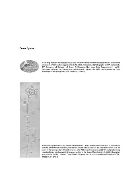
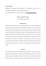
- Page 1: ISSN 0120-4157BiomédicaRevista del
- Page 5: Proceedings of theX International C
- Page 8 and 9: Financial CommiteeLavive Rebaje de
- Page 10 and 11: • Henrique L. Lenzi. Departamento
- Page 12 and 13: United States of America• Beatriz
- Page 15 and 16: Scholarship WinnersTraveling Award
- Page 17 and 18: EditorialIn 1971 the Pan American H
- Page 19 and 20: Scientific Program15:00-18:00 Arriv
- Page 21 and 22: Symposium 3. Virulence factors and
- Page 23 and 24: 16:20-16:4016:40-17:0017:00-17:2017
- Page 25 and 26: 14:20-14:4014:40-15:0015:00-15:2015
- Page 27 and 28: Symposia IndexPage434344Symposium 1
- Page 29 and 30: 91CDC42p controls yeast-cell shape
- Page 31 and 32: Poster Index1 Clinical Aspects / Ca
- Page 33 and 34: 2-02p 1552-03p 1562-04p 1562-05p 15
- Page 35 and 36: 4-20p 1744-21p 1754-22p 1754-23p 17
- Page 37 and 38: 5-10p 1915-11p 1925-12p 1925-13p 19
- Page 39 and 40: 6-06p 2076-07p 2086-08p 2086-09p 20
- Page 41: Symposium1P. brasiliensis:Genomics
- Page 44 and 45: Post-genomic contribution to the kn
- Page 46 and 47: quence. The first domain (aa 1-1073
- Page 49 and 50: Experimental animal models and thei
- Page 51 and 52: CD4 + CD25 + FoxP3 + T cells were d
- Page 53 and 54:
Pattern recognition receptors in th
- Page 55:
A strategy for the generation of a
- Page 59 and 60:
Histoplasma virulence mechanisms wi
- Page 61 and 62:
Virulence insights from the P. bras
- Page 63:
certain antifungal drugs. The effec
- Page 67 and 68:
Microbicidal mechanisms exerted by
- Page 69 and 70:
depletion induces a more severe and
- Page 71 and 72:
did not restore the lymphoprolifera
- Page 73 and 74:
Historical accountsCoccidioidomycos
- Page 75:
In South America, from Venezuela to
- Page 79 and 80:
B-1 cells modulate the kinetics of
- Page 81 and 82:
mechanisms can lead to permissivene
- Page 83 and 84:
of mice (PBS-untreated as negative
- Page 85:
Symposium6Fungal cellular biology A
- Page 88 and 89:
wall construction, mating and osmo-
- Page 90 and 91:
heat shock proteins, processes of h
- Page 92 and 93:
Differential analysis of extracellu
- Page 95 and 96:
The evolving public health importan
- Page 97:
Several difficulties have been iden
- Page 101 and 102:
The impact of histoplasmosis in HIV
- Page 103 and 104:
The conjunct data from 66 cases pre
- Page 105 and 106:
Although drug serum levels are an o
- Page 107 and 108:
Historical accountsHistoplasmosisHi
- Page 109:
Histoplasmosis in the United States
- Page 113 and 114:
The value and the promise of molecu
- Page 115 and 116:
need to consider the P. brasiliensi
- Page 117 and 118:
Lymphomononuclear cells of 29 patie
- Page 119:
Symposium10Advances on antifungal A
- Page 122 and 123:
and Cryptococcus neoformans. MICs f
- Page 124 and 125:
sue assay, revelead that P10-PLGA c
- Page 127 and 128:
P. brasiliensis growth and infectiv
- Page 129 and 130:
Influence of alternating coffee and
- Page 131 and 132:
in Arábica and 0.9% Robusta variet
- Page 133:
Symposium12Evolutive Biology of P.
- Page 136 and 137:
Is Paracoccidioides brasiliensis an
- Page 138 and 139:
matter of conjecture. As a global i
- Page 141 and 142:
1-01Clinical epidemiology of paraco
- Page 143 and 144:
1-05Epidemiological and clinical fe
- Page 145 and 146:
1-09Acute severe paracoccidioidomyc
- Page 147 and 148:
1-13Nested-PCR, immunoblotting and
- Page 149 and 150:
1-17Scintigraphic evaluation of ent
- Page 151 and 152:
1-21Paracoccidioidomycosis in a ren
- Page 153:
Poster Session2Diagnosis
- Page 156 and 157:
2-03Combined use of Paracoccidioide
- Page 159:
Poster Session3Epidemiology / Ecolo
- Page 162 and 163:
3-03Spatial distribution of chronic
- Page 165 and 166:
4-01Towards a molecular genetic sys
- Page 167 and 168:
4-05Evidence for positive selection
- Page 169 and 170:
4-09Formamidase of Paracoccidioides
- Page 171 and 172:
4-13Mating type genes MAT1-1 and MA
- Page 173 and 174:
4-17Autophagy in the human pathogen
- Page 175 and 176:
4-21Transcription regulation of the
- Page 177 and 178:
4-25Changes in the transcription le
- Page 179 and 180:
4-29The high affinity copper transp
- Page 181 and 182:
4-33Analysis and identification of
- Page 183:
4-37The heath shock factor of Parac
- Page 187 and 188:
5-01Phenotypic and functional chara
- Page 189 and 190:
5-05Polymorphisms analysis of CTLA-
- Page 191 and 192:
5-09Alterations in pulmonary mechan
- Page 193 and 194:
5-13The influence of ß2 integrin i
- Page 195 and 196:
5-17Prostaglandin inhibits Paracocc
- Page 197 and 198:
5-21IL-10 deficiency determines a b
- Page 199 and 200:
5-25Granulomatous imflammation and
- Page 201:
5-29Modulation of experimental para
- Page 205 and 206:
6-01Timing of itraconazole therapy
- Page 207 and 208:
6-05Amphotericin B-PLGA-DMSA nanopa
- Page 209 and 210:
6-09Antagonistic effect of farnesol
- Page 211 and 212:
6-13Therapeutic DNA vaccine in BALB
- Page 213:
Poster Session7Other Mycoses
- Page 216 and 217:
7-03Study of genotypic and phenotyp
- Page 218 and 219:
7-07Detection of DNA in peripheral
- Page 220 and 221:
Catão E. 193Cavalcante RS. 103, 14
- Page 222 and 223:
Molano VM. 143Molina RFS. 199Montoy
- Page 224 and 225:
Trinca LA. 167Tristão FSM. 71Urán
- Page 226 and 227:
Personal ContributorsDrs. Karl V. a