Biological and Synthetic Dioxygen Carriers
Biological and Synthetic Dioxygen Carriers
Biological and Synthetic Dioxygen Carriers

You also want an ePaper? Increase the reach of your titles
YUMPU automatically turns print PDFs into web optimized ePapers that Google loves.
4<br />
<strong>Biological</strong> <strong>and</strong> <strong>Synthetic</strong><br />
<strong>Dioxygen</strong> <strong>Carriers</strong><br />
GEOFFREY B. JAMESON<br />
Department of Chemistry<br />
Georgetown University<br />
JAMES A. IBERS<br />
Department of Chemistry<br />
Northwestern University<br />
I. INTRODUCTION: BIOLOGICAL DIOXYGEN TRANSPORT SYSTEMS<br />
Most organisms require molecular oxygen in order to survive. The dioxygen is<br />
used in a host of biochemical transformations, although most is consumed in<br />
the reaction<br />
(4.1)<br />
that is the terminal (or primary) step of oxidative phosphorylation (Chapters 5<br />
<strong>and</strong> 6). For some small animals <strong>and</strong> for plants, where the surface-to-volume<br />
ratio is large, an adequate supply of dioxygen can be obtained from simple<br />
diffusion across cell membranes. The dioxygen may be extracted from air or<br />
water; for plants that produce dioxygen in photosynthesis, it is also available<br />
endogenously. For other organisms, particularly those with non-passive lifestyles,<br />
from scorpions to whales, diffusion does not supply sufficient dioxygen<br />
for respiration.<br />
An elegant three-component system has evolved to transport dioxygen from<br />
regions of high abundance-water (at least if free of pollutant reductants) <strong>and</strong><br />
air-to regions of relatively low abundance <strong>and</strong> high dem<strong>and</strong>-the interior cells<br />
of the organism. This process is illustrated in Figure 4.1. 1 - 3 The central component<br />
is a dioxygen-carrier protein. In the three chemically distinct carriers that<br />
have evolved <strong>and</strong> are found today, the dioxygen-binding site in the protein, that<br />
is, the so-called "active site," is a complex either of copper or of iron. 4 - 6 For<br />
hemoglobins, the most widely distributed family of dioxygen carriers, the active 167
I. INTRODUCTION: BIOLOGICAL DIOXYGEN TRANSPORT SYSTEMS 171<br />
ygen binding <strong>and</strong> release are summarized first. The nonbiological chemistry of<br />
iron <strong>and</strong> copper in the presence of dioxygen is described briefly to elucidate the<br />
key role that the protein plays in supporting oxygen transport by preventing<br />
irreversible oxidation of the binding site or of its lig<strong>and</strong>s. The macroscopic<br />
behavior of the biological systems is related to the microscopic picture that has<br />
been developed over the last 30 years from x-ray crystallographic studies <strong>and</strong> a<br />
miscellany of spectroscopic probes of the oxygen-binding site. Relationships<br />
between the geometry <strong>and</strong> charge distribution in the metal-dioxygen moiety <strong>and</strong><br />
the nature of the interactions between this moiety <strong>and</strong> its surroundings are examined.<br />
Nonbiological dioxygen carriers have proved particularly useful in providing<br />
precise <strong>and</strong> accurate structural information as well as thermodynamic <strong>and</strong><br />
kinetic data against which the corresponding data from biological oxygen carriers<br />
can be contrasted.<br />
The bioinorganic chemistry of the hemoglobin family of oxygen binders is<br />
particularly amenable to study by means of small-molecule model systems: four<br />
of the five lig<strong>and</strong>s that make up the active site are provided by a square-planar<br />
tetradentate lig<strong>and</strong>, the protoporphyrin IX dianion (Figure 4.2). One axial lig<strong>and</strong><br />
in hemoglobin, imidazole from a histidine residue, is provided by the protein,<br />
<strong>and</strong> the remaining sixth coordination site is available for the exogenous lig<strong>and</strong>,<br />
e.g., dioxygen or carbon monoxide. Thus a model system that approximates the<br />
stereochemistry of the active site in hemoglobin may be assembled from an<br />
iron(II) porphyrin <strong>and</strong> a lig<strong>and</strong>, such as imidazole or pyridine. On the other<br />
h<strong>and</strong>, in hemocyanin <strong>and</strong> hemerythrin most of the lig<strong>and</strong>s are supplied by the<br />
protein. Thus the assembly of a model system that provides appropriate lig<strong>and</strong>s<br />
correctly disposed around the pair of metal atoms poses a major synthetic challenge,<br />
especially for hemocyanin, where details on the number, type, <strong>and</strong> arrangement<br />
of lig<strong>and</strong>s have been difficult to establish. Many aspects of the physical,<br />
inorganic, <strong>and</strong> structural chemistry underlying biological oxygen transport<br />
<strong>and</strong> utilization (Chapter 5) have been clarified through model systems.<br />
A. Requirements for Effective Oxygen <strong>Carriers</strong><br />
In order for dioxygen transport to be more efficient than simple diffusion through<br />
cell membranes <strong>and</strong> fluids, it is not sufficient that a metalloprotein merely binds<br />
dioxygen. Not only is there an optimal affinity of the carrier for dioxygen, but<br />
also, <strong>and</strong> more importantly, the carrier must bind <strong>and</strong> release dioxygen at a<br />
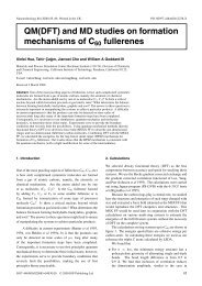
rapid rate. These thermodynamic <strong>and</strong> kinetic aspects are illustrated in Figure<br />
4.3, a general diagram of energy vs. reaction coordinate for the process<br />
(4.2)<br />
where M is an oxygen carrier, for example hemocyanin or a simple nonbiological<br />
metal complex. Thermodynamic or equilibrium aspects are summarized
172<br />
free<br />
energy<br />
(G)<br />
r= =<br />
M + 02<br />
reaction coordinate (M - 02 separation)<br />
M0 2<br />
o<br />
r= 1.9 A<br />
Figure 4.3<br />
Schematic diagram of energy changes in dioxygen binding.<br />
by !1G in Figure 4.3. As illustrated there, !1G is negative, <strong>and</strong> thus the forward<br />
reaction, dioxygen binding, is spontaneous. The equilibrium constant (K) is given<br />
by<br />
K = _a-:(_M_O..=2)_<br />
a(M)a(02) ,<br />
(4.3)<br />
where a is the activity (crudely, concentration) of the component. The equilibrium<br />
constant is related to the change in free energy by<br />
!1GO = - RTln K. (4.4)<br />
The rate of the forward reaction (k f ) is related to !1G 1*; the rate of the reverse<br />
reaction (L 1) is related to !1G!.]. Provided that oxygen binding is effectively a<br />
single-step process, then<br />
K (4.5)<br />
Usually the rates of the forward <strong>and</strong> reverse reactions are related by the empirical<br />
Arrhenius expression to quantities termed the activation energies (El<strong>and</strong><br />
E _ 1) of the reactions, where<br />
k] = A] exp (-E/RT) <strong>and</strong> L] = A-I exp (-E_J/RT). (4.6)<br />
These quantities are experimentally accessible through the change in rate as<br />
a function of temperature.
1. Thermodynamic factors 19-20<br />
I. INTRODUCTION: BIOLOGICAL DIOXYGEN TRANSPORT SYSTEMS 173<br />
The equilibrium constant K was defined in Equation (4.3) in terms of the<br />
activity ai of component i. The ai may be expressed as a function of concentration<br />
as<br />
(4.7)<br />
where for species i, )'i is its activity coefficient <strong>and</strong> [i] is its concentration (strictly<br />
molality, but usually as molarity in mol L-I). At infinite dilution )'i = 1. Provided<br />
that the charge <strong>and</strong> size of species M <strong>and</strong> M02 are similar <strong>and</strong> that O 2<br />
forms an ideal solution, then the activities of Equation (4.3) may be approximated<br />
by concentrations to give the expression<br />
(4.8)<br />
However, Equation (4.8) does not permit a direct comparison of the oxygenbinding<br />
behavior of one species in some solvent with that of a second in some<br />
other solvent. First, for a given partial pressure of dioxygen, the concentration<br />
of O 2 in the solution varies considerably with temperature <strong>and</strong> from one solvent<br />
to another. Second, reliable measurements of oxygen solubilities are not always<br />
available, <strong>and</strong> it is only relatively recently that oxygen electrodes have been<br />
developed to measure directly oxygen concentrations (strictly, activities). However,<br />
oxygen-binding measurements are normally made with a solution of M in<br />
equilibrium with gaseous dioxygen. At equilibrium the molar Gibbs' free energies<br />
(chemical potentials) of the dissolved <strong>and</strong> gaseous dioxygen are identicalif<br />
they are not, gaseous O 2 would dissolve, or dissolved O 2 would be released.<br />
Thus the solvent-dependent quantity [02] in Equation (4.8) may be replaced by<br />
the solvent-independent quantity P(02), the partial pressure of dioxygen. Under<br />
almost all experimental conditions the quantity P(02) is a very good approximation<br />
to the gas-phase activity (fugacity) of dioxygen; hence we obtain for the<br />
equilibrium constant*<br />
(4.9)<br />
It is very convenient to express the affinity as the partial pressure of dioxygen<br />
required for half-saturation of the species M, Pl/2C02). Under such conditions,<br />
[M] = [M0 2 ], one obtains<br />
(4.10)<br />
* There has been considerable discussion as to whether K c (4.8) or K p (4.9) should be used to compare<br />
dioxygen binding under different solvent conditions 21 - 23 We believe that the latter is more appropriate, since<br />
for a system al equilibrium, the chemical potential of gaseous O2 must be identical with that of dissolved 0 2 19<br />
On the other h<strong>and</strong>, the concentration of O 2 varies from one solvent to another.
174 4 / BIOlOGICAl AND SYNTHETIC DIOXYGEN CARRIERS<br />
where P li2(02) is usually given in Torr or mm Hg.* As will be detailed shortly,<br />
values for Plii02) are typically in the range 0.5 to 40 Torr.<br />
The dioxygen affinity is composed of enthalpic (Mf) <strong>and</strong> entropic (AS)<br />
components, with<br />
AGO = -RTln K (4.11)<br />
Within a family of oxygen carriers the values of AS o <strong>and</strong> Mfo are usually similar.<br />
Large deviations (such as a change of sign) are therefore indicative of a<br />
change in the nature of the oxygen-binding process.<br />
a. Non-cooperative <strong>Dioxygen</strong> Binding If the oxygen-binding sites Mare<br />
mutually independent <strong>and</strong> noninteracting, as in moderately dilute solutions of<br />
monomeric molecules, then the concentration of species M02 as a function of<br />
the partial pressure of O 2 is generally well fit by a Langmuir isotherm. 20 Here a<br />
plot of the fractional saturation of dioxygen binding sites, 0, where<br />
o = [M02]<br />
[M] + [M02]<br />
(4.12)<br />
versus P(02) gives the hyberbolic curve labeled "non-cooperative" in Figure<br />
4.4A. 9 Altematively,24 a plot of log (O/(l - 0)) versus log (P(02)), the socalled<br />
"Hill plot," gives a straight line with a slope of unity <strong>and</strong> an intercept<br />
of -log P 1I2(02) (Figure 4.4B). A differential form is shown as the dotted line<br />
in Figure 4.4C. Such binding, where the dioxygen sites are independent of each<br />
other, is termed non-cooperative.<br />
b. Cooperative <strong>Dioxygen</strong> Binding Many dioxygen-binding proteins are not<br />
independent monomers, with only one dioxygen-binding site, but oligomeric<br />
species with the protein comprising two or more similar subunits. The subunits<br />
may be held together by van der Waals' forces or by stronger interactions, such<br />
as hydrogen bonds or salt bridges, or even by covalent bonds. For example,<br />
most mammalian hemoglobins are tetramers, consisting of two pairs [af3h of<br />
myoglobin-like subunits denoted as a <strong>and</strong> f3. Either none, one, two, three, or<br />
all four sites may be occupied by dioxygen. This situation is illustrated schematically<br />
in Figure 4.5, which also shows the statistical weighting of each level<br />
of saturation, treating the a <strong>and</strong> f3 subunits as identical. Thus the binding or<br />
release of dioxygen at one site may affect the affinity <strong>and</strong> kinetics of lig<strong>and</strong><br />
binding <strong>and</strong> release at a neighboring site. As a result, the saturation curve becomes<br />
sigmoidal in shape, as illustrated in Figure 4.4A. The dioxygen binding<br />
is cooperative. When cooperativity is positive, the affinity of a vacant site is<br />
increased by occupancy of an adjacent one.<br />
This behavior, where the binding of one molecule influences the binding of<br />
successive molecules of the same kind, is referred to as a homotropic allosteric<br />
* Many authors use the symbcl P so (corresponding to 50% saturation) for P'/2.
176<br />
saturated<br />
unsaturated<br />
weight = 1<br />
1 or 3 molecules<br />
of 02 bound<br />
weight = 4<br />
2 molecules<br />
of 02 bound<br />
weight = 6<br />
Figure 4.5<br />
Diagram of tetrameric hemoglobin, showing statistical weights<br />
of different saturations (see text).<br />
interaction. A heterotropic allosteric interaction occurs when the interaction with<br />
the protein of a second unlike molecule, for instance, an organic polyphosphate<br />
for human hemoglobins, influences the binding of the first molecule (e.g., dioxygen).<br />
Such molecules are often termed allosteric effectors. A commonly observed<br />
heterotropic allosteric interaction is the Bohr effect, named after the biologist<br />
Christian Bohr, father of physicist Niels Bohr. This effect, which relates<br />
the change in partial pressure of O 2 to a change in pH at constant saturation of<br />
binding sites (0), is related thermodynamically to the Haldane effect, which<br />
relates the number of protons released (#H +) with a change in 0 at constant<br />
pH (Equation 4.13). A very large Bohr effect, where O2 affinity decreases sharply<br />
with pH, is often called the Root effect. 25a It is physiologically important for
I. INTRODUCTION: BIOLOGICAL DIOXYGEN TRANSPORT SYSTEMS 177<br />
fish such as trout, probably in maintaining buoyancy, but its molecular basis in<br />
trout hemoglobin IV remains to be discovered. 25b<br />
(4.13)<br />
The degree of cooperativity can be characterized in a number of ways. By means<br />
of a Hill plot of log (O/(l - 0)) versus log (P(02)), the limiting slopes (which<br />
should be unity) at high O2 pressure <strong>and</strong> low O2 pressure may be extrapolated<br />
as shown in Figure 4.4B to log «(}/(l 0)) = 0, where 0 = 0.5. Two limiting<br />
values for PI/2(02) are obtained, one characterizing the regime of high partial<br />
pressure of dioxygen, where the O2 affinity is high (for the case illustrated of<br />
positive cooperativity). The other PI/2(02) value characterizes the regime of low<br />
partial pressure of dioxygen, where affinity is relatively low. This difference in<br />
affinities can be converted into a difference between the free-energy change<br />
upon O 2 binding in the low-affinity state (K/ ) <strong>and</strong> the high-affinity state (K/)<br />
[the designations T <strong>and</strong> R will be described in subsection d]:<br />
- RTIn (K/ /K/). (4.14)<br />
A second way to characterize cooperativity involves fitting the oxygen-binding<br />
data at intermediate saturation (0.2 < 0 < 0.8)-that is, about the inflection<br />
point in a Hill plot-to the Hill equation<br />
or<br />
O/(l - 0)<br />
log (O/(l - 0))<br />
K p pn(02)<br />
-log (P1/2(02)) + n log (P(02))' (4.15)<br />
The Hill coefficient (n) is an empirical coefficient that has a value of unity for<br />
non-cooperative binding, where Equation (4.15) reduces to the Langmuir isotherm,<br />
Equation (4.12). Any number greater than unity indicates positive cooperativity.<br />
If O2 binding is an all-or-nothing affair, where dioxygen binding sites<br />
are either all occupied or all vacant, n equals the number of subunits in the<br />
molecule. The fit is only approximate, since the Hill plot is only approximately<br />
linear about the inflection point, as may be seen in Figure 4.4B. A more precise<br />
value of n may be obtained by plotting the slope in the Hill plot (n') as a<br />
function<br />
n'<br />
d[log (O/(l - 0))]<br />
d[log (P(02))<br />
(4.16)<br />
of log (P(02)) (Figure 4.4C). The maximum value of n' is taken as the Hill<br />
coefficient n. 9 Note that the maximum in this first-derivative plot of the binding<br />
curve will occur at P \/2(02) only if the Hill plot is symmetric about its inflection<br />
point. For tetrameric hemoglobins, a maximum Hill coefficient of around 3.0 is<br />
seen, <strong>and</strong> for hemocyanins n may be as high as 9. These values, like PI/2(02)<br />
values, are sensitive to the nature <strong>and</strong> concentrations of allosteric effectors.
I. INTRODUCTION: BIOLOGICAL DIOXYGEN TRANSPORT SYSTEMS 179<br />
ciency is only 22.5 percent. Consider now a noncooperative oxygen carrier with<br />
a much higher affinity, PI/2(02) = 1.0 Torr (Figure 4.6, curve b). If we assume<br />
the same ambient pressure of O2 in the tissues, the fractional saturation is 97.6<br />
percent. Note that at 100 Torr of O2 the carrier is 99.0 percent saturated. In<br />
other word:(>, only about 1.4 percent of the available oxygen is delivered.<br />
With a, oligomeric protein that binds dioxygen cooperatively, the problem<br />
of inefficient <strong>and</strong> inflexible oxygen delivery disappears. For example, the tetrameric<br />
protein hemoglobin has a mean affinity for O2 of P I /2(02) = 26 Torr at<br />
3TC <strong>and</strong> pH 7.4. If hemoglobin bound O2 noncooperatively, then the hyberbolic<br />
binding curve (c) in Figure 4.6 would represent the O 2 binding. Instead,<br />
the observed binding follows curve (d). Since the partial pressure of dioxygen<br />
in the lungs <strong>and</strong> arterial blood of vertebrates is around 100 Torr, but in the<br />
tissues <strong>and</strong> venous blood it is around 40 Torr, then at these pressures a typical<br />
myoglobin (PI/2(02) = 1 Torr) remains effectively saturated. On the other h<strong>and</strong>,<br />
about 25 percent of the available dioxygen can be delivered, even in the absence<br />
of myoglobin. With venous blood remaining 75 percent oxygenated, hemoglobin<br />
has substantial capacity to deliver more O2 at times of exertion or stress<br />
when P(02) in the tissues falls below 40 Torr.<br />
The net result is that whole blood, which contains about 15 g of hemoglobin<br />
per 100 mL, can carry the equivalent of 20 mL of O 2 (at 760 Torr) per 100 mL,<br />
whereas blood plasma (no hemoglobin) has a carrying capacity of only 0.3 mL<br />
of O2 per 100 mL. 9<br />
Oxygen binding in vivo is modulated by allosteric effectors that through<br />
interaction with the protein change the affinity <strong>and</strong> degree of cooperativity. For<br />
hemoglobin A (adult human hemoglobin), naturally occurring allosteric effectors<br />
include the proton, carbon dioxide, <strong>and</strong> 2,3-diphosphoglycerate (2,3-DPG).<br />
Increasing concentrations of these species progressively lower the affinity of<br />
free hemoglobin A, thereby enhancing the release of coordinated O 2 (Figure<br />
4.6, curve e). For example, 2,3-DPG is part of a subtle mechanism by which<br />
dioxygen is transferred from mother to fetus across the placenta. The subunits<br />
comprising fetal hemoglobin <strong>and</strong> adult hemoglobin are slightly different. In the<br />
absence of allosteric effectors (referred to as stripped hemoglobin), the oxygenbinding<br />
curves are identical. However, 2,3-DPG binds less strongly to fetal<br />
hemoglobin than to adult hemoglobin. Thus fetal hemoglobin has a slightly higher<br />
affinity for dioxygen, thereby enabling dioxygen to be transferred. The proton<br />
<strong>and</strong> carbon dioxide are part of a short-term feedback mechanism. When O 2<br />
consumption outpaces O 2 delivery, glucose is incompletely oxidized to lactic<br />
acid (instead of CO 2 ). The lactic acid produced lowers the pH, <strong>and</strong> O 2 release<br />
from oxyhemoglobin is stimulated (Figure 4.6, curve e). The CO 2 produced in<br />
respiration forms carbamates with the amino terminals, preferentially of deoxy<br />
hemoglobin.<br />
R-NH 2 + CO 2 :;;::=:::: R-NH-COO - + H +<br />
Thus hemoglobin not only delivers O2 but also facilitates removal of CO2 to the<br />
lungs or gills, where CO 2 is exhaled.
182 4 I BIOLOGICAL AND SYNTHETIC DIOXYGEN CARRIERS<br />
rized by these three parameters, L o, K R , <strong>and</strong> K T . The Adair constants may be<br />
expressed in terms of these parameters:<br />
K(I) = (l + LoC)KT K(2) (l + L OC 2 )KT 1 + L o<br />
1 + LoC<br />
K(3) (l + L OC 3 )KT K(4) (l + L OC 4 )KT 1 + L OC 2<br />
1 + L OC 3<br />
where C = K R / KT' The fractional saturation is given as<br />
a(l + a)3 + LoaC(l + aC)3<br />
() = -----,---------.,-<br />
(l + a)4 + LoaC(l + aC)4 '<br />
(4.20)<br />
(4.21)<br />
where a = KT[X], <strong>and</strong> [X] is the concentration of the free lig<strong>and</strong> (e.g., O2) in<br />
the same units (M or Torr) in which KT is expressed. Figure 4.7B illustrates<br />
how the allosteric parameters, C = KR/K T <strong>and</strong> L 0 = [Ro]/ [T 0], are extracted<br />
from a plot of saturation (as log [()/(l - ()))) versus partial pressure of dioxygen<br />
(as log [P(02)])' Notice how the two-state model (Figure 4.7B) matches very<br />
closely the form of the binding curve for hemoglobin (Figure 4.4B). Equations<br />
(4.20) <strong>and</strong> (4.21) may be generalized to an oligomer with n subunits. In the<br />
case of hemoglobin, Perutz <strong>and</strong> coworkers,1I through the determination of the<br />
crystal structures of a variety of hemoglobin derivatives, have given subsequently<br />
a sound structural basis to the MWC model of two basic quaternary<br />
states (see below).<br />
A more exact treatment of lig<strong>and</strong>-binding data would allow for different<br />
affinities for different binding sites (called subunit heterogeneity) <strong>and</strong> different<br />
intrinsic affinities for lig<strong>and</strong> binding to the R-state conformation compared with<br />
the T-state conformation, for each level of lig<strong>and</strong> saturation-that is, for tertiary<br />
structure change within subunits upon ligation. This more exact treatment requires<br />
25 separate equilibrium constants. Statistical thermodynamical approaches<br />
exist. 29 These explicitly incorporate the different types of subunit interactions<br />
that structural studies have revealed, <strong>and</strong> give improved fits to oxygenbinding<br />
data <strong>and</strong> to the Bohr effect. The key element of two basic quaternary<br />
states is preserved, at least for dioxygen binding. 29b<br />
For some modified hemoglobins, for example [a-Fe(II)h[j3-Mn(III)h, where<br />
in the j3 subunits the heme iron is replaced by Mn(III) , there is now strong<br />
evidence for three quaternary states,29c with the singly <strong>and</strong> several of the doubly<br />
ligated species having an energy state intermediate between the T (unlig<strong>and</strong>ed)<br />
<strong>and</strong> R (fully, triply, <strong>and</strong> the other doubly lig<strong>and</strong>ed) states.<br />
2. Kinetic factors<br />
It is of little benefit to the organism if its dioxygen carrier, such as hemoglobin,<br />
binds <strong>and</strong> releases O2 at such slow rates that O2 is not delivered faster
I. INTRODUCTION: BIOLOGICAL DIOXYGEN TRANSPORT SYSTEMS 183<br />
than it would be by simple diffusive processes. Thus, a binding rate within a<br />
couple of orders of magnitude of the rate of diffusion, together with the high<br />
carrying capacity of O2 that high concentrations of oxygen carrier enable (noted<br />
earlier), <strong>and</strong> a pumping system ensure adequate O2 supplies under all but the<br />
most physiologically stressful conditions.<br />
Whereas measurements of equilibrium give little or no molecular information,<br />
rather more molecular information may be inferred from kinetic data. The<br />
processes of binding <strong>and</strong> release can be examined by a variety of techniques,<br />
with timescales down to the picosecond range. The temperature behavior of the<br />
rates gives information on the heights of energy barriers that are encountered as<br />
dioxygen molecules arrive at or depart from the binding site. The quantitative<br />
interpretation of kinetic data generally requires a molecular model of some sort.<br />
It is because of this multibarrier pathway that the equilibrium constant measured<br />
as kIlL 1 (Equation 4.5) may differ substantially from the thermodynamically<br />
measured value (Equation 4.3).<br />
The simple Adair scheme outlined above is readily adapted to cater to kinetic<br />
data.<br />
3. <strong>Dioxygen</strong> reactions<br />
Most biological conversions involving dioxygen require enzymatic catalysis.<br />
It is reasonable then that metals found in the proteins involved in the transport<br />
<strong>and</strong> storage of O 2 also frequently appear, with minor modification of lig<strong>and</strong>s, in<br />
enzymes that incorporate oxygen from dioxygen into some substrate. <strong>Dioxygen</strong>,<br />
in this case, is not only coordinated, but also activated <strong>and</strong> made available to<br />
the substrate. In the family of proteins with heme groups, hemoglobin is a dioxygen<br />
carrier <strong>and</strong> cytochrome P-450 is an oxygenase. A similar differentiation in<br />
function is also found for hemocyanin <strong>and</strong> tyrosinase from the family of proteins<br />
with a dinuclear copper complex at the active site. Note that not all enzymes<br />
that mediate the incorporation of oxygen from O2 into some substrate coordinate<br />
<strong>and</strong> activate dioxygen. For example, lipoxygenase probably catalyzes the conversion<br />
of a 1,4-diene to a 1,3-diene-4-hydroxyperoxy species by activation of<br />
the organic substrate. The active site does not resemble that of any known oxygen-carrier<br />
protein. This topic is discussed more fully in Chapter 5.<br />
B. <strong>Biological</strong> Oxygen <strong>Carriers</strong><br />
As noted earlier, three solutions to the problem of dioxygen transport have evolved:<br />
hemoglobin (Hb), hemocyanin (Hc), <strong>and</strong> hemerythrin (Hr). Their remarkable<br />
distribution over plant <strong>and</strong> animal kingdoms is shown in Figure 4.8. 15 The<br />
hemoglobins <strong>and</strong> myoglobins found in plants, snails, <strong>and</strong> vertebrates all appear<br />
to share a common, very ancient ancestor. There is some evidence now for a<br />
common ancestral hemocyanin. 42c The appearance of hemerythrin in a few annelid<br />
worms is an evolutionary curiosity. These few words <strong>and</strong> the diagram will
I. INTRODUCTION: BIOLOGICAL DIOXYGEN TRANSPORT SYSTEMS 185<br />
ily available from the blood of human donors.* In some invertebrate hemoglobins,<br />
especially those of annelids, aggregates may contain as many as 192 binding<br />
sites, to give a molecular weight of about 3 x 10 6 Dalton. These <strong>and</strong> other<br />
high-molecular-weight hemoglobins of arthropods are often referred to as erythrocruorins<br />
(Er). In a few annelid worms, the otherwise ubiquitous heme b or<br />
protoheme is replaced by chloroheme (see Figure 4.2) to give chlorocruorins<br />
(Ch), which tum green upon oxygenation (chloros, Greek for green). Some<br />
organisms, for example the clam Scapharca equivalvis, feature a dimeric hemoglobin.<br />
The only known anomalous hemoglobin is Hb Ascaris, which comes from<br />
a parasitic nematode found in the guts of pigs. It has a molecular weight of<br />
about 39 kDa per heme; this value is not a multiple of the myoglobin building<br />
block. 31 Moreover, presumably in response to the low availability of O 2 in pigs'<br />
guts, Hb Ascaris has an extraordinarily high affinity for dioxygen, in large part<br />
owing to an extremely slow rate of dioxygen release. 32 Leghemoglobin is another<br />
carrier with a high affinity for dioxygen, in this case because of a high<br />
rate of O 2 binding. Since O 2 is a poison for the nitrogenase enzyme, yet the<br />
nodules also require dioxygen, diffusion of O2 is facilitated, but the concentration<br />
of free dioxygen in the vicinity of nitrogen-fixing sites is minimized. 33<br />
Kinetic <strong>and</strong> thermodynamic data for dioxygen binding <strong>and</strong> release from a<br />
variety of hemoglobins are summarized in Table 4.2. 9 ,10,3I.34-36 Notice that for<br />
the hemoglobin tetramer, which comprises two pairs of slightly dissimilar subunits,<br />
the a <strong>and</strong> {3 chains bind O 2 with significantly different affinities <strong>and</strong> rate<br />
constants, especially in the T state. Isolated chains behave like monomeric<br />
vertebrate hemoglobins, such as whale myoglobin, which have affinities close<br />
to those of R-state hemoglobin. The chlorocruorins have a low affinity compared<br />
to other erythrocruorins. Especially for proteins that bind O 2 cooperatively, a<br />
range of values is specified, since affinities <strong>and</strong> rates are sensitive to pH, ionic<br />
strength, specific anions <strong>and</strong> cations (allosteric effectors), <strong>and</strong> laboratory. For<br />
example, as we noted above, the O 2 affinity of hemoglobin A is sensitive to the<br />
concentration of 2,3-DPG <strong>and</strong> to pH (Bohr effect). Trout hemoglobin I is insensitive<br />
to these species, whereas a second component of trout blood, trout hemoglobin<br />
IV, is so sensitive to pH (Root effect) that at pH < 7 trout hemoglobin<br />
IV is only partially saturated at P(02) = 160 Torr. 4 Note that O 2 affinities span<br />
five orders of magnitude. Since heme catabolism produces carbon monoxide,<br />
<strong>and</strong> since in some environments CO is readily available exogenously, selected<br />
data for CO binding are also presented.<br />
2. The hemocyanin family<br />
Hemocyanins (Hc), the copper-containing dioxygen carriers, are distributed<br />
erratically in two large phyla, Mollusca (for example, octopi <strong>and</strong> snails) <strong>and</strong><br />
* Blood from human donors is also a source for a variety of abnonnal hemoglobins, the most famous of<br />
which is HbS, the hemoglobin giving rise to sickle-cell anemia, It was Pauling <strong>and</strong> coworkers 30 who first<br />
found that HbS differs from HbA through the single substitution of valine for glutamic acid in each of two of<br />
the four subunits comprising Hb, Sickle-cell anemia was the first condition to be denoted a "molecular<br />
disease. "
ex><br />
--.J<br />
Hemocyanins a<br />
Molluscan Hc<br />
Helix pomatia R<br />
Helix pomatia T<br />
Levantina<br />
hierosohimia R<br />
Levantina<br />
hierosohimia T<br />
Arthropod Hc<br />
Panulirus<br />
interruptus R b<br />
P. interruptus<br />
monomer<br />
Leirus quinquestris<br />
R<br />
Leirus quinquestris<br />
T<br />
Hemerythrins<br />
Phascolopsis<br />
gouldii<br />
Themiste zostericola<br />
8-mer<br />
T. zostericola<br />
monomer<br />
2.7 -11.5 -12.6 3.8 10.<br />
55. -15.4 - 31.1 1.3 300.<br />
3.8 7.5 -1.8<br />
18. +3.1 +31.<br />
1.0 31. 60.<br />
9.3 57. 100.<br />
1.7 -7.4 O.<br />
117. +3.1 +27.<br />
2.0<br />
6.0<br />
2.2<br />
- 12.4 - 18. 7.4<br />
Solubility of O 2 in water: 1.86 x 10 .. 6 M/Torr<br />
Solubility of CO in water: 1.36 x 10 -6 M/Torr<br />
a 10 mM Ca 2 + added: necessary for cooperativity.<br />
b CO binding at pH 9.6.<br />
7.5<br />
78.<br />
56.<br />
82.<br />
315.<br />
10. -13.5 -24. 0.66 70.<br />
CO binding noncooperative since not measurable<br />
720.<br />
-6.0 -2.7 4.1<br />
CO binding noncooperative<br />
not known to bind CO<br />
8100.
190 4 I BIOLOGICAL AND SYNTHETIC DIOXYGEN CARRIERS<br />
The structure of hemerythrin in a variety of derivatives (oxy, azido, met,<br />
<strong>and</strong> deoxy) is now well-characterized. With three bridging lig<strong>and</strong>s, a distinctive<br />
cofacial bioctahedral stereochemistry is seen (Figure 4.10).45-48<br />
(A)<br />
o A<br />
Figure 4.10<br />
Structure of hemerythrin: (A) The tertiary structure of octameric hemerythrin 46b with four (Xhelices<br />
(A, B, C, D) of one of the eight subunits. The filled half-circles denote anion binding<br />
sites (e.g., CI0 4 -); the filled circle the Fe2 site; <strong>and</strong> the cross-hatched oval the N 3 - <strong>and</strong> SCNbinding<br />
sites (FeIIlh <strong>and</strong> the O 2 binding sites (Fellh. Reproduced with permission from R. E.<br />
Stenkamp, L. C. Sieker, <strong>and</strong> L. H. Jensen, J. Mol. Bioi. 126 (1978), 457-466. (B) The structure<br />
of the active site of metazidomyohemerythrin,48 showing the cofacial bioctahedral stereochemistry.<br />
The structure of oxyhemerythrin is very similar, including the orientations of the<br />
(H)0211Iig<strong>and</strong>.45 Reproduced with permission from S. Sheriff, W. A. Hendrickson, <strong>and</strong> J. L.<br />
Smith, 1. Mol. Bioi. 197 (1987), 273-296.
C. Hazards of Life with <strong>Dioxygen</strong><br />
II. SELECTED CHEMISTRY OF DIOXYGEN, IRON, COPPER, AND COBALT 191<br />
The binding of dioxygen is normally a reversible process:<br />
(4.22)<br />
Under some circumstances, such as in the presence of added nucleophiles <strong>and</strong><br />
protons, coordinated dioxygen is displaced as the superoxide anion radical,<br />
O 2 -', leaving the metal center oxidized by one electron <strong>and</strong> unreactive to dioxygen:<br />
49,50<br />
(4.23)<br />
For hemoglobin there exists a flavoprotein reductase system, comprising a reduced<br />
pyridine nucleotide (e.g., NADH), cytochrome b 5 reductase, <strong>and</strong> cytochrome<br />
b 5 , that reduces the ferric iron back to the ferrous state, so that it may<br />
coordinate dioxygen again. 1,51 In addition, all aerobically respiring organisms<br />
<strong>and</strong> many air-tolerant anaerobes contain a protein, superoxide dismutase, that<br />
very efficiently catalyzes the dismutation of superoxide ion to dioxygen <strong>and</strong><br />
hydrogen peroxide: 52-54<br />
(4.24)<br />
However, the physiological effects of the superoxide moiety remain controversial.<br />
53,54 Finally, there is a third enzyme, the hemoprotein catalase, that converts<br />
the toxic hydrogen peroxide into water <strong>and</strong> dioxygen: I<br />
This topic is discussed further in Chapter 5.<br />
II. SELECTED CHEMISTRY OF DIOXYGEN, IRON, COPPER,<br />
AND COBALT<br />
(4.25)<br />
<strong>Dioxygen</strong> is a powerful oxidant, capable of oxidizing all but the noble metals<br />
<strong>and</strong> of converting many low-valent metal complexes to higher-valent states. As<br />
will be detailed in this section, the binding of dioxygen to metals is most usefully<br />
considered as an oxidative addition process. The nature of the interaction<br />
is determined by the metal, its oxidation state, <strong>and</strong> its lig<strong>and</strong>s that modulate the<br />
redox properties of the metal center. In biological <strong>and</strong> nonbiological oxygen<br />
carriers, several factors allow reversible binding of O2 to occur, even though<br />
this process is metastable with respect to (irreversible) oxidation of the metal,<br />
or its lig<strong>and</strong>s, or other species that may be present. Later in this section the<br />
bioinorganic chemistry of iron, copper, <strong>and</strong> cobalt is described. For a wider<br />
perspective on the coordination chemistry of these metals, see comprehensive<br />
texts on inorganic chemistry. 56-58
192 4 / BIOLOGICAL AND SYNTHETIC DIOXYGEN CARRIERS<br />
Many techniques have been used to probe the metal-dioxygen moiety. A<br />
summary of these techniques, key concepts, <strong>and</strong> results is presented in Table<br />
4.3. 59 - 61 UV-visible spectroscopy usually characterizes the oxidation state of the<br />
metal <strong>and</strong> in favorable cases the number, geometry, <strong>and</strong> lig<strong>and</strong> field strength of<br />
lig<strong>and</strong>s. The 0-0 <strong>and</strong> M-O stretching modes may be investigated with infrared<br />
spectroscopy, provided that the complex is not a centrosymmetric dimer,<br />
for then the 0-0 stretch for the JL-dioxygen species is infrared-inactive. Resonance<br />
Raman techniques complement infrared spectroscopy. Not only are the<br />
selection rules different in Raman spectroscopy, but a suitable choice of the<br />
irradiating wavelength (to coincide approximately with an M-L electronic transition)<br />
can amplify those vibrational modes that are coupled, or in resonance,<br />
with the electronic transition. This technique is particularly suited as a probe of<br />
the metal-lig<strong>and</strong> environment of metalloproteins, since the many solely protein<br />
vibrational modes disappear into background noise. Geometric information on<br />
the orientation of the CO moiety with respect to the heme normal has been<br />
obtained by examining polarization behavior of infrared b<strong>and</strong>s following photolysis<br />
of the Fe-CO bond by linearly polarized light.<br />
Spin <strong>and</strong> oxidation states of mononuclear iron-porphyrin systems may be<br />
assigned directly from magnetic susceptibility measurements <strong>and</strong> indirectly from<br />
Mossbauer spectroscopy. Variable temperature susceptibility measurements are<br />
particularly useful for detecting dinuclear systems that share at least one lig<strong>and</strong><br />
in common if there is antiferromagnetic (or ferromagnetic) coupling of the electron<br />
spin of one metal center with that of a second.<br />
Definitive characterization of the stereochemistry is usually provided by xray<br />
diffraction data when single crystals are available. In general, the level of<br />
resolution <strong>and</strong> precision available from protein crystal structures leads to tantalizing<br />
uncertainties over the geometry of the M-02 species <strong>and</strong> of the structural<br />
changes occurring on oxygenation that are the origin of cooperativity. Precise<br />
structural data are more readily obtained from small-molecule model systems.<br />
The relevance of these to biological systems is established through congruence<br />
of spectroscopic <strong>and</strong> functional properties. X-ray diffraction techniques also provide<br />
important information on the environment beyond the immediate surroundings<br />
of the metal center: this information is usually unobtainable from other<br />
techniques, although recent developments in two-dimensional NMR spectroscopy<br />
can provide this information for diamagnetic systems. Limited information<br />
may be obtained with the use of spin labels or, if the metal center is paramagnetic,<br />
with EPR techniques.<br />
Two other techniques that selectively probe the immediate environment of<br />
the metal center are EXAFS (Extended X-ray Absorption Fine Structure) 60 <strong>and</strong><br />
XANES (X-ray Absorption Near-Edge Structure). 61 The former may yield information<br />
on the number <strong>and</strong> type of bonded atoms <strong>and</strong> their radial separation<br />
from the metal center. The latter technique may reveal the oxidation state <strong>and</strong>,<br />
in principle, may yield geometric information, although in its present state of<br />
development some interpretations are contentious. Both techniques have the ad-
II. SELECTED CHEMISTRY OF DIOXYGEN, IRON, COPPER, AND COBALT 193<br />
Table 4.3<br />
Techniques used to probe the active sites of oxygen carriers.<br />
Technique Abbrev. Description of technique Description of results<br />
Nuclear NMR Quantized orientation of nuclear Identification of histidine by deutermagnetic<br />
spin in a magnetic field. Energy ium exchange (N-H vs. N-D) at<br />
resonance separations sampled with radio- or near metal, especially if paramagfrequency<br />
radiation. netic.<br />
Electron EPR Quantized orientation of elec- Location of unpaired electron density<br />
paramagnetic tron spin in a magnetic field. from hyperfine splitting by metals or<br />
resonance Energy separations sampled atoms with nuclear spin.<br />
with X- or Q-b<strong>and</strong> microwave<br />
radiation.<br />
Magnetic Strength of interaction of sam- Identification of spin state, spinsusceptibility<br />
pIe with magnetic field. Solid equilibria, <strong>and</strong> spin coupling (ferrostate<br />
or solution state by Evans' or antiferromagnetic); identification<br />
NMR method. of FellI-O-Fe llI moiety.<br />
Infrared IR Vibrational modes involving Classification of 0-0 moiety (suspectroscopy<br />
change in dipole moment. peroxo vs. peroxo). Identification of<br />
v (M-O) <strong>and</strong> v(M-O-M)<br />
modes, etc.<br />
Raman <strong>and</strong> R,RR Vibrational modes involving a Complementary to IR. v(0-0) <strong>and</strong><br />
resonance change in polarizability. For v(M-O) especially in metallo-<br />
Raman RR enhancement of modes cou- proteins. In porphyrins, oxidation<br />
pled with electronic transition <strong>and</strong> spin state.<br />
excited by laser light source.<br />
UV-visible UV-Vis Valence electron transitions. Electronic state of metal from d-d<br />
spectroscopy transitions. Identification of unusual<br />
lig<strong>and</strong>s, e.g., Cu(II}-SR,<br />
FellI-OPh, FellI-O-Fe llI . Single<br />
crystals <strong>and</strong> polarized light give geometrical<br />
information.<br />
X-ray XPS Inner-shell electron transitions. Oxidation state of metal.<br />
photoelectron (ESCA)<br />
spectroscopy<br />
Mossbauer Excitation of nuclear spin by y Oxidation <strong>and</strong> spin state. Antiferspectroscopy<br />
rays. romagnetic coupling (Fe only).<br />
X-ray single- Fourier transform of diffraction Precise three-dimensional structure,<br />
crystal dif- data reveals location of electron bond distances <strong>and</strong> angles for small<br />
fraction density. molecules. Lower resolution <strong>and</strong><br />
precision for proteins.<br />
Extended EXAFS Backscattering of x-rays pro- Number, type, <strong>and</strong> radial distance of<br />
x-ray absorp- duces interference fringes on lig<strong>and</strong> donor atoms bonded to the<br />
tion fine absorption curve at energies metal.<br />
structure just greater than metal absorption<br />
edge (Kf3 transition)<br />
X-ray XANES Similar to EXAFS except that As for EXAFS. May give geometric<br />
absorption absorption is monitored at ener- information.<br />
near-edge gies near <strong>and</strong> below the absorpstructure<br />
tion edge.
194 4 / BIOLOGICAL AND SYNTHETIC DIOXYGEN CARRIERS<br />
vantage of not requiring crystalline material. The structural information is more<br />
reliable if definitive model systems are available for comparison.<br />
X-ray (<strong>and</strong>, less frequently, neutron) diffraction techniques on single crystals<br />
give absolute structural information* <strong>and</strong> thus provide the basis for interpretation<br />
of data obtained from these other techniques that yield relative structural<br />
information.<br />
A. General Aspects of the Chemistry of <strong>Dioxygen</strong><br />
1. Redox chemistry of free molecular dioxygen<br />
<strong>Dioxygen</strong> has a rich redox chemistry that is not explicitly exploited in the<br />
oxygen carriers, but which is central to enzymes that coordinate <strong>and</strong> activate<br />
dioxygen for subsequent reaction with a substrate. On reduction of dioxygen by<br />
one electron, the superoxide anion radical O 2 -. is formed. Concomitant with a<br />
reduction in bond order from 2.0 to 1.5 is an increase in bond length from 1.21<br />
to 1. 30 A. A second reduction step produces the peroxide anion 0 2 2 -; the bond<br />
order is one, <strong>and</strong> the 0-0 separation is 1.49 A. Each of these reduced species,<br />
O 2 -. <strong>and</strong> 0/-, has a characteristic 0-0 stretching vibration in the infrared<br />
region. The free-energy changes <strong>and</strong> electrochemical potentials for the<br />
reduction of dioxygen at unit activity, pH = 1 (EO), are different from those at<br />
pH 7.0 (EO'), as shown in Figure 4.11. 58 • 62 The values at pH 7.0 are more<br />
relevant to physiological conditions. Note that the superoxide anion may function<br />
as either an oxidant or a reductant.<br />
o<br />
2.5<br />
(1.77)<br />
1.36<br />
(1.23)<br />
0.82<br />
(-0.32)<br />
-0.73<br />
o I'. Go'<br />
kcal/mol<br />
Figure 4.11<br />
Free-energy changes in the aqueous redox chemistry of dioxygen. St<strong>and</strong>ard state P(02) = 1.<br />
Electrode potentials are at pH 7; those in parentheses are at unit activity.<br />
* In favorable situations, sophisticated NMR techniques have been applied successfully to detennine the<br />
polypeptide folding (e.g., in metallothionein).55<br />
-50
II. SELECTED CHEMISTRY OF DIOXYGEN, IRON, COPPER, AND COBALT 195<br />
2. Geometry <strong>and</strong> electronic structure of coordinated dioxygen<br />
In coordinating to metals, dioxygen shows a great variety of geometries <strong>and</strong><br />
two formal oxidation states. Many complexes have v(O-O) values in the range<br />
740 to 930 cm -1, <strong>and</strong>, where known, an 0-0 separation in the range lAO to<br />
1.50 A. By analogy with the peroxide anion, these species are designated peroxo,<br />
OZl1-. Similarly, the designation superoxo Oz I- is applied to those complexes<br />
where v(O-O) values are in the range 1075 to 1200 cm -1, <strong>and</strong> the<br />
0-0 separation is around 1. 30 A.63 Although such 0-0 separations <strong>and</strong><br />
vibrations are consistent with coordinated peroxide or superoxide moieties, the<br />
net amount of charge transferred onto the dioxygen lig<strong>and</strong> from the metal <strong>and</strong><br />
its other lig<strong>and</strong>s is difficult to measure experimentally <strong>and</strong> is probably variable.<br />
Thus the oxidation state of the dioxygen lig<strong>and</strong> <strong>and</strong> that of the metal are best<br />
considered in a formal sense rather than literally-hence the use of the terminology<br />
OZI to indicate oxidation state I for the Oz moiety as a unit (not each<br />
o atom). Because of the high degree of covalency in the M-O bond, a more<br />
sensible comparison, at least for the peroxo class of compounds, is with organic<br />
peroxides, ROOH or ROOR. The clear separation of coordinated dioxygen into<br />
either the superoxo or the peroxo class is shown in Figure 4.12. 63 - 66 Only those<br />
compounds for which both stretching frequencies (v(O-O» <strong>and</strong> 0-0 separations<br />
(r (0-0» are available are shown; for the purpose of the plot, noncoordinated<br />
anions <strong>and</strong> cations, replacement of ethylenendiamine by two ammonia<br />
lig<strong>and</strong>s, <strong>and</strong> replacement of triphenylphosphine by alkylphenylphosphines<br />
are assumed not to perturb significantly v(O-O) or r(O-O).<br />
1.50<br />
1.40<br />
1.30<br />
1.20<br />
r(O-O)<br />
o<br />
A 1.10<br />
I<br />
I<br />
I<br />
I<br />
I<br />
I<br />
I<br />
I<br />
I<br />
I Hc02 I<br />
I<br />
Na 20 2 I •<br />
I<br />
I<br />
I<br />
I<br />
I<br />
I<br />
I<br />
I HrO,<br />
I -<br />
I<br />
I<br />
I<br />
I Hb02 I<br />
1.00 L--_--L-__--L-_-.l.__---' --L- ----l.L- --'--<br />
700<br />
900<br />
v(O-O) cm- 1<br />
1,000<br />
1,100<br />
Figure 4.12<br />
Scatter diagram showing the distribution of° ° stretching frequencies <strong>and</strong> separations in<br />
ionic superoxides <strong>and</strong> peroxides (6) <strong>and</strong> in coordination compounds. An open circle denotes O 2<br />
coordinated to one metal; a filled circle denotes O2 bridging two metals. The °- ° stretching<br />
frequencies of oxyhemoglobin, oxyhemocyanin, <strong>and</strong> oxyhemerythrin are marked by dashed<br />
lines.<br />
o<br />
1.200
II. SELECTED CHEMISTRY OF DIOXYGEN, IRON, COPPER, AND COBALT 201<br />
the unstable monomeric purple peroxo complex formed by the addition of hydrogen<br />
peroxide to basic aqueous Fe III(EDTA) solutions remains structurally<br />
uncharacterized. 89 ,90<br />
3. Oxidation <strong>and</strong> spin states of iron porphyrins<br />
Iron porphyrins, the active sites of the hemoglobin family, have a rich magnetochemistry.91<br />
Iron porphyrins may be octahedral (two axial lig<strong>and</strong>s), square<br />
pyramidal (one axial lig<strong>and</strong>), or square planar (no axial lig<strong>and</strong>). The metal d<br />
orbitals, now having partial porphyrin 7T* character, are split, as shown in Figure<br />
4.16. The radius of the metal atom is much greater when it is high spin<br />
(5 = 2 for Fe II, 5 = -r for Fe 1II) than when it is low spin (5 = 0 for Fe II,<br />
5 = t for Fe III). This difference influences Fe-N porph separations, porphyrin<br />
conformation, <strong>and</strong> the displacement of the iron center with respect to the porphyrin<br />
plane. For iron(II)-porphyrins, two strong-field axial lig<strong>and</strong>s, such as a<br />
pair of imidazoles or an imidazole <strong>and</strong> carbon monoxide, lead to diamagnetic<br />
complexes (5 = 0) with the six 3d electrons occupying those orbitals of approximate<br />
t2g symmetry. In a classic experiment in 1936, Pauling <strong>and</strong> Coryell<br />
proved that oxyhemoglobin <strong>and</strong> carbonmonoxyhemoglobin are diamagnetic. 92*<br />
d2 2 -x<br />
- y<br />
d 2 2<br />
x -y<br />
- d}<br />
d xy -tt- --+t------- d xy ------+t-<br />
strong field<br />
e.g., NO<br />
weak field<br />
e.g., 1m<br />
d 2 2<br />
x - y .......<br />
d}<br />
----+--<br />
--+t------- d xy ------+t-<br />
§ I<br />
strong field<br />
e.g., 1m 2<br />
1m O 2<br />
weak field<br />
e.g., THF 2<br />
Figure 4.16<br />
d-Orbital splitting in metalloporphyrins as a function of number <strong>and</strong> lig<strong>and</strong>-field strength<br />
of lig<strong>and</strong>s. 91 Orbital occupancy is illustrated for a d 6 species (Fell or COlli).<br />
* There was a considerable flurry of interest when an Italian group, using a SQUID (Superconducting<br />
Quantum Mechanical Interference Device), reported that at room temperature oxyhemoglobin was significantly<br />
paramagnetic. 93 Not surprisingly, several theoretical papers followed that "proved" the existence of low-lying<br />
triplet <strong>and</strong> quintet excited states 94 - 96 Subsequently, the residual paramagnetism was doubted 97 <strong>and</strong> shown to<br />
arise from incomplete saturation of hemoglobin by O 2 ; in other words, small amounts of deoxy hemoglobin<br />
remained 98 Since oxygen affinity increases with decreased temperature, the concentration of paramagnetic<br />
impurity decreased with decreasing temperature.
II. SELECTED CHEMISTRY OF DIOXYGEN, IRON, COPPER, AND COBALT 205<br />
D. General Aspects of the Chemistry of Cobalt<br />
Many parallels exist between the chemistry of Fe II_ <strong>and</strong> Co II-porphyrinato systems.<br />
<strong>Dioxygen</strong> binds to many COil complexes to give mononuclear 1: 1 Co:0 2<br />
complexes with a bent geometry<br />
(4.37)<br />
<strong>and</strong> dinuclear 2: 1 Co:0 2 complexes,64,66 analogous to those described for Fe II<br />
systems in Reactions (4.29a) <strong>and</strong> (4.29b). Indeed, these dinuclear systems were<br />
the first nonbiological oxygen carriers to be isolated. The geometry of the dioxygen<br />
moiety, spanning two metals, may be cis or trans:<br />
/0-0"<br />
Co Co (4.38)<br />
However, whereas these dinuclear cobalt species are invariably octahedral, dinuclear<br />
copper-peroxo species are tetrahedral or distorted square pyramidal. 40,41<br />
In the late 1960s, 1: 1 Co:02 species were first isolated by use of a combination<br />
of low temperatures <strong>and</strong> specific Schiff-base lig<strong>and</strong>s. 104 It was found that<br />
cobalt corrins, such as vitamin B 12n also formed 1:1 dioxygen adducts,105 although<br />
this chemistry is not known to be utilized by living systems. 103 Cobalt(Il)<br />
porphyrins also form 1: 1 adducts but with low O2 affinity, especially in nonpolar,<br />
aprotic solvents. Thus hemoglobin <strong>and</strong> myoglobin may be reconstituted from<br />
a cobaltoheme with preservation not only of dioxygen-binding capabilities but<br />
also of cooperativity. 106 The synthetic 1: 1 Co:02 complexes have proven to be<br />
very useful in increasing our underst<strong>and</strong>ing of factors that determine oxygen<br />
affinity for cobalt systems <strong>and</strong> by extrapolation for iron systems. Two important<br />
differences make COil systems more accessible. First, in contrast to iron systems,<br />
the cleavage reaction (4.29c) <strong>and</strong> redimerization to a fL-OXO species (Reaction<br />
4.29d) do not occur (see Figure 4.15). Thus COli complexes of O 2 are<br />
stable in solution at room temperature without the need for protection illustrated<br />
in Figure 4.14. Second, for CoII-porphyrinato systems, the equilibrium constant<br />
for the addition of a second axial base, such as pyridine or I-methylimidazole,<br />
is small. Thus the disproportionation to four-coordinate <strong>and</strong> six-coordinate species<br />
that occurs for corresponding Fe 11 systems (Reaction 4.33) does not occur.<br />
This difference simplifies the interpretation of spectral changes that are used to<br />
obtain thermodynamic <strong>and</strong> kinetic parameters of which there are now voluminous<br />
examples. 66<br />
Moreover, the 1: 1 Co-02 complexes are paramagnetic. From the small<br />
59CO hyperfine splitting, it is deduced that the single unpaired electron resides<br />
primarily on the dioxygen moiety. 104a,105 From other experiments 107 it is apparent<br />
that net transfer of electron density from the metal onto the dioxygen varies
206 4 / BIOLOGICAL AND SYNTHETIC DIOXYGEN CARRIERS<br />
considerably, from about O.le- to about 0.8e-. For example, it is found for a<br />
given COIl Schiff base, Co(bzacen), that the redox potential of the cobalt-Schiffbase<br />
center LCo, measured by cyclic voltammetry, E1/2 ,<br />
B 2LCo 11I + e - ( ) B 2LCo"<br />
B = substituted pyridine<br />
(4.39)<br />
is a linear function of log K(02) as the axial base B is varied. The more easily<br />
the COIl center may be oxidized, the higher is the O 2 affinity, 103 as illustrated in<br />
Figure 4.18A. The dioxygen affinity also increases as the basicity of the axial<br />
nitrogenous lig<strong>and</strong> increases. 104a This effect is illustrated in Figure 4.18B. Because<br />
of differing steric requirements, dimethylformamide (DMF) , substituted<br />
imidazole, <strong>and</strong> piperidine (pip) lig<strong>and</strong>s do not fall on the correlation defined by<br />
the series of substituted pyridine species. Note the synergistic nature of dioxygen<br />
binding: in general, the more electron density that is pumped onto the metal<br />
by the axial base, the more electron density is available for donation into the<br />
7T* orbitals of the dioxygen lig<strong>and</strong>. El/2 <strong>and</strong> log K(02) are also correlated, although<br />
more weakly, for a number of hemoglobins (Figure 4.18C).108 Here the<br />
porphyrin <strong>and</strong> axial base remain constant, but presumably the surroundings of<br />
the heme group <strong>and</strong> O2 binding site vary in a manner that is less well-defined<br />
than in the model systems of Figure 4.18A <strong>and</strong> B. Notwithst<strong>and</strong>ing these various<br />
perturbations to the metal center, the 0-0 stretch occurs at about<br />
1140 cm -1, placing all 1: 1 cobalt <strong>and</strong> iron-dioxygen complexes of nitrogenous<br />
<strong>and</strong> other hard lig<strong>and</strong>s into the superoxo class.*<br />
Cobalt(II) porphyrins <strong>and</strong> their adducts with diamagnetic molecules invariably<br />
have spin S = i. (See Figure 4.16, but add one electron.) Thus the structural<br />
changes are less pronounced than for corresponding iron(II) systems. 110,111<br />
From the similarities in geometries <strong>and</strong> differences in electronic structures between<br />
cobalt- substituted <strong>and</strong> native hemoglobins <strong>and</strong> their models, many insights<br />
have been gained about the factors that determine oxygen affinity as well<br />
* Because the 0-0 stretch may be coupled with other lig<strong>and</strong> modes, 109 its value should not be used to<br />
estimate superoxo character, although in a series of fL-superoxo <strong>and</strong> fL-peroxo complexes of carefully controlled<br />
stereochemistry, small changes in 1'(0-0) have been correlated with the pK a of the suite of lig<strong>and</strong>s 66<br />
Figure 4,18 (facing page)<br />
Linear free-energy relationships: (A) Correlation of the O 2 affinity at 21°C of Co(bzacen)L<br />
with the COllI ;;::::: COil cyclic voltammetric wave of Co(bzacen)L 2 species,I04a (B) Correlation of<br />
lig<strong>and</strong> affinity with pKa- Squares (0) pertain to the binding of L to Co(PPIX) at 23°e. Circles<br />
(0) pertain to the O 2 affinity at - 45°C of Co(PPIX)L species, Filled shapes pertain to substi<br />
tuted pyridines; the least-squares lines shown are calculated from these data only, 104a (C) Correlation<br />
of EO I<br />
(from cyclic voltammetry) with affinity for a miscellany of hemoglobins: 108 1,<br />
Aphysia limacina Mb; 2, Physeter macrocephalus Mb; 3, C<strong>and</strong>ida mycoderma Hb; 4, Chironomus<br />
thummi Hb; 5, HbM-Hyde Park; 6, HbM-Iwate; 7, Hb a chain; 8, Hb f3 chain; 9, Hb F; 10,<br />
Hb Y chain; 11, HbA; 12, Glycera dibranchiata; 13, IegHb-a; 14, IegHb-c, Reproduced with<br />
permission from A, W. Addison <strong>and</strong> S. Burman, Biochim. Biophys. Acta 828 (1985), 362-368.
208 4 / BIOLOGICAL AND SYNTHETIC DIOXYGEN CARRIERS<br />
as how cooperativity might, or might not, work at the molecular level. 112,113<br />
The mechanism of cooperativity has also been probed by the substitution of<br />
other metalloporphyrins into the globin: for example, zinc porphyrins have been<br />
used for their excited triplet-state properties,114 manganese porphyrins for their<br />
EPR activity, 115 <strong>and</strong> ruthenium porphyrins as a member of the iron triad. 116<br />
E. Other Lig<strong>and</strong>s for <strong>Biological</strong> Oxygen <strong>Carriers</strong><br />
As noted above, a variety of other a-donor or 7T-acceptor lig<strong>and</strong>s will bind to<br />
the active sites of biological oxygen carriers.<br />
1, Carbon monoxide<br />
As documented in Table 4.2, carbon monoxide (CO) generally binds more<br />
strongly to hemoglobin than does dioxygen, hence causing carbon-monoxide<br />
poisoning. In addition to being readily available from car exhausts <strong>and</strong> tobacco<br />
smoke to convert oxyhemoglobin to carbonmonoxyhemoglobin, CO is produced<br />
in the catabolism of heme molecules. 1I7 Thus under even the most favorable of<br />
conditions, about 3 percent of human hemoglobin is in the carbonmonoxy form.<br />
When CO binds to a single metal atom in nonbiological systems, without exception<br />
it does so through the carbon atom <strong>and</strong> in a linear manner: 56<br />
Fe-C=O
2. Oxyhemoglobin<br />
II. SELECTED CHEMISTRY OF DIOXYGEN, IRON, COPPER, AND COBALT 213<br />
The 0-0 stretch that is observed by difference infrared techniques at around<br />
1105 cm - I for oxyhemoglobin <strong>and</strong> oxymyoglobin ]49 clearly categorizes the<br />
dioxygen moiety as a superoxo species; that is, the order of the 0-0 bond is<br />
about 1.5. Considerable ink has been spilled about the nature of the Fe-02<br />
fragment since Pauling's original suggestion 150 in 1948 that dioxygen binds to<br />
iron in an end-on bent fashion:<br />
/<br />
Fe-O<br />
o<br />
(4.46)<br />
He subsequently reaffirmed this geometry, <strong>and</strong> proposed that hydrogen bonding<br />
between the coordinated dioxygen <strong>and</strong> the distal imidazole H- N group was<br />
important in stabilizing the Fe-02 species. 137 In an alternative model Weiss<br />
proposed that a low-spin Fe III center (5 = !) was very strongly antiferromagnetically<br />
coupled to a superoxide anion radical (5 = !).151 A triangular peroxo<br />
mode has also been advanced. 152 ,153 The problem has been how to resolve the<br />
observed diamagnetism of oxyhemoglobin 92,98 with UV-visible, x-ray absorption,<br />
<strong>and</strong> resonance Raman spectroscopic characteristics 154 that are distinctly<br />
different from those of Fell systems (such as carbonmonoxyhemoglobin <strong>and</strong> lowspin<br />
six-coordinated hemochromes, such as Fe(Porph)(Pyh) <strong>and</strong> from unambiguously<br />
Fe III systems (such as chloromethemoglobin or cyanomethemoglobin).<br />
Any adequate theoretical treatment must also explain how iron-porphyrin<br />
systems can bind not only O2, but also CO, NO, alkyl isocyanides, <strong>and</strong> alkylnitroso<br />
moieties. A simple qualitative model presented by Wayl<strong>and</strong> <strong>and</strong><br />
coworkers 129,155 conveniently summarizes lig<strong>and</strong>-binding geometries of cobalt<br />
<strong>and</strong> iron porphyrins. Although a reasonable quantitative theoretical consensus<br />
exists for 1: 1 cobalt-dioxygen species, the same cannot be said yet for irondioxygen<br />
systems.<br />
3. A simple model for the electronic structure of Iig<strong>and</strong>ed<br />
hemoglobins<br />
Why does dioxygen bind to iron <strong>and</strong> cobalt porphyrins in an end-on bentbond<br />
fashion as in (4.37) <strong>and</strong> (4.46)? Why does carbon monoxide bind in a<br />
linear manner (Equation 4.40)? Why are six-coordinate dioxygen <strong>and</strong> carbonmonoxide<br />
adducts more stable than five-coordinate ones? A unified picture of<br />
lig<strong>and</strong> binding that addresses these questions is important in underst<strong>and</strong>ing properly<br />
the specific case of dioxygen binding to hemoglobin <strong>and</strong> related systems.<br />
The splitting of the metal d orbitals for a four-coordinate metalloporphyrin<br />
is shown in the center of Figure 4.22. These orbitals contain some porphyrin<br />
character <strong>and</strong> are antibonding with respect to metal-porphyrin bonds. As shown<br />
in Figure 4.16, the primary effect of a single a-donor axial lig<strong>and</strong>, such as<br />
pyridine or 1-methylimidazole, is to elevate the energy of the antibonding dz 2<br />
<strong>and</strong> lower the energy of the d x 2 _ y2 orbital <strong>and</strong> hence lead to a high-spin species
214 4 I BIOLOGICAL AND SYNTHETIC DIOXYGEN CARRIERS<br />
in place of the intermediate-spin four-coordinate one. Thus, for simplicity in<br />
highlighting interaction of the metal center with the diatomic o--donor: 7T-acid<br />
lig<strong>and</strong>s CO, NO, <strong>and</strong> O2 , the perturbations wrought by primarily o--donor lig<strong>and</strong>s,<br />
such as 1-methylimidazole, are omitted. For the corresponding cobalt(II)<br />
compound, there is an additional electron. The diatomic lig<strong>and</strong>s of interest share<br />
a qualitatively similar molecular orbital scheme. The filling of orbitals for CO<br />
is shown on the left-h<strong>and</strong> side. <strong>Dioxygen</strong>, which is shown on the right-h<strong>and</strong><br />
side, has two more electrons than CO; these occupy the doubly degenerate 7T*<br />
orbitals. Quantitative calculations show that the energy of the 7T* orbitals decreases<br />
monotonically from CO to NO to O2 , indicating increasing ease of reduction<br />
of the coordinated molecule, a feature that has not been included in the<br />
diagram. Only those interactions of molecular orbitals that have appropriate<br />
symmetry <strong>and</strong> energy to interact significantly with the metal d orbitals are shown.<br />
Two extremes are shown in Figure 4.22 for the interaction of a diatomic<br />
molecule A-B with the metal center: a linear geometry on the left <strong>and</strong> a bent<br />
geometry on the right. A side-on geometry is omitted for the binding of O 2 to<br />
a COIl or Fell porphyrin, since this would lead to either an MIll side-on superoxo<br />
or an M 1V peroxo species; both these modes of coordination to these metals are<br />
currently without precedent.<br />
Linear diatomic metal bonding maximizes the metal-d 7T to lig<strong>and</strong>-p7T* bonding.<br />
When a lig<strong>and</strong> coordinates in a bent manner, axial symmetry is destroyed,<br />
<strong>and</strong> the degeneracy of the lig<strong>and</strong> P7T* orbitals is lifted. One P7T* orbital is now<br />
oriented to combine with the metal dz2 orbital to form a 0- bond, <strong>and</strong> the other<br />
is oriented to combine with dxz <strong>and</strong> dyz orbitals to form a 7T bond. A bent ge<br />
ometry for the diatomic molecule will result when either or both of the metal<br />
dz2 or the lig<strong>and</strong> P7T* orbitals are occupied, since this geometry stabilizes the<br />
occupied dz2 orbital in the five-coordinate complex. Thus O2 binds in a strongly<br />
bent manner to COIl <strong>and</strong> Fell porphyrins; NO binds in a strongly bent manner to<br />
COIl porphyrins; CO binds in a linear fashion to Fell porphyrins.<br />
The interaction of NO with Fell porphyrins <strong>and</strong> CO with COIl porphyrinsthe<br />
resultant species are formally isoelectronic-is more complicated. The degree<br />
of bending seen in Fell(TPP)(NO) is midway between the two extremes. III<br />
For CO the higher-energy P7T* orbitals lead to a greater mismatch in energy<br />
between the dz 2 <strong>and</strong> P7T* orbitals, <strong>and</strong> less effective 0- bonding. In EPR experiments<br />
the odd electron is found to be localized in a molecular orbital with about<br />
0.87 metal d z 2 character for the five-coordinate Co-CO adduct, as expected<br />
for a nearly linear geometry. 129 On the other h<strong>and</strong>, for the Fe-NO adduct the<br />
metal d z 2 character of the odd electron is about 0.4 to 0.5; 155 a somewhat bent<br />
geometry (140°) is observed in the crystal structure of Fe(TPP)(NO). Because<br />
the CO lig<strong>and</strong> is a very weak 0- donor, the Co-CO species exists only at low<br />
temperatures.<br />
Only qualitative deductions can be made from this model about the extent<br />
of electron transfer, if any, from the metal onto the diatomic lig<strong>and</strong>, especially<br />
for dioxygen. The higher in energy the metal d z 2 orbital is with respect to the<br />
dioxygen P7T* orbitals, the closer the superoxo lig<strong>and</strong> comes to being effectively<br />
a coordinated superoxide anion. With an additional electron, the dioxygen li-
216 4 / BIOLOGICAL AND SYNTHETIC DIOXYGEN CARRIERS<br />
The other (distal) side of the heme plane is more or less open to accommodate<br />
a small sixth lig<strong>and</strong> (see Figure 4.2).<br />
For hemerythrin <strong>and</strong> hemocyanin the requirements of the protein chain are<br />
more severe. In contrast to the hemoglobin family, all but two of the lig<strong>and</strong>s<br />
are provided by the protein chain, <strong>and</strong> in addition the metal ions are encapsulated<br />
as a pair. The exogenous lig<strong>and</strong>s for hemerythrin are a fL-(hydr)oxo moiety<br />
<strong>and</strong> dioxygen or anions (depending on oxidation state); for hemocyanin, the<br />
identity of the second exogenous lig<strong>and</strong>, if there is one at all, is still unclear.<br />
Although hemerythrin has a distinctive cofacial bioctahedral structure (Figure<br />
4.10)46-48 that would appear to be very difficult to assemble in the absence of<br />
the protein, it turns out that with a variety of tridentate lig<strong>and</strong>s the (fL-OXO)bis(fLcarboxylato)diiron(III)<br />
core may be assembled rather easily.82,156,157 Thus, this<br />
core appears to be a thermodynamically very stable structural motif. Such a<br />
synthesis has been termed "self-assembly" <strong>and</strong> appears to be a common phenomenon<br />
in biological systems. 158 The low-temperature assembly of bis-copper(II)-fL-peroxo<br />
complexes (models for oxyhemocyanin) from mononuclear<br />
copper(l) compounds provides other examples of this phenomenon. 103f ,g<br />
2. Protection of the metal-dioxygen moiety<br />
The immobilization of the heme group inside the protein prevents (i) the<br />
bimolecular contact of an Fe02 species with an Fell species (Reaction 4.29b),<br />
the key step in the irreversible oxidation of Fe II porphyrins; (ii) the facile access<br />
of nucleophiles that would cause autoxidation (Reactions 4.30 <strong>and</strong> 4.31); (iii)<br />
the oxygenase activity (Reaction 4.32) that is the normal function of other hemoproteins,<br />
such as cytochrome P-450, horseradish peroxidase, catalase, etc.; <strong>and</strong><br />
(iv) the self-oxygenase activity that has been observed in some iron(II) systems<br />
that bind dioxygen, activating it for destruction of the lig<strong>and</strong> itself. Avoiding<br />
these last two fates also appears to be very important in the active site of hemocyanin.<br />
Finally (v), the globin chain serves to restrain the binding of the distal<br />
histidine to give a six-coordinate hemochrome (Reaction 4.33), at least at room<br />
temperature. 159 Thus, unoxygenated hemoglobin is held in a five-coordinate state,<br />
allowing a rapid rate of oxygen binding <strong>and</strong> greater oxygen affinity-hemochromes<br />
such as Fe(TPP)(Py)z are impervious to oxygenation <strong>and</strong> subsequent<br />
oxidation.<br />
3. Modulation of lig<strong>and</strong>-binding properties<br />
The protein chain in hemoglobin may place restraints on the iron-to-proximal<br />
histidine bond. On the other side of the heme, the distal histidine <strong>and</strong> occluded<br />
water molecules may hydrogen-bond to the coordinated dioxygen <strong>and</strong><br />
force lig<strong>and</strong>s to adopt geometries that are different from those observed in the<br />
absence of steric hindrances. The conformation of the porphyrin skeleton may<br />
also be perturbed by the protein chain. Clearly, it is the protein chain that bestows<br />
the property of cooperativity on oligomeric oxygen carriers.
III. STRUCTURAL BASIS OF LIGAND AFFINITIES OF OXYGEN CARRIERS 219<br />
ious "capped" porphyrins,166 provide binding sites with steric hindrance even<br />
to small diatomic molecules.<br />
In the next section the structures of various derivatives of hemoglobin <strong>and</strong><br />
its models are presented, <strong>and</strong> the relationship of structure to lig<strong>and</strong>-binding<br />
properties is examined. Although there is now a wealth of thermodynamic data<br />
available from model systems, attention is focused primarily on those for which<br />
structural data are also available.<br />
III. STRUCTURAL BASIS OF LIGAND AFFINITIES<br />
OF OXYGEN CARRIERS<br />
The interaction of lig<strong>and</strong>s, such as dioxygen, with metal complexes, such as<br />
iron-porphyrinato systems, <strong>and</strong> the means by which this interaction is characterized,<br />
have been covered in broad outline in the previous sections. As noted<br />
earlier, the affinities of hemoglobins for carbon monoxide <strong>and</strong> dioxygen span a<br />
wide range (see Table 4.2 <strong>and</strong> Figure 4.24). In this section the active site is<br />
examined in much finer detail than before in order to develop relationships between<br />
perturbations in structure <strong>and</strong> affinity (<strong>and</strong> hence function)-so called<br />
structure-function relationships. The reference point is the somewhat hypothetical<br />
situation where the dioxygen binder is in the gas phase <strong>and</strong> independent of<br />
interactions with solvent molecules, solute molecules, <strong>and</strong> itself, <strong>and</strong> where<br />
dioxygen, carbon monoxide, <strong>and</strong> other small molecules may bind without steric<br />
constraints-in other words, a state where intrinsic affinity is measured. In this<br />
section attention is focused exclusively on the hemoglobin family <strong>and</strong> on iron<strong>and</strong><br />
cobalt-porphyrinato systems. In recent years structural data on hemoglobin,<br />
myoglobin, <strong>and</strong> their derivatives have become available with a precision that<br />
permits meaningful comparison with the more precisely determined model or<br />
synthetic systems. In addition, the various hemoglobins <strong>and</strong> myoglobins, <strong>and</strong><br />
especially the naturally occurring mutants of hemoglobin A (human Hb), have<br />
provided a sort of poor man's site-directed mutagenesis. Now the techniques of<br />
molecular biology permit the site of mutation to be selected, the altered gene to<br />
be inserted into E. coli, <strong>and</strong> the mutant protein to be expressed in large (mg)<br />
quantities. With the conditions for crystallization of hemoglobins now well-established,<br />
we can discover quite rapidly what structural perturbations are caused<br />
by the substitution of one amino acid for another, <strong>and</strong> can relate these to the<br />
perturbations in properties, such as cooperativity, dioxygen affinity, <strong>and</strong> kinetics<br />
of lig<strong>and</strong> binding.<br />
The principles enunciated here are applicable generally to hemerythrin <strong>and</strong><br />
hemocyanin; however, we currently lack the thermodynamic <strong>and</strong> especially<br />
structural data we would like to have for these systems.<br />
Figure 4.23 (facing page)<br />
A selection of synthetic porphyrins used to probe the dioxygen-binding process.
220 4 / BIOLOGICAL AND SYNTHETIC DIOXYGEN CARRIERS<br />
A. Lig<strong>and</strong> Affinities in Hemoglobins <strong>and</strong> Their Models<br />
The O 2 affinities in biological carriers span five orders of magnitude, which at<br />
room temperature corresponds to a difference in the free energy of oxygen binding<br />
(4.47)<br />
of about 6.0 kcallmol. This wide range of O2 <strong>and</strong> CO affinities has not yet been<br />
paralleled in synthetic systems; the values for O 2 affinity do not exceed those<br />
for R-state human hemoglobin. A selection of values from model systems is<br />
given in Table 4.5. 23 ,31,160-]65 For the flat-open porphyrin system (Figure 4.23)<br />
the dioxygen ostensibly binds in an unconstrained manner, but is actually subject<br />
to solvent influences. In order to obtain thermodynamic constants on these<br />
"unhindered" systems, one must gather data at several low temperatures <strong>and</strong><br />
then extrapolate to room temperature, or obtain them from kinetic measurements,<br />
K = kon/koff, at room temperature.<br />
For the picket-fence porphyrins, dioxygen binds in a protected pocket that<br />
is deep enough to accommodate it <strong>and</strong> to prevent the dimerization that leads to<br />
irreversible oxidation, provided that there is a slight excess of base to ensure<br />
full saturation of the coordination sites on the unprotected face of the porphyrin.<br />
72 Thus the picket-fence, the capped, <strong>and</strong> the bis-pocket porphyrins reversibly<br />
bind dioxygen at room temperature with little oxidation over many cycles.<br />
This stability facilitated isolation of crystals of a synthetic iron-dioxygen<br />
species of the picket-fence porphyrin. The capped porphyrin offers a more highly<br />
protected site. The low affinity these latter systems have for dioxygen indicates<br />
that the binding cavity is so small that repulsive steric interactions between<br />
coordinated dioxygen <strong>and</strong> the cap are unavoidable. The left-h<strong>and</strong> side of Figure<br />
4.24 depicts on a logarithmic scale the range of O2 affinities. Each power of 10<br />
corresponds to around 1.2 kcallmol at 25°C.<br />
The right-h<strong>and</strong> side of Figure 4.24 illustrates the range of affinities for CO<br />
binding. For many synthetic systems the CO affinities are orders of magnitude<br />
greater than in the biological systems that have an O 2 affinity similar to the<br />
synthetic; for example, see the entries for the picket-fence porphyrin. Comparison<br />
of the left- <strong>and</strong> right-h<strong>and</strong> sides of Figure 4.24 reveals that the strongest O 2<br />
binder, hemoglobin Ascaris, is one of the weakest CO binders. The O 2 affinity<br />
of the picket-fence porphyrins is very similar to that of myoglobin, but, as will<br />
be detailed shortly, one cannot infer from this that the binding sites are strictly<br />
comparable. Indeed, similar affinities have been observed with a non-porphyrin<br />
iron complex. 121 ,162 Moreover, if the CO affinity of myoglobin paralleled that<br />
of the picket-fence porphyrins, some 20 percent of myoglobin (<strong>and</strong> hemoglobin)<br />
would be in the carbonmonoxy form (in contrast to the approximately 3 percent<br />
that occurs naturally), a level that could render reading this section while chewing<br />
gum physically taxing. 117
III. STRUCTURAL BASIS OF LIGAND AFFINITIES OF OXYGEN CARRIERS 223<br />
There is a convenient index to summarize the extent to which CO (or O 2 )<br />
binding is discriminated against for a given iron-porphyrin system. M is defined<br />
as the ratio of O 2 affinity (as P1/2) to CO affinity for a particular system <strong>and</strong><br />
experimental conditions:<br />
(4.48)<br />
From Figure 4.24 <strong>and</strong> from Tables 4.2 <strong>and</strong> 4.5 the M values calculated may be<br />
somewhat arbitrarily divided into three classes: those where M > 2 X 10 4 (good<br />
CO binder); those where 2 x 10 2 < M < 2 X 10 4 ; <strong>and</strong> those where<br />
M < 2 X 10 2 (good O 2 binder). An analogous parameter, N, may be defined<br />
to summarize the differences in the O 2 affinity between an iron-porphyrin system<br />
<strong>and</strong> its cobalt analogue:<br />
(4.49)<br />
For the picket-fence porphyrins <strong>and</strong> for vertebrate hemoglobins N is in the range<br />
10 to 250, whereas for the flat-open porphyrins <strong>and</strong> for some hemoglobins that<br />
lack a distal histidine (e.g., hemoglobin Glycera <strong>and</strong> hemoglobin Aplysia) , N is<br />
at least an order of magnitude larger, indicating for these latter species that the<br />
cobalt analogue binds O 2 relatively poorly 167,168 (see Table 4.6).<br />
Note that whereas the O2 binding of the picket-fence porphyrins is similar<br />
to that for myoglobin, the kinetics of the process are very different; the synthetic<br />
system is more than an order of magnitude faster in k l <strong>and</strong> k_ 1 (often also<br />
referred to as k on <strong>and</strong> k off )' On the other h<strong>and</strong>, O2 binding to the pocket porphyrin<br />
is similar to that for the biological system. The factors by which lig<strong>and</strong><br />
affinities are modulated, generally to the benefit of the organism, are subtle <strong>and</strong><br />
varied, <strong>and</strong> their elucidation requires the precise structural information that is<br />
currently available only from x-ray diffraction experiments. Figure 4.25 shows<br />
the structural features of interest that will be elaborated upon in the next subsections.<br />
110 ,169<br />
B. General Structural Features that Modulate Lig<strong>and</strong> Affinity<br />
There are many ways in which lig<strong>and</strong> affinity may be perturbed (Figure 4.25).<br />
It is convenient to divide these into two groups, referred to as distal <strong>and</strong> proximal<br />
effects. 163 Proximal effects are associated with the stereochemistry of the<br />
metalloporphyrinato moiety <strong>and</strong> the coordination of the axial base, <strong>and</strong> thus<br />
their influence on O 2 <strong>and</strong> CO affinity is indirect. Distal effects pertain to noncovalent<br />
interactions of the metal-porphyrinato skeleton <strong>and</strong> the sixth lig<strong>and</strong><br />
(0 2 , CO, etc.) with neighboring solvent molecules, with substituents, such as<br />
pickets or caps, on the porphyrin, <strong>and</strong> with the surrounding protein chain. The<br />
distal groups that hover over the Orbinding site engender the most important
Table 4.6<br />
Relative affinities (M) of iron-porphyrinato systems for O 2 <strong>and</strong> CO, <strong>and</strong> relative affinities (N) for O 2 of iron <strong>and</strong><br />
cobalt-porphyrinato systems.<br />
P,dFe-CO)<br />
M<br />
PI/2(Fe-0 2)<br />
N<br />
PI/2(Co-02 )<br />
Compound Torr Torr P,dFe-0 2)IP,nCFe-CO) Torr P,nCCo-02)IPI/2CFe-0 2)<br />
H 20, pH 7<br />
Whale Mb 0.018 0.51 28 57 110<br />
Whale Mb 0.0049 6.2 1,300<br />
(E7His---3>Gly)<br />
Aplysia Mb 0.013 2.7 200 50 x CoMb >1,000<br />
Glycera Mb 0.00089 5.2 5,800 50 x CoMb >1,000<br />
Fe(PPIX-Im) 0.002 1.0 500<br />
Toluene/Benzene<br />
Fe(PF-Im)/<br />
Co(PF) (I-Melm) 0.000022 0.58 27,000 140 240<br />
M(PF)(I, 2-Me2Im) 0.0089 38 4,300 900 24<br />
M(Bis-Poc)-<br />
(I, 2-Me2Im) 0.0091 508 55,800<br />
Fe(PPIX-Im)/<br />
Co(PPIX) (I-Melm) 0.00025 5.6 22,000 18,000 3,200<br />
M(CrCap)( I-Melm) 0.0054 23 4,200 140,000 6,100
III. STRUCTURAL BASIS OF LIGAND AFFINITIES OF OXYGEN CARRIERS 227<br />
example, for Fe(TPP)(l-Melmh, the Fe-N 1m bond length is 2.016(5) A,IIO<br />
whereas for Fe(TPP)(l-Melm)(NO) it is 2.180(4) A.ll1 For sterically active lig<strong>and</strong>s,<br />
such as 2-methylimidazole compared to I-methylimidazole (4.34), the<br />
longer Co-N 1m bond occurs for the 2-Melm lig<strong>and</strong> because of steric clash<br />
between the 2-methyl group <strong>and</strong> the porphyrin. III<br />
It is possible that combinations of intrinsic bonding <strong>and</strong> steric factors may<br />
give rise to a double minimum <strong>and</strong> two accessible axial lig<strong>and</strong> conformations<br />
(see Figure 4.26). This situation seems to occur in the solid state for Fe(PF)(2-<br />
/0<br />
° 11.90 A<br />
Fe<br />
I 2.11<br />
N CH 3<br />
CY "' H<br />
/0<br />
°11.75 A<br />
Fe<br />
I 2.25<br />
N CH 3<br />
CY"' CH 3<br />
Fe(PF) (2-Melm) (02) Fe(PF) (1, 2-Me 2 Im) (02)<br />
'EtOH<br />
Figure 4.26<br />
Two arrangements of axial lig<strong>and</strong>s. The right-h<strong>and</strong> side features<br />
a short L 2 <strong>and</strong> long L I; the left-h<strong>and</strong> side the opposite.<br />
Melm)(02)' EtOH, where a short Fe-N1m <strong>and</strong> a long Fe-O bond are observed<br />
both from the structure revealed by single-crystal x-ray diffraction methods<br />
<strong>and</strong> by EXAFS data. On the other h<strong>and</strong>, for solvate-free Fe(PF)(2-MeIm)(02)<br />
<strong>and</strong> for Fe(PF)(l,2-Me2Im)(02)' the EXAFS patterns are interpreted in terms of<br />
a short Fe-O <strong>and</strong> long Fe-1m bond.l?l<br />
d. The Angle
228 4 / BIOLOGICAL AND SYNTHETIC DIOXYGEN CARRIERS<br />
approximately bisects the imidazole C-N-C bond angle, as illustrated III<br />
Figure 4.25. 110 ,172<br />
2. Distal effects<br />
Distal effects arise from noncovalent interactions of the coordinated dioxygen,<br />
carbon monoxide, or other lig<strong>and</strong> with its surroundings. The protein matrix,<br />
the pickets, <strong>and</strong> the caps are functionally equivalent to an anisotropic solvent<br />
matrix that contains a variety of solutes. The limits of this simplification<br />
are illustrated in the following example. The electronically similar cobalt meso-,<br />
deutero-, <strong>and</strong> protoporphyrin IX complexes bind dioxygen with similar affinities<br />
under identical solvent conditions. When they are embedded in globin, larger<br />
differences in affinity <strong>and</strong> changes in cooperativity are observed. 170 These effects<br />
are attributed to the slightly different nestling of the porphyrin molecules<br />
in the cleft in hemoglobin or, in the generalization introduced, to slightly different<br />
solvation effects.<br />
Interaction of the coordinated O2 or CO molecule with solvent molecules or<br />
with the protein has a profound influence on kinetics <strong>and</strong> thermodynamics (see<br />
Figure 4.24, <strong>and</strong> Tables 4.2 <strong>and</strong> 4.5). As discussed earlier, there is accumulation<br />
of negative charge on the dioxygen lig<strong>and</strong>. The possibility then arises for<br />
stabilization of coordination through hydrogen bonding or dipolar interactions<br />
with solute molecules,175 porphyrin substituents (such as amide groups in the<br />
picket-fence porphyrins 176 <strong>and</strong> some species of strapped porphyrins 161), or with<br />
protein residues* (such as histidine). 167.177-179<br />
Destabilization of coordinated lig<strong>and</strong>s <strong>and</strong> lowered affinity can result if the<br />
coordinated lig<strong>and</strong> is unable, through steric clash, to achieve its optimum stereochemistry<br />
or if the closest neighboring groups are electronegative, as are the<br />
ether <strong>and</strong> ester linkages on capped porphyrins. 31 ,180 We will describe in detail<br />
in the next subsection (III. C) the fascinating variety of means by which lig<strong>and</strong><br />
binding is modulated by distal amino-acid residues.<br />
3. Approximate contributions of proximal <strong>and</strong> distal effects to lig<strong>and</strong><br />
affinity<br />
Dissimilar systems may show similar affinities for a lig<strong>and</strong> as a result of a<br />
different mix of the proximal <strong>and</strong> distal effects enumerated above. These effects<br />
are not all of equal magnitude, <strong>and</strong> an attempt is made here to show the increment<br />
in free energy that occurs if the effect is manifest in the deoxy or lig<strong>and</strong>ed<br />
state of Figure 4.3. Increasing the free energy of the deoxy state while holding<br />
that of the lig<strong>and</strong>ed state constant leads to an increase in affinity. The reference<br />
state is gaseous Fe(TPP)(I-Me1m). The magnitude <strong>and</strong> sign of these effects are<br />
shown in Figure 4.27. For the coordination of alkylisocyanide molecules to<br />
* For Glycera CoMb0 2 no change in EPR parameters occurs on substituting D 20 for H 20.!6S No hydrogen<br />
bond between O2 <strong>and</strong> a distal group comparable in strength to that in whale CoMb0 2 was inferred,
free<br />
energy<br />
(G)<br />
Compress Fe - N'm<br />
M + 02(CO)<br />
deoxy Fe-L, nonoptimal<br />
Fe-L 2 nonoptimal<br />
Stretch Fe -N 1m<br />
reaction coordinate<br />
M0 2<br />
(MCO)<br />
oxy (carbonyl)<br />
Hydrogen bonding<br />
(oxyonly)<br />
Figure 4.27<br />
Proximal <strong>and</strong> distal effects on the lig<strong>and</strong> affinities.<br />
t<br />
Destabilize<br />
M0 2 (MCO)<br />
Stabilize<br />
M0 2 (MCO)<br />
t<br />
hemoglobin, the steric effects of different alkyl groups have been quantified. 35<br />
Lowered affinity occurs with increasing alkyl chain length, with the exception<br />
of methyl isocyanide.<br />
C. Detailed Structures of Hemoglobins <strong>and</strong> Model Systems<br />
With thermodynamic background <strong>and</strong> general structural features relevant to lig<strong>and</strong><br />
affinity enumerated, attention may now be turned to the detailed structural<br />
aspects of the active site <strong>and</strong> its surroundings. As was shown crudely in Figure<br />
4.3, the lig<strong>and</strong> affinity of an iron porphyrin may be perturbed either by modulating<br />
the structure of the deoxy material or by modulating the structure <strong>and</strong><br />
surroundings of the lig<strong>and</strong>ed material or both. The model systems provide the<br />
reference points against which the protein structures may be compared.<br />
1. Structures relevant to deoxy hemoglobins<br />
The structure of the picket-fence porphyrin compound, Fe(PF)(2-MeIm), is<br />
shown in Figure 4.28. 172 Minus the pickets, it is essentially a magnified view<br />
of the active site of deoxymyoglobin, shown in Figure 4.29. 181 Some metrical<br />
details of these structures, of a very similar unsubstituted tetraphenylporphyrin,<br />
I 10 <strong>and</strong> of several other deoxyhemoglobins I Ie, 182-185 are listed in Table<br />
4.7. In general they are all similar, but important differences exist.<br />
In all structures, except deoxyerythrocruorin,183 the iron atom is displaced<br />
about 0.4 to 0.5 A from the plane of the porphyrin toward the axial base. For<br />
deoxyerythrocruorin the displacement is less than half this, perhaps because the<br />
water molecule is weakly coordinated to the iron center.<br />
229
230<br />
(6)<br />
Figure 4.28<br />
(A) ,,,,,,OO'ag,,'''' of th< ,wetu", of P
Figure 4.29<br />
Structure of metmyoglobin <strong>and</strong> deoxymyoglobin at 2.0 Aresolution near the heme. \8\a Solid<br />
bonds are for metmyoglobin; open bonds for deoxymyoglobin. Note the water molecule coordinated<br />
to the iron center <strong>and</strong> hydrogen bonded to the distal imidazole group in metmyoglobin.<br />
Reproduced with permission from T. Takano, J. Mol. Biol. 110 (1977), 569-584.<br />
An imidazole group from a histidine residue-the distal histidine E7 in position<br />
7 on helix labeled E-hovers over the binding site for most vertebrate<br />
hemoglobins, except for genetically engineered mutants of human hemoglobin<br />
(j3E7His -? Gly), pathological mutant hemoglobins, such as hemoglobin Zurich<br />
(j3E7His -? Arg), <strong>and</strong> some others, such as elephant hemoglobin. Long believed<br />
to be noncoordinating, this distal histidine may, in fact, coordinate weakly<br />
to the Fe center at low temperature. 159 In the a chains of human deoxyhemoglo-<br />
Table 4.7<br />
Metrical details of deoxyhemoglobins <strong>and</strong> their models a<br />
Resol. Fe;N p Fe ... Porp DOl!Jing Fe-N hn Tilt<br />
Compound (;\) (A) (;\) (A) (;\) (deg) (deg)<br />
Fe(PF)(2-Melm) 2.072(5) 0.43 0.03 2.095(6) 22.8 9.6<br />
Fe(TPP)(2-Melm) 2.086(6) 0.40 0.13 2.161(5) 7.4 10.3<br />
Mb 1.4 2.03(10) 0.42 0.08 2.22 19 11<br />
Er' .. H 2O 1.4 2.02 0.17 -0.06 2.25 7 3<br />
HbA (a' .. H2O) 1.74 2.08(3) 0.40(5) 0.16(6) 2.16(6) 18(1 ) 12(2)<br />
(f3) 2.05(3) 0.36(5) 0.10(6) 2.09(6) 24(1) 11(2)<br />
CoHbb 2.5 0.14(5) 0.13 2.24(6)<br />
Co(TPP)(I-Melm) 1.977(6) 0.13 0.01 2.157(3) 3.8 0<br />
Co(TPP)(I, 2-Me2Im) 1.985(3) 0.15 0.05 2.216(2) 10<br />
a See Figure 4.25 for definition of symbols.<br />
b From a difference refinement of CoHb vs. Hb, where the difference in metal-to-porphyrin-plane separation was<br />
0.24(2);\ <strong>and</strong> the difference in M-N 1m was 0.13(4) A. Doming is similar to Hb.<br />
231
232 4 / BIOLOGICAL AND SYNTHETIC DIOXYGEN CARRIERS<br />
bin, hemoglobin A, a water molecule is found in the binding cavity. 182 For<br />
many years the binding cavity has been referred to as the hydrophobic pocketliterally,<br />
water-hating. Although many hydrophobic groups, such as valine, leucine,<br />
isoleucine, <strong>and</strong> phenylalanine are positioned over the porphyrin, the immediate<br />
environment around the binding site is, in fact, polar, with the distal<br />
histidine <strong>and</strong> associated water molecules, as well as the heme group itself. As<br />
will be shown in the next section, the label "hydrophobic pocket" becomes<br />
more misleading when the interaction of coordinated lig<strong>and</strong>s with distal groups<br />
is examined.<br />
The orientation of the axial base, angle 1>1, is similar for Fe(PF)(2-MeIm)<br />
<strong>and</strong> for several vertebrate deoxyhemoglobins. On the other h<strong>and</strong>, Fe(TPP)(2<br />
Melm) <strong>and</strong> deoxyerythrocruorin have a similar eclipsed axial-base orientation.<br />
At least for five-coordinate species, where the iron center is substantially out of<br />
the porphyrin plane, orientation of the axial base does not invariably induce<br />
structural perturbations, e.g., doming, in the porphyrin skeleton.<br />
The conformation of the protein chain is such that the proximal histidine in<br />
deoxyhemoglobin coordinates in a slightly tilted manner,182,186 comparable to<br />
the tilt that the sterically active 2-methyl substituent induces in the synthetic<br />
systems. 172 Clearly, coordination of the histidine to the heme in a symmetric<br />
manner, as would be expected in the absence of the protein constraints, does<br />
not produce the conformation of lowest free energy for the whole molecule.<br />
2. Structures relevant to lig<strong>and</strong>ed hemoglobins<br />
a. Stereochemistry of the Active Site Before the advent of techniques that<br />
enabled the preparation <strong>and</strong> stabilization of oxyhemoglobin crystals, key information<br />
on the probable structure of oxyhemoglobin <strong>and</strong> thence on the mechanism<br />
of cooperativity was extrapolated from structures of methemoglobin<br />
derivatives II <strong>and</strong> from various five- <strong>and</strong> six-coordinate cobalt-porphyrinato<br />
complexes. llO ,112,113 The structures of these met derivatives have proved to be<br />
similar to that of oxyhemoglobin, at least in the stereochemistry of the metalloporphyrinato<br />
species <strong>and</strong> for the protein tertiary <strong>and</strong> quaternary structure as<br />
well.<br />
Tw
Figure 4.30<br />
Molecular structure of Fe(TPP)(py)(CO), a<br />
model for unhindered coordination of CO. 118<br />
ical oxygen carriers have been determined.183.188-191 Although the dioxygen moiety<br />
is usually ordered, the precision is tantalizingly just less than that needed to<br />
decide whether the apparently more-linear geometry seen for oxyerythrocruorin 183<br />
<strong>and</strong> oxyhemoglobin 190 is significantly different from that for oxymyoglobin 188<br />
<strong>and</strong> therefore attributable to the water molecule or imidazole that is hydrogenbonded<br />
to the coordinated dioxygen lig<strong>and</strong>. Nonetheless, several interesting differences<br />
emerge.<br />
The axial base in oxymyoglobin <strong>and</strong> oxyhemoglobin is almost eclipsed; that<br />
is, 1 = 0°. The axial base has moved from a tilted position in deoxyhemoglobin<br />
to a symmetric one in oxyhemoglobin. In the absence of steric constraints,<br />
the iron atom is essentially in the center of the porphyrin plane for Fe(PF)(1<br />
MeIm)(02), oxymyoglobin, <strong>and</strong> oxyhemoglobin. For the 2-methyl analogue,<br />
Fe(PF)(2-MeIm)(02), the iron remains significantly out of the plane, as also<br />
appears to occur for oxyerythrocruorin.<br />
In the structure of Fe(TPP)(Py)(CO), Figure 4.30, a model for carbonyl<br />
hemoglobins, the iron atom is in the plane <strong>and</strong> the Fe-C == 0 bond is linear<br />
<strong>and</strong> perpendicular, as expected. 118 Not so for carbonyl hemoglobins, where the<br />
blob of electron density that is identified with the coordinated carbon monoxide<br />
lies substantially off the normal to the porphyrin. We return to this point shortly.<br />
In general, with the exception of the coordinated lig<strong>and</strong>, the structures of sixcoordinate<br />
low-spin hemoglobins, whether FeU or Fe III, are similar. Indeed, the<br />
refined structures of oxy- <strong>and</strong> carbonmonoxyhemoglobin are superimposable within<br />
experimental uncertainties, except in the immediate vicinity of the diatomic lig<strong>and</strong>.<br />
Some metrical details are given in Table 4.8. llc,118,121.172.183,187,188,190-192<br />
233
III. STRUCTURAL BASIS OF LIGAND AFFINITIES OF OXYGEN CARRIERS 235<br />
b. Interactions of Coordinated Lig<strong>and</strong>s with Distal Groups Without exception<br />
to date (but see footnote 1 in Reference 168), in structurally characterized<br />
oxyhemoglobins, the coordinated dioxygen lig<strong>and</strong> is hydrogen-bonded to<br />
the distal histidine or to a water molecule--even though theoretical calculations<br />
show that hydrogen bonding would destabilize M-02 moieties. 192 This universal<br />
observation of hydrogen bonding in these biological systems is consistent<br />
with notions that electron density accumulates on the dioxygen molecule upon<br />
coordination. Given the errors associated with atomic positions (at best, ± 0.20 A)<br />
the x-ray crystallographic evidence could be equivocal, since hydrogen atoms<br />
on the distal imidazole are not observed. There are at least three lines of evidence<br />
that support the existence of a specific O2 ... HN interaction. First, the<br />
EPR spectrum of cobalt oxyhemoglobin indicates that the coordinated dioxygen<br />
is hydrogen-bonded to something. 177 - 179 Second, <strong>and</strong> more directly, in the neutron-diffraction<br />
structure of oxymyoglobin,189 where hydrogen <strong>and</strong> especially<br />
deuterium nuclides scatter strongly, the imino hydrogen or deuteron was located<br />
on the nitrogen atom closest to the coordinated dioxygen, as illustrated in Figure<br />
4.31A. In contrast, in the neutron-diffraction structure of carbonmonoxymyoglobin,<br />
the alternative imidazole tautomer was observed (Figure 4.3IB). 125,189<br />
The absence of hydrogen bonding of the distal imidazole residue with the coordinated<br />
CO molecule is consistent with other lines of evidence that there is<br />
little accumulation of electron density on the carbonyl lig<strong>and</strong>. Third, but less<br />
directly, genetically engineered mutants have been produced in which the distal<br />
histidine has been replaced by glycine-sperm whale Mb E7His-....? Gly, <strong>and</strong> HbA<br />
Q'E7His-....?Gly <strong>and</strong> HbA /3E7His-....?Gly.35b,192 For the myoglobin mutant, the O2<br />
binding rate constant at room temperature increases by an order of magnitude,<br />
but the dissociation rate constant increases by two orders of magnitude, leading<br />
to a decrease in affinity of more than an order of magnitude, as derived from<br />
kon/koff' This leads to an estimate of the free energy associated with hydrogen<br />
bonding of<br />
flG<br />
[ PlIi0 RTiog<br />
2)-Native] 15k I<br />
= . ca /mol.<br />
P 1d 0 2)-Mutant<br />
In addition, this mutant myoglobin autooxidizes rapidly compared to the native<br />
one. On the other h<strong>and</strong>, the affinity for CO is greatly increased, leading to a<br />
value of M for the mutant of 1300, compared to 16 for the native. Thus the<br />
distal histidine stabilizes a coordinated O2 lig<strong>and</strong> by hydrogen bonding <strong>and</strong> destabilizes<br />
a coordinated CO lig<strong>and</strong> by steric clash.<br />
A similar discrimination is seen for the Q' chains of the hemoglobin mutant<br />
in the binding of the fourth O2 or CO molecule. For the /3 chains little difference<br />
is seen relative to the native protein: hydrogen bonding between the distal histidine<br />
<strong>and</strong> the coordinated dioxygen lig<strong>and</strong> appears to be much weaker in /3<br />
chains, as evidenced by longer N(H) ... 0 separations than those seen in the<br />
Q' chains. Comparison of the crystal structures of the native <strong>and</strong> mutant<br />
Q'2(/3E7His-....? GlYh structures reveals negligible changes in the distal environ-
236<br />
(A) Mb 02<br />
distal<br />
histidine<br />
proximal<br />
histidine<br />
(C)<br />
H<br />
I<br />
N<br />
L!; N.<br />
(8) Mb CO<br />
@,WRT 00<br />
Figure 4.31<br />
(A) The hydrogen bonding interaction between coordinated dioxygen <strong>and</strong> the distal histidine.<br />
(B) The interaction of coordinated carbon monoxide <strong>and</strong> the distal histidine, showing the absence<br />
of hydrogen bonding. (C) Stereodiagram of a neutron-difference density map for oxymyoglobin.<br />
The refined structure showing the FeOz <strong>and</strong> distal histidine moieties is superimposed.<br />
The imidazole deuterium atom is arrowed. 189 Reproduced with permission from S. E. V. Phillips<br />
<strong>and</strong> B. P. Schoenborn, Nature 292 (1981), 81-82.<br />
ment, except for that occasioned by the replacement of -CHZ-C3NzH 3 (histidine<br />
side chain) by -H (glycine side chain).<br />
Studies of hemoglobin mutants where the nonpolar distal residue ,BValEll<br />
(-CH(CH 3h) is replaced by alanine (-CH 3 ), isoleucine (-CH(CH3)CHzCH3),<br />
<strong>and</strong> leucine (-CHzCH(CH 3h) reveal that this valine offers steric hindrance to<br />
oxygen binding in the T state.<br />
o<br />
o
240 4 / BIOLOGICAL AND SYNTHETIC DIOXYGEN CARRIERS<br />
For the cobalt-dioxygen derivative, the putative hydrogen bonding between<br />
the dioxygen <strong>and</strong> the amide groups of the pickets assumes greater importance<br />
because the coordinated dioxygen is substantially more negative. Again the picketfence<br />
porphyrin, being structurally characterized, is the reference system. Although<br />
no Co picket-fence porphyrin structures have been determined, the structures<br />
may be predicted with confidence from the iron analogues together with<br />
related structures of COlI <strong>and</strong> COllI tetraphenylporphyrinato systems.*<br />
2. Structural changes in low-affinity systems<br />
The 2-methyl substituent on 2-methylimidazole is not sterically active in the<br />
five-coordinate structures Fe(PF)(2-MeIm) <strong>and</strong> Fe(TPP)(2-MeIm), since the iron<br />
atom is displaced from the plane of the porphyrin by the expected amount <strong>and</strong><br />
the Fe-N1m bond is unstretched <strong>and</strong> similar to that in deoxyhemoglobin (low<br />
O2 affinity) <strong>and</strong> deoxyMb (higher O2 affinity). Moreover, resonance Raman<br />
measurements also indicate little strain t in this bond. 200 In other words, there is<br />
no "tension at the heme," a key concept in early discussions of cooperativity<br />
before structures on model systems <strong>and</strong> high-resolution, refined protein structures<br />
became available. IIa On moving into the plane of the porphyrin upon oxygenation,<br />
the 2-methyl substituent prevents the Fe-imidazole group from achieving<br />
its optimum geometry with the iron at the center of the porphyrin hole, as<br />
seen in the structure of Fe(PF)(l-MeIm)(02)' Thus, the sterically active 2-methyl<br />
substituent leads to lowered O2 (<strong>and</strong> CO) affinity relative to the I-methyl analogue.<br />
In metrical terms the lowered affinity is reflected in an increase in the<br />
sum of the axial bond lengths from 1.75 + 2.07 = 3.82 A to<br />
1.90 + 2.11 = 4.01 A.<br />
In the crystal structure of Fe(C2Cap)(l-MeIm)(CO) the cap is about 5.6 A<br />
from the porphryin plane. I2Id Hence, in the crystal structures of the free base<br />
H2(C2Cap)201a <strong>and</strong> FeCI(C2Cap)20Ib species, in which the cap is screwed down<br />
to approximately 4.0 A from the porphyrin plane, considerable conformational<br />
rearrangement of the cap <strong>and</strong> the four chains attaching it to the porphyrin is<br />
needed to provide room for a small lig<strong>and</strong> such as CO. This is even more<br />
pronounced in a Co(C3Cap) complex where the cap is only 3.49 A from the<br />
mean porphyrin plane. 202 Thus not only is affinity for CO lowered, but some<br />
additional discrimination against it is induced, since a linear, perpendicular coordination<br />
creates considerable strain energy elsewhere in the molecule.<br />
For the pocket porphyrin (Figure 4.23), structural data are available on the<br />
* For CoHb02 , single-crystal EPR spectra have been interpreted in terms of a nearly triangularly coordi<br />
nated O2 ,'97 although a crystal structure of CoMb02 shows a bent CoOO group.'98 There is no precedent for<br />
this triangular arrangement in any Colll-superoxo (02 '<br />
) system, whereas there are many for angularly coordinated<br />
O2 in electronically not dissimilar square-planar Schiff-base systems 66 Regardless of geometry, the<br />
picket amide ... O2 contacts do not change substantially.<br />
t Shortly (10- 9 _10- 12 s) after a lig<strong>and</strong> dissociates, a large difference in v(Fe-N,m) between R<strong>and</strong> T<br />
structures is observed, prior to relaxation to the equilibrium R<strong>and</strong> T conformations. 199
III. STRUCTURAL BASIS OF LIGAND AFFINITIES OF OXYGEN CARRIERS 241<br />
carbonyl adduct. 12lb The CO lig<strong>and</strong> is unable to achieve the linear perpendicular<br />
geometry seen in the high-affinity picket-fence porphyrin derivative, Fe(PF)(l<br />
MeIm)(CO), IIO <strong>and</strong> distortion of the porphyrin core is greater. In the pocketporphyrin<br />
system, O2 affinity is unaffected, but CO affinity is lowered.<br />
The crystal structure of partially oxygenated hemoglobin, [a-Fe02h[t3-Feh, 191a<br />
reveals that the quaternary structure, except in the immediate vicinity of the a<br />
hemes, which have O2 coordinated, resembles that of T-state deoxyhemoglobin<br />
rather than R-state lig<strong>and</strong>ed hemoglobin. In accord with the low affinity of Tstate<br />
hemoglobin, the Fe--N lm bonds for the six-coordinate a-hemes at 2.37 A<br />
are significantly longer than those in fully oxygenated R-state oxyhemoglobin,<br />
[a-Fe02h[l3-Fe02h in the notation above, (1.94 (a-hemes) <strong>and</strong> 2.06 A(13 hemes»<br />
<strong>and</strong> that found in oxymyoglobin (2.07 A). In contrast to the R-state structure<br />
<strong>and</strong> oxyMb, the a-hemes are folded as seen in the deoxy parent, leaving the Fe<br />
still substantially displaced (0.2 A) from the plane of the four pyrrole nitrogen<br />
atoms. The deoxyhemoglobin T-state quaternary structure also has been observed<br />
in two other partially lig<strong>and</strong>ed hybrid hemoglobins, [a-FeCOh<br />
[f3-Mn(II)h 203 <strong>and</strong> [a-Nih[f3-FeCOh- 191d Again, structural changes upon coordination<br />
do not propagate beyond the immediate vicinity of the lig<strong>and</strong>ed heme<br />
to the critical alf32 interfaces.<br />
Note that although the crystal structure of hemoglobin A reveals that access<br />
to the binding site for the 13 chains is blocked by groups at the entrance to the<br />
cavity above the iron center, this does not prevent facile access to the binding<br />
site; the rate of O2 binding is slowed by a factor of only five. A similar situation<br />
occurs also for vertebrate myoglobins.<br />
The large structural differences that exist between deoxy (n <strong>and</strong> oxy (R)<br />
hemoglobin <strong>and</strong> the much smaller differences between deoxy (n <strong>and</strong> partially<br />
lig<strong>and</strong>ed (n hybrid hemoglobin are shown in Figure 4.32. 203<br />
Because of the steric hindrance afforded by the distal histidine, all biological<br />
systems have low affinity for CO relative to the picket-fence porphyrins,<br />
with the exception of mutants where the distal histidine has been replaced by<br />
glycine. Thus low affinity to CO is associated primarily with the inability of the<br />
Fe-CO group to achieve its preferred linear geometry perpendicular to the porphyrin.<br />
Low-affinity O2binding in the hemoglobins appears to be associated with the<br />
inability of the Fe-proximal histidine unit to move into the plane of the porphyrin<br />
<strong>and</strong> less so to distal effects, such as a cavity too small to accommodate<br />
the coordinated lig<strong>and</strong>. The blocked access to the site affects the kinetics but<br />
not necessarily the thennodynamics of lig<strong>and</strong> binding, as evidenced by the structure<br />
of T-state [a-Nih[f3-FeCOh- 19Id Some similarities between the structures <strong>and</strong><br />
properties of partially oxygenated (T-state) [a-Fe02h[f3-Feh hemoglobin <strong>and</strong><br />
Fe(PF)(2-MeIm)(02) are provided in Table 4.10. In the synthetic systems low<br />
O2 affinity can be induced by 2-methyl substituents-a restraint on the movement<br />
of the Fe-imidazole moiety analogous to that provided by the protein chain.<br />
A second means is by distal effects, such as caps <strong>and</strong> straps.
242<br />
(A)<br />
(B)<br />
Figure 4.32<br />
The effects of changes in ligation <strong>and</strong> in quaternary structure on stereochemistry in the vicinity<br />
of a hemes: 203 (A) Stereodiagram of the large structural differences between deoxy T-state<br />
(heavy lines) <strong>and</strong> oxy R-state hemoglobin (light lines). (B) Stereodiagram of the small structural<br />
differences between deoxy T-state (heavy lines) <strong>and</strong> partially lig<strong>and</strong>ed T-state la-FeCOhll3-Mnh<br />
hemoglobin (light lines). Reproduced with permission from A. Arnone et al., J. Mol. Biol. 188<br />
(1986),693-706.
246 4 I BIOLOGICAL AND SYNTHETIC DIOXYGEN CARRIERS<br />
(3) Can one design a "high-affinity" model system? What will this tell us<br />
about the largely ill-defined high-affinity systems that are found in Nature?<br />
There remain many intriguing questions about biological molecular-oxygen<br />
carriers, questions that will be answered by complementary studies on the biological<br />
<strong>and</strong> model systems. To make <strong>and</strong> study such model systems is an example<br />
of the challenge <strong>and</strong> excitement of this aspect of bioinorganic chemistry.<br />
V. REFERENCES<br />
1. C. K. Mathews <strong>and</strong> K. E. van Holde, Biochemistry, Benjamin/Cummings, 1990.<br />
2a. M. Nikinmaa, Vertebrate Red Blood Cells: Adaptations ofFunction to Re.lpiratory Requirements, Springer<br />
Verlag, 1990.<br />
2b. D. Hershey, ed., Blood Oxygenation, Plenum, 1970.<br />
2c. D. W. LUbbers, H. Acker, E. Leniger-Follert, <strong>and</strong> T. K. Goldstick, eds., Oxygen Transport to Tissues<br />
V, Plenum, 1984.<br />
2d. F. Kreuzer, S. M. Cain, Z. Turek, <strong>and</strong> T. K. Goldstick, eds., Oxygen Transport to Tissues VII,<br />
Plenum, 1984.<br />
3a. P. Astrup <strong>and</strong> M. R0rth, eds., Oxygen Affinity ofHemoglobin <strong>and</strong> Red Cell Acid Base Status, Munksgaard,<br />
1972.<br />
3b. G. L. Eichhorn, ed., Inorganic Biochemistry, Elsevier, 2 vols., 1973.<br />
4. M. Brunori, A. Coletta, <strong>and</strong> B. Giardina, in P. M. Harrison, ed., Topics in Molecular <strong>and</strong> Structural<br />
Biology, Verlag Chemie, 7, Part 2 (1985), 263-331.<br />
5. A. G. Sykes, in A. G. Sykes, ed., Advances in Inorganic <strong>and</strong> Bioinorganic Mechanisms, Academic<br />
Press,1 (1985), 121-178.<br />
6. M. Brunori, B. Giardina, <strong>and</strong> H. A. Kuiper, in H. A. O. Hill, ed., Inorganic Biochemistry. Royal<br />
Society of Chemistry 3 (1982), 126-182.<br />
7. R. E. Dickerson <strong>and</strong> 1. Geis, Hemoglobin: Structure. Function. Evolution. <strong>and</strong> Pathology, Benjamin/<br />
Cummings, 1983.<br />
8. H. F. Bunn, B. G. Forget, <strong>and</strong> H. M. Ranney, Human Hemoglobins, Saunders, 1977.<br />
9. K. Imai, Allosteric Effects in Haemoglobin, Cambridge University Press, 1982.<br />
10. E. Antonini <strong>and</strong> M. Brunori, Hemoglobin <strong>and</strong> Myoglobin in Their Reactions with Lig<strong>and</strong>s, North<br />
Holl<strong>and</strong>, 1971.<br />
Ila. M. F. Perutz, Nature 228 (1970), 726-739.<br />
lib. M. F. Perutz, Annu. Rev. Biochem. 48 (1979), 327-386.<br />
Ilc. M. F. Perutz et al., Ace. Chem. Res. 20 (1987), 309-321.<br />
12. M. C. M. Chung <strong>and</strong> H. D. Ellerton, Prog. Biophys. Mol. Bioi. 35 (1979), 53-102.<br />
13. J. Lamy <strong>and</strong> J. Lamy, eds., Invertebrate Oxygen-Binding Proteins: Structure. Active Site <strong>and</strong> Function,<br />
Dekker, 1981.<br />
14. H. D. Ellerton, N. F. Ellerton, <strong>and</strong> H. A. Robinson, Prog. Biophys. Mol. Bioi. 41 (1983), 143-248.<br />
15a. K. E. van Holde <strong>and</strong> K. I. Miller, Quart. Rev. Biophys. 15 (1982), 1-129.<br />
15b. K. D. Karlin <strong>and</strong> J. Zubieta, eds., <strong>Biological</strong> <strong>and</strong> Inorganic Chemistry of Copper, Academic Press,<br />
2 vols., 1986.<br />
16. P. C. Wilkins <strong>and</strong> R. G. Wilkins, Coord. Chem. Rev. 79 (1987), 195-214; in this see references to<br />
the original literature.<br />
17. I. M. Klotz <strong>and</strong> D. M. Kurtz, Jr., Ace. Chem. Res. 17 (1984), 16-22.<br />
18. J. S<strong>and</strong>ers Loehr <strong>and</strong> T. M. Loehr, Adv. Inorg. Blochem. 1 (1979), 235-252.<br />
19. K. Denbigh, The Principles of Chemical Equilibrium, Cambridge University Press, 4th ed., 1981.<br />
20. P. W. Atkins, Physical Chemistry, Freeman, 3d ed., 1986.<br />
21. J. P. Collman, J. I. Brauman, <strong>and</strong> K. M. Doxsee, Proc. Natl. Acad. Sci. USA 76 (1979), 6035-6039.<br />
22. D. Lexa et al., Inorg. Chem. 25 (1986), 4857-4865.<br />
23. K. S. Suslick, M. M. Fox, <strong>and</strong> T. J. Reinert, J. Am. Chem. Soc. 106 (1984), 4522-4525.<br />
24. A. V. Hill, J. Physiol. 40 (1910), iv-vii.
V. REFERENCES 247<br />
25a. R. W. Root, Bioi. Bull. (Woods Hole, Mass.) 61 (1931), 427-456.<br />
25b. G. G. Dodson et al., J. Mol. Bioi. 211 (1990), 691-692.<br />
26. G. S. Adair,1. Bioi. Chem. 63 (1925),529-545.<br />
27. Reference 9, 114.<br />
28. J. Monod, J. Wyman, <strong>and</strong> J.-P. Changeux, J. Mol. Bioi. 12 (1965), 88-118.<br />
29a. M. L. Johnson, B. W. Turner, <strong>and</strong> G. K. Ackers, Proc. Natl. Acad. Sci. USA 81 (1984), 1093-1097.<br />
29b. M. Straume <strong>and</strong> M. L. Johnson, Biochemistry 27 (1988), 1302-1310.<br />
29c. G. K. Ackers <strong>and</strong> F. R. Smith, Annu. Rev. Biophys. Chem. 16 (1987), 583-609.<br />
30. L. Pauling et al., Science, 110 (1949),543-548.<br />
31. G. B. Jameson <strong>and</strong> J. A. Ibers, Comments Inorg. Chem. 2 (1983), 97-126; in this see references to<br />
the original literature.<br />
32. Q. H. Gibson <strong>and</strong> M. H. Smith, Proc. Roy. Soc., Ser. B, Bioi. Sci. 163 (1965), 206-214.<br />
33a. T. Imamura, A. Riggs, <strong>and</strong> Q. H. Gibson, 1. Bioi. Chem. 247 (1972), 521-526.<br />
33b. J. B. Wittenberg, C. A. Appleby, <strong>and</strong> B. A. Wittenberg, 1. Bioi. Chem. 247 (1972), 527-531.<br />
34. L. J. Parkhurst, Annu. Rev. Phys. Chem. 30 (1979), 503-546; in this see references to the original<br />
literature.<br />
35a. M. P. Mims et al., 1. Bioi. Chem. 258 (1983), 14219-14232; in this see references to the original<br />
literature.<br />
35b. J. S. Olson et al., Nature 336 (1988),265-266.<br />
36. G. D. Armstrong <strong>and</strong> A. G. Sykes, Inorg. Chem. 25 (1986),3135-3139.<br />
37. R. Lontie <strong>and</strong> R. Winters, in H. Sigel, ed., Metal Ions in <strong>Biological</strong> Systems, Dekker, 13 (1981),<br />
229-258.<br />
38a. M. Brunori et al., in Reference 13, 693-701.<br />
38b. E. Antonini et al., Biophys. Chem. 18 (1983),117-124.<br />
39a. B. Richey, H. Decker, <strong>and</strong> S. J. Gill, Biochemistry 24 (1985),109-117.<br />
39b. M. Brunori et al., 1. Mol. Bioi. 153 (1981),1111-1123.<br />
39c. H. Decker et al., Biochemistry 27 (1988), 6901-6908.<br />
40a. G. L. Woolery et al., 1. Am. Chem. Soc. 106 (1984), 86-92.<br />
40b. J. M. Brown et al., 1. Am. Chem. Soc. 102 (1980), 4210-4216.<br />
41. M. S. Co. et al., J. Am. Chem. Soc. 103 (1981), 984-986.<br />
42a. W. P. J. Gaykema et aI., Nature 309 (1984),23-29.<br />
42b. W. P. J. Gaykema, A. Volbeda, <strong>and</strong> W. G. J. Hoi, J. Mol. BioI. 187 (1985), 255-275.<br />
42c. B. Linzen et al., Science 229 (1985),519-524.<br />
43a. H. A. DePhillips, Jr., Arch. Biochem. Biophys. 144 (1971), 122-126.<br />
43b. D. E. Richardson, R. C. Reem, <strong>and</strong> E. 1. Solomon, 1. Am. Chem. Soc. 105 (1982), 7780-7781.<br />
44. D. J. A. de Waal <strong>and</strong> R. G. Wilkins, 1. BioI. Chem. 251 (1976), 2339-2343.<br />
45. R. E. Stenkamp et al., Proc. Natl. Acad. Sci. USA 82 (1985), 713-716.<br />
46a. R. E. Stenkamp, L. C. Sieker, <strong>and</strong> L. H. Jensen, 1. Am. Chem. Soc. 106 (1984), 618-622.<br />
46b. Ibid., J. Mol. Bioi. 126 (1978), 457-466.<br />
47. S. Sheriff et al., Proc. Natl. Acad. Sci. USA 82 (1985), 1104-1107.<br />
48. S. Sheriff, W. A. Hendrickson, <strong>and</strong> J. L. Smith, J. Mol. Bioi. 197 (1987), 273-296.<br />
49. H. P. Misra <strong>and</strong> 1. Fridovich, 1. Bioi. Chem. 247 (1972), 6960-6962.<br />
50. W. J. Wallace, J. C. Maxwell, <strong>and</strong> W. S. Caughey, FEBS Lett. 43 (1974),33-36; Biochem. Biophys.<br />
Res. Comm. 57 (1974),1104-1110.<br />
51a. D. E. Hultquist, L. J. Sannes, <strong>and</strong> D. A. Juckett, Curro Top. Cell. Regul. 24 (1984),287-300.<br />
51 b. M. R. Mauk <strong>and</strong> A. G. Mauk, Biochemistry 21 (1982), 4730-4734.<br />
52. 1. Fridovich, Ace. Chem. Res. 5 (1972), 321-326.<br />
53. D. T. Sawyer <strong>and</strong> J. S. Valentine, Ace. Chem. Res. 14 (1981),393-400.<br />
54. 1. Fridovich vs. D. T. Sawyer <strong>and</strong> J. S. Valentine, Ace. Chem. Res. 15 (1982),200 (correspondence).<br />
55. W. Braun et al., 1. Mol. Bioi. 187 (1986),125-129.<br />
56. F. A. Cotton <strong>and</strong> G. Wilkinson, Advanced Inorganic Chemistry: A Comprehensive Text, Wiley, 4th<br />
ed., 1980.<br />
57. A. F. Wells, Structural Inorganic Chemistry, Oxford University Press, 5th ed., 1984.<br />
58. J. E. Huheey, Inorganic Chemistry: Principles of Structure <strong>and</strong> Reactivity, Harper <strong>and</strong> Row, 3rd ed.,<br />
1983.<br />
59. S. A. Fairhurst <strong>and</strong> L. H. Sutcliffe, Prog. Biophys. Mol. Bioi. 34 (1978), ]-79.<br />
60. B. K. Teo, EXAFS: Basic Principles <strong>and</strong> Data Analysis, Springer-Verlag, ]986.<br />
61. A. Bianconi et al., Phys. Rev. B 26 (1982), 6502-6508.
248 4 / BIOLOGICAL AND SYNTHETIC DIOXYGEN CARRIERS<br />
62. H<strong>and</strong>book of Chemistry <strong>and</strong> Physics, CRC Press, 68th ed., 1987-88, DI51-DI55.<br />
63. L. Vaska, Acc. Chem. Res. 9 (1976), 175-183; in this see references to the original literature.<br />
64. R. D. Jones, D. A. Summerville, <strong>and</strong> F. Basolo, Chem. Rev. 79 (1979), 139-179; in this see references<br />
to the original literature.<br />
65. W. R. Scheidt <strong>and</strong> Y. J. Lee, Structure <strong>and</strong> Bonding 64 (1987), 1-70; in this see references to the<br />
original literature.<br />
66. E. C. Niederhoffer, J. H. Timmons, <strong>and</strong> A. E. Martell, Chem. Rev. 84 (1984), 137-203; in this see<br />
references to the original literature.<br />
67. J. O. Alben et al., Biochemistry 7 (1968),624-635.<br />
68. G. S. Hammond <strong>and</strong> c.-S. Wu, Adv. Chem. Ser. 77 (1968), 186-207.<br />
69. D.-H. Chin et al., J. Am. Chem. Soc. 99 (1977),5486-5488.<br />
70. J. E. Penner-Hahn et al., J. Am. Chem. Soc. 108 (1986),7819-7825.<br />
71. A. L. Balch et al., 1. Am. Chem. Soc. 106 (1984),7779-7785.<br />
72. J. P. Collman, Acc. Chem. Res. 10 (1977), 265-272.<br />
73. J. H. Wang, J. Am. Chem. Soc. 80 (1958),3168-3169.<br />
74. E.-I. Ochiai, Inorg. Nucl. Chem. Lett. 10 (1974), 453-457.<br />
75. C. C. Winterbourn <strong>and</strong> J. K. French, Biochem. Soc. Trans. 5 (1977), 1480-1481; J. K. French, C.<br />
C. Winterbourn, <strong>and</strong> R. W. Carrell, Biochem. J. 173 (1978), 19-26.<br />
76. B. C. Antanaitis <strong>and</strong> P. Aisen, Adv. Inorg. Biochem. 5 (1983), 111-136.<br />
77. B.-M. Sjoberg <strong>and</strong> S. A. Graslund, Adv. Inorg. Biochem. 5 (1983),87-110.<br />
78a. H. Toftlund et al., J. Chem. Soc. Chem. Comm. (1986), 191-192.<br />
78b. M. P. Woodl<strong>and</strong> <strong>and</strong> H. Dalton, J. Bioi. Chem. 259 (1984),53-59.<br />
79a. J. LeGall et al., Biochemistry 27 (1988), 1636-1642.<br />
79b. E. C. Theil, Adv. Inorg. Biochem. 5 (1983), 1-38.<br />
80. K. S. Murray, Coord. Chem. Rev. 12 (1974), 1-35.<br />
81. P. Gomez-Romero, G. C. DeFotis, <strong>and</strong> G. B. Jameson, J. Am. Chem. Soc. 108 (1986), 851-853.<br />
82. W. H. Armstrong et al., 1. Am. Chem. Soc. 106 (1984),3653-3667.<br />
83. C. C. au et al., J. Am. Chem. Soc. 100 (1978), 2053-2057.<br />
84. W. M. Reiff, G. J. Long, <strong>and</strong> W. A. Baker, Jr., J. Am. Chem. Soc. 90 (1968),6347-6351.<br />
85. J. A. Bertr<strong>and</strong> <strong>and</strong> P. G. Eller, Inorg. Chem. 13 (1974), 927-934.<br />
86. B. F. Anderson et al., Nature 262 (1976),722-724.<br />
87a. R. S. Czernuszewicz, J. E. Sheats, <strong>and</strong> T. G. Spiro, Inorg. Chem. 26 (1987),2063-2067.<br />
87b. B. A. Averill et al., 1. Am. Chem. Soc. 109 (1987), 3760-3767.<br />
88a. P. Chaudhuri et al., Angew. Chem. Inti. Ed. Engl. 24 (1985),778-779.<br />
88b. J. A. R. Hartman et al., J. Am. Chem. Soc. 109 (1987),7387-7396.<br />
89. C. Bull, G. J. McClune, <strong>and</strong> J. A. Fee, 1. Am. Chem. Soc. 105 (1983), 5290-5300.<br />
90a. R. E. Hester <strong>and</strong> E. M. Nour, J. Raman Spectrosc. 11 (1981), 35-38.<br />
90b. S. Ahmad et al., Inorg. Chem. 27 (1988),2230-2233.<br />
91. W. R. Scheidt <strong>and</strong> C. A. Reed, Chem. Rev. 81 (1981), 543-555; in this see references to the original<br />
literature.<br />
92. L. Pauling <strong>and</strong> C. D. Coryell, Proc. Natl. Acad. Sci. USA 22 (1936), 210-216.<br />
93. M. Cerdonio et al., Proc. Natl. Acad. Sci. USA 74 (1977), 398-400.<br />
94. Z. S. Herman <strong>and</strong> G. H. Loew, 1. Am. Chem. Soc. 102 (1980), 1815-1821.<br />
95. A. Dedieu, M.-M. Rohmer, <strong>and</strong> A. Veillard, in B. Pullman <strong>and</strong> N. Goldblum, eds., Metal Lig<strong>and</strong><br />
Interactions in Organic Chemistry <strong>and</strong> Biochemistry, Part 2, Reidel, 1977, 101-130.<br />
96. W. A. Goddard III <strong>and</strong> B. D. Olafson, Ann. N. Y. Acad. Sci. 367 (1981), 419-433.<br />
97. B. Boso et al., Biochim. Biophys. Acta 791 (1984), 244-251.<br />
98. J. P. Savicki, G. Lang, <strong>and</strong> M. Ikeda-Saito, Proc. Natl. Acad. Sci. USA 81 (1984), 5417-5419.<br />
99. P. E. Ellis, Jr., R. D. Jones, <strong>and</strong> F. Basolo, J. Chem. Soc. Chem. Comm. (1980), 54-55.<br />
100. M. Rougee <strong>and</strong> D. Brault, Biochemistry 14 (1975), 4100-4106.<br />
101. J. S. Thompson, T. J. Marks, <strong>and</strong> J. A. Ibers, J. Am. Chem. Soc. 101 (1979), 4180-4192.<br />
102. M. G. Burnett et al., 1. Chem. Soc. Chem. Comm. (1980),829-831; M.G. Burnett, V. McKee, <strong>and</strong><br />
S. M. Nelson, loc. cit. (1980), 599-601.<br />
l03a. J. S. Thompson, J. Am. Chem. Soc. 106 (1984),8308-8309.<br />
1mb. c. L. Merrill et al., 1. Chem. Soc., Dalton Trans. (1984),2207-2221.<br />
l03c. L. Casella, M. S. Silver, <strong>and</strong> J. A. Ibers, Inorg. Chem. 23 (1984), 1409-1418.<br />
l03d. Y. Nishida et al., Inorg. Chim. Acta 54 (1981), Ll03-Ll04.<br />
103e. K. D. Karlin et al., J. Am. Chem. Soc. 110 (1988),1196-1207.
V.REFERENCES 249<br />
100f. R. R. Jacobson et al., J. Am. Chem. Soc. 110 (1988),3690-3692.<br />
100g. N. Kitajima et al., 1. Am. Chem. Soc. 114 (1992), 1277-1291.<br />
104a. F. Basolo, B. M. Hoffman, <strong>and</strong> J. A. Ibers, Acc. Chon. Res. 8 (1975),384-392; in this see references<br />
to the original literature.<br />
104b. G. A. Rodley <strong>and</strong> W. T. Robinson, Nature 235 (1972), 438-439.<br />
105. T. D. Smith <strong>and</strong> J. R. Pilbrow, Coord. Chem. Rev. 39 (1981), 295-383; in this see references to the<br />
original literature, <strong>and</strong> a critical reanalysis of results in References 107a <strong>and</strong> 197.<br />
106. B. M. Hoffman <strong>and</strong> D. H. Petering, Proc. Natl. Acad. Sci. USA 67 (1970), 637-643.<br />
107a. B. S. Tovrog, D. J. Kitko, <strong>and</strong> R. S. Drago, J. Am. Chem. Soc. 98 (1976), 5144-5153.<br />
107b. R. S. Drago <strong>and</strong> B. B. Corden, Acc. Chem. Res. 13 (1980), 353-360.<br />
108. A. W. Addison <strong>and</strong> S. Burman, Biochim. Biophys. Acta 828 (1985), 362-368.<br />
109. L. M. Proniewicz, K. Nakamoto, <strong>and</strong> J. R. Kincaid, J. Am. Chem. Soc. 110 (1988), 4541-4545.<br />
110. J. L. Hoard, in K. M. Smith, ed., Porphyrins <strong>and</strong> Metalloporphyrins, Elsevier, 1975,317-380.<br />
Ill. W. R. Scheidt, Acc. Chem. Res. 10 (1977),339-345.<br />
112. R. G. Little <strong>and</strong> J. A. Ibers, J. Am. Chem. Soc. 96 (1974), 4452-4463.<br />
113. J. L. Hoard <strong>and</strong> W. R. Scheidt, Proc. Natl. Acad. Sci. USA 70 (1973), 3919-3922, <strong>and</strong> 71 (1974),<br />
1578.<br />
114. S. E. Peterson-Kennedy et al., 1. Am. Chem. Soc. 108 (1986), 1739-1746.<br />
115. N. V. Blough <strong>and</strong> B. M. Hoffman, 1. Am. Chem. Soc. 104 (1982), 4247-4250.<br />
116a. D. R. Paulson et al., 1. Bioi. Chem. 254 (1979),7002-7006.<br />
116b. T. S. Srivastava, Biochim. Biophys. Acta 491 (1977), 599-604.<br />
117. R. B. Frydman <strong>and</strong> B. Frydman, Acc. Chem. Res. 20 (1987), 250-256.<br />
118. S.-M. Peng <strong>and</strong> J. A. Ibers, 1. Am. Chem. Soc. 98 (1976), 8032-8036.<br />
119. V. L. Goedken <strong>and</strong> S.-M. Peng, 1. Am. Chem. Soc. 96 (1974),7826-7827.<br />
120. V. L. Goedken et al., J. Am. Chem. Soc. 98 (1976),8391-8400.<br />
121a. D. H. Busch et al., Proc. Natl. Acad. Sci. USA 78 (1981), 5919-5923.<br />
121b. K. Kim et al., 1. Am. Chem. Soc. 111 (1989), 403-405.<br />
12lc. X.-Y. Li <strong>and</strong> T. G. Spiro, 1. Am. Chem. Soc. 110 (1988), 6024-6033.<br />
121d. K. Kim <strong>and</strong> J. A. Ibers, J. Am. Chem. Soc. 113 (1991),6077-6081.<br />
122. J. Kuriyan et al., 1. Mol. Bioi. 192 (1986),133-154.<br />
123. J. M. Baldwin, J. Mol. Bioi. 136 (1980), 103-128.<br />
124. E. J. Heidner, R. C. Ladner, <strong>and</strong> M. F. Perutz, J. Mol. Bioi. 104 (1976),707-722.<br />
125a. J. C. Norvell, A. C. Nunes, <strong>and</strong> B. P. Schoenborn, Science 190 (1975),568-570.<br />
125b. J. C. Hanson <strong>and</strong> B. P. Schoenborn, J. Mol. Bioi. 153 (1981), 117-146.<br />
126. E. A. Padlan <strong>and</strong> W. E. Love, J. Bioi. Chem. 249 (1974), 4067-4078.<br />
127. A. Bianconi et al., Nature 318 (1985), 685-687.<br />
128. A. Bianconi et al., Biochim. Biophys. Acta 831 (1985), Il4-119.<br />
129. B. B. Wayl<strong>and</strong>, J. V. Minkiewicz, <strong>and</strong> M. E. Abd-Elmageed, J. Am. Chem. Soc. 96 (1974),2795-<br />
2801.<br />
130. B. S. Tovrog <strong>and</strong> R. S. Drago, J. Am. Chem. Soc. 96 (1974), 6765-6766.<br />
131. B. M. Hoffman, T. Szymanski, <strong>and</strong> F. Basolo, J. Am. Chem. Soc. 97 (1975), 673-674.<br />
132. W. R. Scheidt <strong>and</strong> D. K. Geiger, lnorg. Chem. 21 (1982),1208-1211.<br />
133. J. C. Maxwell <strong>and</strong> W. S. Caughey, Biochemistry 15 (1976), 388-396.<br />
134. R. C. C. St. George <strong>and</strong> L. Pauling, Science 114 (1951),629-634.<br />
135. G. B. Jameson <strong>and</strong> J. A. Ibers, lnorg. Chem. 18 (1979), 1200-1208.<br />
136. J.-M. Bassett et al., J. Chem. Soc. Chem. Comm. (1977), 853-854.<br />
137. L. Pauling, Nature 203 (1964), 182-183.<br />
138. L. S. Liebeskind et al., 1. Am. Chem. Soc. 100 (1978),7061-7063.<br />
139. D. Mansuy et al., J. Am. Chem. Soc. 105 (1983), 455-463.<br />
140. T. B. Freedman, J. S. Loehr, <strong>and</strong> T. M. Loehr, J. Am. Chem. Soc. 98 (1976), 2809-2815.<br />
141. T. J. Thamann, J. S. Loehr, <strong>and</strong> T. M. Loehr, J. Am. Chem. Soc. 99 (1977), 4187-4189.<br />
142. D. M. Kurtz, Jr., D. F. Shriver, <strong>and</strong> 1. M. Klotz, J. Am. Chem. Soc. 98 (1976),5033-5035.<br />
143. L. Y. Fager <strong>and</strong> J. O. Alben, Biochemistry 11 (1972), 4786-4792.<br />
144. M. Munakata, S. Kitagawa, <strong>and</strong> K. Goto, 1. lnorg. Biochem. 16 (1982),319-322.<br />
145. R. R. Gay <strong>and</strong> E. 1. Solomon, 1. Am. Chem. Soc. 100 (1978), 1972-1973.<br />
146. A. K. Shiernke, T. M. Loehr, <strong>and</strong> J. S<strong>and</strong>ers-Loehr, 1. Am. Chem. Soc. 108 (1986),2437-2443.<br />
147a. J. W. Dawson et al., Biochemistry 11 (1972), 461-465.<br />
147b. M. J. Maroney et al., J. Am Chem. Soc. 108 (1986), 6871-6879.
250 4 / BIOLOGICAL AND SYNTHETIC DIOXYGEN CARRIERS<br />
148. J. M. Nocek et al., J. Am. Chem. Soc. 107 (1985), 3382-3384.<br />
149. C. H. Barlow et al., Biochem. Biophys. Res. Comm. 55 (1973), 91-95.<br />
150. L. Pauling, Stanford Med. Bull. 6 (1948), 215-222.<br />
151. J. J. Weiss, Nature 202 (1964), 83-84.<br />
152. J. S. Griffith, Proc. Roy. Soc. A 235 (1956), 23-36.<br />
153. H. B. Gray, Adv. Chem. Ser. 100 (1971), 365-389.<br />
154. T. G. Spiro, in A. B. P. Lever <strong>and</strong> H. B. Gray, eds., Iron Porphyrins, Addison-Wesley (1983), Part<br />
II, 89-159.<br />
155. B. B. Wayl<strong>and</strong> <strong>and</strong> L. W. Olson, 1. Am Chem. Soc. 96 (1974),6037-6041.<br />
156. K. Wieghardt, K. Pohl, <strong>and</strong> W. Gebert, Angew. Chem. Inti. Ed. Engl. 22 (1983),727.<br />
157. P. Gomez-Romero et al., 1. Am. Chem. Soc. 110 (1988), 1988-1990.<br />
158. J. A. Ibers <strong>and</strong> R. H. Holm, Science 209 (1980), 223-235.<br />
159. A. Levy <strong>and</strong> J. M. Rifkind, Biochemistry 24 (1985), 6050-6054.<br />
160. K. S. Suslick <strong>and</strong> M. M. Fox, 1. Am. Chem. Soc. 105 (1983), 3507-3510.<br />
161a. M. Momenteau et al., 1. Chem. Soc. Chem. Comm. (1983),962-964.<br />
161b. 1. Mispelter et al., 1. Am. Chem. Soc. 105 (1983), 5165-5166.<br />
162. N. Herron et al., J. Am. Chem. Soc. 105 (1983), 6585-6596.<br />
163. T. G. Traylor, Ace. Chem. Res. 14 (1981),102-109.<br />
164. T. G. Traylor, N. Koga, <strong>and</strong> L. A. Deardurff, J. Am Chem. Soc. 107 (1985), 6504-6510.<br />
165. J. P. Collman et al., J. Am. Chem. Soc. 105 (1983), 3052-3064.<br />
166. J. Almog et al., J. Am. Chem. Soc. 97 (1975),226-227.<br />
167. M. Ikeda-Saito M. Brunori, <strong>and</strong> T. Yonetani, Biochim. Biophys. Acta 533 (1978), 173-180.<br />
168. M. Ikeda-Saito et al., J. Bioi. Chem. 252 (1977), 4882-4887.<br />
169. G. B. Jameson, W. T. Robinson, <strong>and</strong> J. A. Ibers, in C. Ho, ed., Hemoglobin <strong>and</strong> Oxygen Binding,<br />
Elsevier, 1982, 25-35.<br />
170. T. Yonetani, H. Yamamoto, <strong>and</strong> G. V. Woodrow III, 1. Bioi. Chem. 249 (1974), 682-690.<br />
171. G. L. Woolery et al., J. Am. Chem. Soc. 107 (1985), 2370-2373.<br />
172. G. B. Jameson et al., 1. Am. Chem. Soc. 102 (1980), 3224-3237.<br />
173. W. Byers et al., Inorg. Chem. 25 (1986), 4767-4774.<br />
174. M. M. Doeff, D. A. Sweigart, <strong>and</strong> P. O'Brien, Inorg. Chem. 22 (1983), 851-852.<br />
175. R. S. Drago, J. P. Cannady, <strong>and</strong> K. A. Leslie, 1. Am. Chem. Soc. 102 (1980), 6014-6019.<br />
176. G. B. Jameson <strong>and</strong> R. S. Drago, J. Am. Chem. Soc. 107 (1985), 3017-3020.<br />
177. F. A. Walker <strong>and</strong> J. Bowen, J. Am. Chem. Soc. 107 (1985), 7632-7635.<br />
178. T. Yonetani, H. Yamamoto, <strong>and</strong> T. lizuka, 1. Bioi. Chem. 249 (1974),2168-2174.<br />
179. T. Kitagawa et al., Nature 298 (1982), 869-871.<br />
180. J. E. Linard et al.. 1. Am. Chem. Soc. 102 (1980), 1896-1904.<br />
181a. T. Takano, J. Mol. Bioi. 110 (1977),569-584.<br />
181b. Deoxymyoglobin at 1.6 A resolution has been determined (S. E. V. Phillips). See Reference 182.<br />
182. G. Fermi et al., J. Mol. Bioi. 175 (1984),159-174.<br />
183. W. Steigemann <strong>and</strong> E. Weber, 1. Mol. Bio!. 127 (1979),309-338.<br />
184. G. Fermi et al., J. Mol. Bioi. 155 (1982), 495-505.<br />
185. E. A. Padlan, W. A. Eaton, <strong>and</strong> T. Yonetani, J. Bioi. Chem. 250 (1975), 7069-7073.<br />
186. J. Baldwin <strong>and</strong> C. Chothia, 1. Mol. Bioi. 129 (1979), 175-220.<br />
187. G. B. Jameson et al., Inorg. Chem. 17 (1978), 850-857.<br />
188. S. E. V. Phillips, 1. Mol. Bioi. 142 (1980),531-554.<br />
189. S. E. V. Phillips <strong>and</strong> B. P. Schoenborn, Nature 292 (1981),81-82.<br />
190. B. Shaanan, J. Mol. Bioi. 171 (1983), 31-59.<br />
191a. A. Brzozowski et al., Nature 307 (1984),74-76.<br />
191b. Z. Derewenda et al., 1. Mol. Bioi. 211 (1990), 515-519.<br />
19lc. R. Liddington et al., Nature 331 (1988), 725-728.<br />
191d. B. Luisi et al., 1. Mol. Bioi. 214 (1990), 7-14.<br />
192. K. Nagai et al., Nature 329 (1987), 858-860.<br />
193. A. Dedieu et al., J. Am. Chem. Soc. 98 (1976),3717-3718.<br />
194a. J. C. Stevens et al., J. Am. Chem. Soc. 102 (1980), 3283-3285.<br />
194b. P. J. Jackson et al., Inorg. Chem. 25 (1986), 4015-4020.<br />
195. L. Powers et al., Biochemistry 23 (1984), 5519-5523.<br />
196a. D. Braunstein et al., Proc. Natl. Acad. Sci. USA 85 (1988),8497-8501.<br />
196b. P. Ormos et al., Proc. Natl. Acad. Sci. USA 85 (1988),8492-8496.
VI. ABBREVIATIONS 251<br />
196c. J. N. Moore, P. A. Hansen, <strong>and</strong> R. M. Hochstrasser, Proc. Natl. Acad. Sci. USA 85 (1988), 5062<br />
5066.<br />
197. J. C. W. Chien <strong>and</strong> L. C. Dickinson, Proc. Natl. Acad. Sci. USA 69 (1972), 2783-2787; L. C.<br />
Dickinson <strong>and</strong> J. C. W. Chien, Proc. Natl. Acad. Sci. USA 77 (1980), 1235-1239.<br />
198. G. A. Petsko et al., in P. L. Dutton, J. S. Leigh, <strong>and</strong> A. Scarpa, eds., Frontiers in Bioenergetics,<br />
Academic Press, 1978, 1011-1017.<br />
199. E. W. Findsen et al., Science 229 (1985), 661-665.<br />
200. K. Nagai <strong>and</strong> T. Kitagawa, Proc. Natl. Acad. Sci. USA 77 (1980), 2033-2037.<br />
201a. G. B. Jameson <strong>and</strong> J. A. Ibers, 1. Am. Chern. Soc. 102 (1980), 2823-2831.<br />
201b. M. Sabat <strong>and</strong> J. A. Ibers, J. Am. Chern. Soc. 104 (1982),3715-3721.<br />
202. J. W. Sparapany et al., 1. Am. Chern. Soc. 110 (1988), 4559-4564.<br />
203. A. Arnone et al., 1. Mol. Bioi. 188 (1986),693-706.<br />
204. E. G. Arutyunyan et al., Sov. Phys. Crystallogr. 25 (1980), 43-58.<br />
205. D. L. Ollis et al., Austral. 1. Chern. 36 (1983), 451-468.<br />
206a. K. Shikama <strong>and</strong> A. Matsuoka, Biochemistry 25 (1986), 3898-3903.<br />
206b. M. Bolognesi et al., J. Mol. Bioi. 213 (1990), 621-625.<br />
207. S. J. Lippard, Angew. Chern. Inti. Ed. Engl. 27 (1988), 344-361.<br />
208. M. F. Perutz <strong>and</strong> F. S. Mathews, 1. Mol. Bioi. 21 (1966), 199-202.<br />
209. J. C. Kendrew et al., Nature 185 (1960), 422-427.<br />
210. GBJ gratefully acknowledges support from the National Institutes of Health (DK 37702) <strong>and</strong> sabbaticalleave<br />
support from the Department of Chemistry <strong>and</strong> Biochemistry, Massey University, Palmerston<br />
North, New Zeal<strong>and</strong>. JAI is pleased to acknowledge the support of the National Institutes of Health<br />
(HL 13157).<br />
VI. ABBREVIATIONS<br />
I-Melm<br />
1,2-Me2Im<br />
2-Melm<br />
2,3-DPG<br />
3,4-Mer Py<br />
4-t-Bu-Py<br />
4-CN-Py<br />
4-NHr Py<br />
Gj<br />
Arg<br />
B<br />
Ch<br />
E t , E_ 1<br />
EDTA<br />
EO<br />
EO'<br />
Er<br />
EtOH<br />
EXAFS<br />
G<br />
H<br />
Hi6,6-CP)<br />
H 2(Poc-PF)<br />
H 2Amide-Im<br />
1-methylimidazole<br />
I, 2-dimethylimidazole<br />
2-methylimidazole<br />
2,3-diphosphoglycerate<br />
3,4-dimethylpyridine<br />
4-t-butylpyridine<br />
4-cyanopyridine<br />
4-aminopyridine<br />
activity of component i<br />
arginine<br />
general axial base lig<strong>and</strong> that binds to a metalloporphyrin<br />
chlorocruorin<br />
activation energy<br />
ethylenediaminetetraacetic acid<br />
electrochemical potential at unit activity <strong>and</strong> fugacity<br />
electrochemical potential at physiological pH (7.4) <strong>and</strong> unit pressure<br />
(l atm = 760 Torr)<br />
erythrocruorin<br />
ethanol<br />
extended x-ray absorption fine structure<br />
Gibbs free energy<br />
enthalpy<br />
cyclophane-strapped porphyrin (see Figure 4.23)<br />
pocket picket-fence porphyrin<br />
basket-h<strong>and</strong>le porphyrin: imidazole base covalently attached to porphyrin<br />
with amide straps; amide straps on other side of porphyrin<br />
(see Figure 4.23)
252 4 I BIOLOGICAL AND SYNTHETIC DIOXYGEN CARRIERS<br />
H2Bis-Poc<br />
H 2bzacen<br />
H2C 2Cap<br />
H2Ether-Py<br />
H2MPIX-Im<br />
H2PPIX<br />
H2PPIX-Im<br />
Hb<br />
HbA<br />
HbF<br />
Hc<br />
His<br />
His 203<br />
Hr<br />
1m<br />
k<br />
K p , K c<br />
L;<br />
M<br />
M<br />
M<br />
Mb<br />
Me<br />
met<br />
OEP<br />
P<br />
THF<br />
TPP<br />
meso-tetrakis (2, 4, 6-triphenylphenyl) porphyrin<br />
N, N-ethylenebis (benzoylacetoninime)<br />
capped porphyrin; see Figure 4.23<br />
basket-h<strong>and</strong>le porphyrin: as for H2Amide-Im except ether straps <strong>and</strong><br />
pyridine base (see Figure 4.23)<br />
mesoporphyrin IX dimethylester with imidazole covalently attached to<br />
porphyrin (see Figure 4.23)<br />
picket fence porphyrin, meso-tetrakis (a, a, a, a-o-pivalamidephenyl)<br />
porphyrin (see Figure 4.23)<br />
protoporphyrin IX dimethylester<br />
protoporphyrin IX dimethylester with imidazole covalently attached to<br />
porphyrin (see Figure 4.23)<br />
hemoglobin<br />
human adult hemoglobin<br />
human fetal hemoglobin<br />
hemocyanin<br />
histidine<br />
position on polypeptide chain (203) of a histidine residue<br />
hemerythrin<br />
imidazole<br />
rate constant<br />
equilibrium constant: concentration of gas expressed in terms of pressure<br />
(P) <strong>and</strong> molarity (M), respectively<br />
allosteric constant: equilibrium constant for conformational change of<br />
protein with i lig<strong>and</strong>s bound<br />
general metalloporphyrinato species<br />
molarity, moles/L<br />
general metal complex<br />
myoglobin<br />
methyl group<br />
oxidized (e.g. met Hb)<br />
2,3,7,8,12, 13, 17, 18-octaethylporphyrinato<br />
pressure, usually in Torr or mm Hg<br />
tetrahydrofuran<br />
5, 10, 15,20-tetraphenylporphyrinato