SUMA NeuroImage - the AFNI/NIfTI Server - National Institutes of ...
SUMA NeuroImage - the AFNI/NIfTI Server - National Institutes of ...
SUMA NeuroImage - the AFNI/NIfTI Server - National Institutes of ...

Create successful ePaper yourself
Turn your PDF publications into a flip-book with our unique Google optimized e-Paper software.
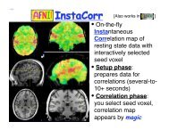
6 Z.S. Saad, R.C. Reynolds / <strong>NeuroImage</strong> xxx (2011) xxx–xxxperforms group seed-based correlations on one or two groups, <strong>the</strong>n performsone- or two-sample t-tests with or without subject-level covariates,and sends all <strong>the</strong> statistical results back for display in <strong>SUMA</strong>. Wedetailed this feature, not only because it is really cool, but also to saythat being able to interactively query and navigate large collections <strong>of</strong>datasets leads to insights into <strong>the</strong> data that are o<strong>the</strong>rwise hard to findwhen looking only at summary results. It remains quite striking towatch resting state patterns shift as <strong>the</strong> cursor is dragged along <strong>the</strong> surface,revealing <strong>the</strong> high level <strong>of</strong> detail present in <strong>the</strong> data; e.g., asreflected in <strong>the</strong> bilateral homology <strong>of</strong> correlations (Jo et al., 2011).Future featuresFor s<strong>of</strong>tware developers, user feedback is mostly <strong>of</strong> <strong>the</strong> negativevariety. If we were to summarize <strong>the</strong> appeal <strong>of</strong> <strong>SUMA</strong> from <strong>the</strong> smallerbut quite positive sample, it would be <strong>the</strong> ease with which multiplesurfaces and associated data are displayed and related to <strong>the</strong>volume, and <strong>the</strong> way group analysis can be carried out. Eye candy imagesare also a big plus. Currently, <strong>SUMA</strong> remains under active development.The authors’ <strong>SUMA</strong>-specific code exceeds 250,000 lines <strong>of</strong> Cfor <strong>the</strong> graphical interface and three dozen command-line programs.Continuing with <strong>the</strong> <strong>the</strong>me <strong>of</strong> ready access to data, we plan to makemore information readily available with minimal interface clutter.For example, beyond parcellation results (FreeSurfer), and variousvolume-based atlas queries currently supported, it would be useful toprovide selective anatomical or functional connectivity information,whe<strong>the</strong>r atlas-based or data-derived. A researcher examining a set <strong>of</strong>SPM blobs would benefit from seeing, after pointing to a region <strong>of</strong> interest,what connectivity or co-activation information exists from that regionto <strong>the</strong> rest <strong>of</strong> <strong>the</strong> brain. Queries could be conducted on datasetspresent on one's computer or more likely from websites containing extensivedatabases such as BrainMap http://www.brainmap.org. The newXML-based connectivity format CIFTI http://www.nitrc.org/projects/ciftiwould help facilitate such information exchanges.Summary<strong>SUMA</strong> is a suite <strong>of</strong> programs for performing surface-based analysisand visualization. It was written to facilitate viewing sets <strong>of</strong> multipledense surfaces while maintaining <strong>the</strong> linkage between <strong>the</strong>m, and toallow for a fine control <strong>of</strong> and simplify <strong>the</strong> once tedious process frommapping volume data to <strong>the</strong> surface domain, through <strong>the</strong> group statisticsstage. <strong>SUMA</strong>'s graphical interface allows for fast, simultaneous, andlinked rendering <strong>of</strong> a large number <strong>of</strong> surfaces, along with compositedisplays <strong>of</strong> multiple datasets with contouring options for representingparcellations or atlas regions and translucent overlaying <strong>of</strong> continuousvalued datasets. The surface displays are intimately connected to <strong>the</strong>volumetric data, allowing for direct verification <strong>of</strong> surface to volume correspondence.The <strong>SUMA</strong> GUI is fully scriptable, making it uniquely suitedto navigate and summarize results from large numbers <strong>of</strong> datasets withminimal effort.AcknowledgmentsMuch <strong>of</strong> <strong>the</strong> utility and speed <strong>of</strong> <strong>SUMA</strong> (and for better or worse, <strong>the</strong>first author's) is due to <strong>the</strong> leveraging <strong>of</strong> <strong>AFNI</strong>'s libraries, interface, andmost importantly <strong>the</strong> vast and generous mind <strong>of</strong> <strong>AFNI</strong>'s creator, RobertW. Cox. The authors are particularly grateful to Michael S. Beauchampfor feedback and support, and to all users with encouraging messagesand thoughtful suggestions. Credit is also due to Edgar A. DeYoe whoseearly ideas for a good surface-mapping interface shaped many <strong>of</strong> <strong>SUMA</strong>'sfeatures years later. This research was supported by <strong>the</strong> NIMH andNINDS Intramural Research Programs <strong>of</strong> <strong>the</strong> NIH.ReferencesAnticevic, A., Dierker, D.L., Gillespie, S.K., Repovs, G., Csernansky, J.G., Van Essen, D.C.,Barch, D.M., 2008. Comparing surface-based and volume-based analyses <strong>of</strong> functionalneuroimaging data in patients with schizophrenia. <strong>NeuroImage</strong> 41, 835–848.Argall, B.D., Saad, Z.S., Beauchamp, M.S., 2006. Simplified intersubject averaging on <strong>the</strong>cortical surface using <strong>SUMA</strong>. Hum. Brain Mapp. 27, 14–27.Ashburner, J., 2007. A fast diffeomorphic image registration algorithm. <strong>NeuroImage</strong> 38,95–113.Biswal, B.B., Mennes, M., Zuo, X.N., Gohel, S., Kelly, C., Smith, S.M., Beckmann, C.F., Adelstein,J.S., Buckner, R.L., Colcombe, S., Dogonowski, A.M., Ernst, M., Fair, D., Hampson, M.,Hoptman, M.J., Hyde, J.S., Kiviniemi, V.J., Kotter, R., Li, S.J., Lin, C.P., Lowe, M.J., Mackay,C., Madden, D.J., Madsen, K.H., Margulies, D.S., Mayberg, H.S., McMahon, K., Monk,C.S., Most<strong>of</strong>sky, S.H., Nagel, B.J., Pekar, J.J., Peltier, S.J., Petersen, S.E., Riedl, V., Rombouts,S.A., Rypma, B., Schlaggar, B.L., Schmidt, S., Seidler, R.D., Siegle, G.J., Sorg, C., Teng, G.J.,Veijola, J., Villringer, A., Walter, M., Wang, L., Weng, X.C., Whitfield-Gabrieli,S., Williamson, P., Windischberger, C., Zang, Y.F., Zhang, H.Y., Castellanos, F.X.,Milham, M.P., 2010. Toward discovery science <strong>of</strong> human brain function. ProcNatl Acad Sci U S A 107, 4734–4739.Cheyne, D., Bakhtazad, L., Gaetz, W., 2006. Spatiotemporal mapping <strong>of</strong> cortical activityaccompanying voluntary movements using an event-related beamforming approach.Hum. Brain Mapp. 27, 213–229.Cohen, A.L., Fair, D.A., Dosenbach, N.U., Miezin, F.M., Dierker, D., Van Essen, D.C., Schlaggar,B.L., Petersen, S.E., 2008. Defining functional areas in individual human brains usingresting functional connectivity MRI. <strong>NeuroImage</strong> 41, 45–57.Cox, R.W., 1996. <strong>AFNI</strong>: s<strong>of</strong>tware for analysis and visualization <strong>of</strong> functional magneticresonance neuroimages. Comput. Biomed. Res. 29, 162–173.Dale, A.M., Fischl, B., Sereno, M.I., 1999. Cortical surface-based analysis. I. Segmentationand surface reconstruction. <strong>NeuroImage</strong> 9, 179–194.Fischl, B., Sereno, M.I., Dale, A.M., 1999a. Cortical surface-based analysis. II: inflation,flattening, and a surface-based coordinate system. <strong>NeuroImage</strong> 9, 195–207.Fischl,B.,Sereno,M.I.,Tootell,R.B.,Dale,A.M.,1999b.High-resolutionintersubjectaveragingand a coordinate system for <strong>the</strong> cortical surface. Hum. Brain Mapp. 8, 272–284.Hagler Jr., D.J., Saygin, A.P., Sereno, M.I., 2006. Smoothing and cluster thresholding forcortical surface-based group analysis <strong>of</strong> fMRI data. <strong>NeuroImage</strong> 33, 1093–1103.JackJr.,C.R.,Bernstein,M.A.,Fox,N.C.,Thompson,P.,Alexander,G.,Harvey,D.,Borowski,B.,Britson, P.J., J, L.W., Ward, C., Dale, A.M., Felmlee, J.P., Gunter, J.L., Hill, D.L., Killiany, R.,Schuff, N., Fox-Bosetti, S., Lin, C., Studholme, C., DeCarli, C.S., Krueger, G., Ward, H.A.,Metzger,G.J.,Scott,K.T.,Mallozzi,R.,Blezek,D.,Levy,J.,Debbins,J.P.,Fleisher,A.S.,Albert,M., Green, R., Bartzokis, G., Glover, G., Mugler, J., Weiner, M.W., 2008. The Alzheimer'sDisease Neuroimaging Initiative (ADNI): MRI methods. J. Magn. Reson. Imaging 27,685–691.Jo, H.J., Saad, Z.S., Gotts, S.J., Glen, D.R., Martin, A., Cox, R.W., 2011. Quantifying contralateralresting-state homology across <strong>the</strong> whole cortex. Organization for Human BrainMapping, Quebec City, Canada.Klein, A., Andersson, J., Ardekani, B.A., Ashburner, J., Avants, B., Chiang, M.C., Christensen,G.E., Collins, D.L., Gee, J., Hellier, P., Song, J.H., Jenkinson, M., Lepage, C., Rueckert, D.,Thompson, P., Vercauteren, T., Woods, R.P., Mann, J.J., Parsey, R.V., 2009. Evaluation<strong>of</strong> 14 nonlinear deformation algorithms applied to human brain MRI registration.<strong>NeuroImage</strong> 46, 786–802.Mueller, S.G., Weiner, M.W., Thal, L.J., Petersen, R.C., Jack, C.R., Jagust, W., Trojanowski,J.Q., Toga, A.W., Beckett, L., 2005. Ways toward an early diagnosis in Alzheimer'sdisease: <strong>the</strong> Alzheimer's Disease Neuroimaging Initiative (ADNI). AlzheimersDement. 1, 55–66.Saad, Z.S., Reynolds, R.C., Argall, B., Japee, S., Cox, R.W., 2004. <strong>SUMA</strong>: an interface forsurface-based intra- and inter-subject analysis with <strong>AFNI</strong>. 2004 IEEE InternationalSymposium on Biomedical Imaging: From Nano to Macro. IEEE, Arlington, VA,pp. 1510–1513.Van Essen, D.C., Maunsell, J.H.R., 1980. Two dimensional maps <strong>of</strong> <strong>the</strong> cerebral cortex. J.Comp. Neurol. 191, 255–281.Van Essen, D.C., Drury, H.A., Dickson, J., Harwell, J., Hanlon, D., Anderson, C.H., 2001. Anintegrated s<strong>of</strong>tware suite for surface-based analyses <strong>of</strong> cerebral cortex.[comment].J. Am. Med. Inform. Assoc. 8, 443–459.Warnking,J.,Dojat,M.,Guerin-Dugue,A.,Delon-Martin,C.,Olympieff,S.,Richard,N.,Chehikian, A., Segebarth, C., 2002. fMRI retinotopic mapping—step by step. <strong>NeuroImage</strong>17, 1665–1683.Please cite this article as: Saad, Z.S., Reynolds, R.C., <strong>SUMA</strong>, <strong>NeuroImage</strong> (2011), doi:10.1016/j.neuroimage.2011.09.016