You also want an ePaper? Increase the reach of your titles
YUMPU automatically turns print PDFs into web optimized ePapers that Google loves.
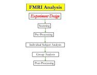
–15–Types of Contrast Used in Brain <strong>FMRI</strong>• T1 contrast at high spatial resolution! Technique: use very short timing between RF shots (smallTR) and use large flip angles! Useful for anatomical reference scans! 5-10 minutes to acquire 256!256!128 volume! 1 mm resolution easily achievableo finer voxels are possible, but acquisition time increases a lot• T2 (spin-echo) and T2* (gradient-echo) contrast! Useful for functional activation studies! 100 ms per 64!64 2D slice $ 2-3 s to acquire whole brain! 4 mm resolutiono better is possible with better gradient system, and/or multiple RFreadout coils–16–What is Functional MRI?• 1991: Discovery that MRI-measurable signal increases afew % locally in <strong>the</strong> brain subsequent to increases inneuronal activity (Kwong, et al.)Cartoon ofMRI signalin an“activated”brain voxel