Trauma Guideline Manual - SUNY Upstate Medical University
Trauma Guideline Manual - SUNY Upstate Medical University
Trauma Guideline Manual - SUNY Upstate Medical University
You also want an ePaper? Increase the reach of your titles
YUMPU automatically turns print PDFs into web optimized ePapers that Google loves.
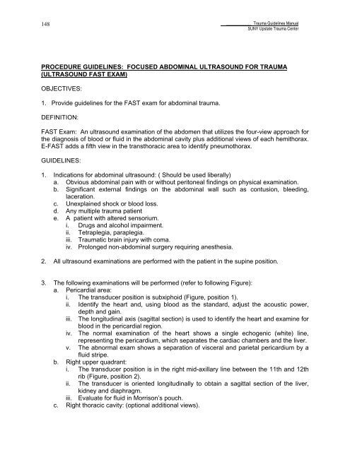
148____________ <strong>Trauma</strong> <strong>Guideline</strong>s <strong>Manual</strong><strong>SUNY</strong> <strong>Upstate</strong> <strong>Trauma</strong> CenterPROCEDURE GUIDELINES: FOCUSED ABDOMINAL ULTRASOUND FOR TRAUMA(ULTRASOUND FAST EXAM)OBJECTIVES:1. Provide guidelines for the FAST exam for abdominal trauma.DEFINITION:FAST Exam: An ultrasound examination of the abdomen that utilizes the four-view approach forthe diagnosis of blood or fluid in the abdominal cavity plus additional views of each hemithorax.E-FAST adds a fifth view in the transthoracic area to identify pneumothorax.GUIDELINES:1. Indications for abdominal ultrasound: ( Should be used liberally)a. Obvious abdominal pain with or without peritoneal findings on physical examination.b. Significant external findings on the abdominal wall such as contusion, bleeding,laceration.c. Unexplained shock or blood loss.d. Any multiple trauma patiente. A patient with altered sensorium.i. Drugs and alcohol impairment.ii. Tetraplegia, paraplegia.iii. <strong>Trauma</strong>tic brain injury with coma.iv. Prolonged non-abdominal surgery requiring anesthesia.2. All ultrasound examinations are performed with the patient in the supine position.3. The following examinations will be performed (refer to following Figure):a. Pericardial area:i. The transducer position is subxiphoid (Figure, position 1).ii.Identify the heart and, using blood as the standard, adjust the acoustic power,depth and gain.iii. The longitudinal axis (sagittal section) is used to identify the heart and examine forblood in the pericardial region.iv. The normal examination of the heart shows a single echogenic (white) line,representing the pericardium, which separates the cardiac chambers and the liver.v. The abnormal exam shows a separation of visceral and parietal pericardium by afluid stripe.b. Right upper quadrant:i. The transducer position is in the right mid-axillary line between the 11th and 12thrib (Figure, position 2).ii.The transducer is oriented longitudinally to obtain a sagittal section of the liver,kidney and diaphragm.iii. Evaluate for fluid in Morrison’s pouch.c. Right thoracic cavity: (optional additional views).


