Trauma Guideline Manual - SUNY Upstate Medical University
Trauma Guideline Manual - SUNY Upstate Medical University
Trauma Guideline Manual - SUNY Upstate Medical University

Create successful ePaper yourself
Turn your PDF publications into a flip-book with our unique Google optimized e-Paper software.
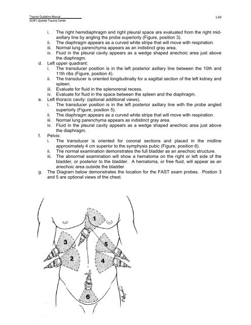
<strong>Trauma</strong> <strong>Guideline</strong> <strong>Manual</strong>______________<strong>SUNY</strong> <strong>Upstate</strong> <strong>Trauma</strong> Center149i. The right hemidiaphragm and right pleural space are evaluated from the right midaxillaryline by angling the probe superiorly (Figure, position 3).ii. The diaphragm appears as a curved white stripe that will move with respiration.iii. Normal lung parenchyma appears as an indistinct gray area.iv. Fluid in the pleural cavity appears as a wedge shaped anechoic area just abovethe diaphragm.d. Left upper quadrant:i. The transducer position is in the left posterior axillary line between the 10th and11th ribs (Figure, position 4).ii. The transducer is oriented longitudinally for a sagittal section of the left kidney andspleen.iii. Evaluate for fluid in the splenorenal recess.iv. Evaluate for fluid in the space between the spleen and the diaphragm.e. Left thoracic cavity: (optional additional views).i. The transducer position is in the left posterior axillary line with the probe angledsuperiorly (Figure, position 5).ii. The diaphragm appears as a curved white stripe that will move with respiration.iii. Normal lung parenchyma appears as indistinct gray area.iv. Fluid in the pleural cavity appears as a wedge shaped anechoic area just abovethe diaphragm.f. Pelvis:i. The transducer is oriented for coronal sections and placed in the midlineapproximately 4 cm superior to the symphysis pubic (Figure, position 6).ii. The normal examination demonstrates the full bladder as an anechoic structure.iii. The abnormal examination will show a hematoma on the right or left side of thebladder, or posterior to the bladder. A hematoma, or free fluid, will appear as ananechoic area outside the bladderg. The Diagram below demonstrates the location for the FAST exam probes. Postion 3and 5 are optional views of the chest.