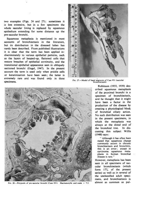
~~~~~~~~~~~~~~~~~~~~~~.228F. WHITWELLThe pre-saccular bronchishow marked polypoid changesin <strong>the</strong> epi<strong>the</strong>lium. In Fig. 25polyposis <strong>of</strong> <strong>the</strong> basal segmentalbronchi can be compared with<strong>the</strong> smooth epi<strong>the</strong>lial lining <strong>of</strong><strong>the</strong> normal apical bronchus.The epi<strong>the</strong>lial lining <strong>of</strong> <strong>the</strong>saccules is flatter than in <strong>the</strong>previous specimen, <strong>and</strong> areas <strong>of</strong>squamous metaplasia are moreextensive. No connexion wasfound between <strong>the</strong> saccules <strong>and</strong>any distal bronchial tree-infact no intact distal bronchi arepresent, though respiratory bronchiolesoccur in <strong>the</strong> parenchyma.The base <strong>of</strong> <strong>the</strong> lobe is wellaerated except for a narrow zone<strong>of</strong> collapse around <strong>and</strong> between<strong>the</strong> saccules. In <strong>the</strong> areas <strong>of</strong> collapse<strong>the</strong> alveoli are flattened concentricallyaround <strong>the</strong> saccules.There is no inflammatoryreaction in <strong>the</strong> parenchyma, <strong>and</strong>only within <strong>the</strong> peribronchial,perisaccular, <strong>and</strong> interlobularsepta is an increase <strong>of</strong> fibroustissue to be found.THE PATHOLOGY OF SACCULARBRONCHIECTASISFIG. 26.-Saccular <strong>bronchiectasis</strong> 5 cm. from hilum (Case 451). Haematoxylin <strong>and</strong> eosin x 1.5. These two examples arethought to be <strong>the</strong> end-resultconspicuous by thickening <strong>of</strong> interlobular fibrous septa. <strong>of</strong> a distinctive t: ;ype <strong>of</strong> <strong>bronchiectasis</strong>. AlthoughWithin <strong>the</strong>se lobules <strong>the</strong>re are some respiratory bron- only 17% <strong>of</strong> s,pecimens show <strong>the</strong>se lesions,chioles, but terminal bronchioles are replaced by fibrous some <strong>of</strong> <strong>the</strong> unclatssifiedlobes are probably earlierscars. stages <strong>of</strong> <strong>the</strong> ssame condition. CharacteristicCASE 451.-An 18-year-old boy was admitted to features are grossloss <strong>of</strong> bronchial structures in <strong>the</strong>hospital because <strong>of</strong> a troublesome cough <strong>and</strong> a single saccules, normaliity <strong>of</strong> <strong>the</strong> alveoli around <strong>the</strong>slight haemoptysis. Bronchitis had occurred during <strong>the</strong>sprevious four winters, but <strong>the</strong>re was no history <strong>of</strong> earlier polyposis" <strong>of</strong> <strong>the</strong> pre-saccular<strong>and</strong>saccules, rrespiratory complaint or infectious diseases. He pro- bronchi,*etain normal supporting tissues.whlch rduced 3 oz. <strong>of</strong> purulent sputum daily, had marked finger The saccules <strong>and</strong> <strong>the</strong> pre-saccular bronchi can beclubbing, <strong>and</strong> bronchograms showed <strong>bronchiectasis</strong> considered separaltely, <strong>the</strong> former being regarded aslimited to <strong>the</strong> left lower lobe. This lobe was excised. <strong>the</strong> primary abno irmality.Macroscopic Appearance <strong>of</strong> Specimen.-The lobe is SACCULES.-Saccules are essentially fibrous struc-epi<strong>the</strong>lium; <strong>the</strong>y have nobulky, aerated, slightly pigmented, <strong>and</strong> has a shiny tures lined by a ciuboidalpleural membrane.elastic tissue, mus,cleor cartilage in <strong>the</strong>ir walls, <strong>and</strong>Sections <strong>of</strong> <strong>the</strong> lobe (Figs. 25 <strong>and</strong> 26) show a normal <strong>the</strong>ir lining memtbranes contain no normal ciliatedapical segment but severe <strong>bronchiectasis</strong> in <strong>the</strong> whole respiratory epi<strong>the</strong>lium.This absence <strong>of</strong> recogniz-saccules has led to speculation<strong>of</strong> <strong>the</strong> base. The segmental basal bronchi <strong>and</strong> <strong>the</strong>ir main able tissues in thiebranches are not thickened or dilated, but <strong>the</strong>y are about <strong>the</strong>ir originpartially occluded by epi<strong>the</strong>lial " polyposis";<strong>and</strong> this subject will be discussedmorelater.distal bronchi are saccular <strong>and</strong> filled with pus. The la er.alveoli <strong>of</strong> <strong>the</strong> basal segments are aerated, except imme- The constant finding <strong>of</strong> areas <strong>of</strong> squamousdiately around <strong>the</strong> saccules where <strong>the</strong>re is some collapse. metaplasia in <strong>the</strong> saccular epi<strong>the</strong>lium is <strong>of</strong> specialMicroscopic Features.-There is obvious similarity interest as this p] ohenomenon is uncommon in any.to <strong>the</strong> previous case, <strong>and</strong> only points <strong>of</strong> difference will <strong>of</strong> <strong>the</strong> o<strong>the</strong>r types <strong>of</strong> <strong>bronchiectasis</strong> examined. Thebe stressed.usual distribution <strong>of</strong> this metaplasia is shown in <strong>the</strong>
_ * _ * ........... r _ r- -[two examples (Figs. 24 <strong>and</strong> 27); sometimes itis less extensive, but in a few specimens <strong>the</strong>whole saccular lining is replaced by squamous Iepi<strong>the</strong>lium extending for some distance up <strong>the</strong>pre-saccular bronchi.Squamous metaplasia is mentioned in most_Xaccounts <strong>of</strong> <strong>bronchiectasis</strong> in <strong>the</strong> literature,but its distribution in <strong>the</strong> diseased lobes has ararely been described. From published illustrationsit is clear that <strong>the</strong> term has been applied indiscriminatelyto various epi<strong>the</strong>lial patterns, suchas <strong>the</strong> layers <strong>of</strong> heaped-up, flattened cells thatrestore breaches <strong>of</strong> epi<strong>the</strong>lial continuity, <strong>and</strong> <strong>the</strong>transitional epi<strong>the</strong>lial appearance seen in obliquely -~ rsectioned bronchi (Engel, 1947). In <strong>the</strong> presentaccount <strong>the</strong> term is used only when prickle cellsor keratinization have been seen; <strong>the</strong> latter isextremely rare <strong>and</strong> was found only in threeFIG. 27.-Model <strong>of</strong> basal segments <strong>of</strong> Case 451 (saccularspecimens.<strong>bronchiectasis</strong>).e§;ev16Robinson (1933, 1939) desei / l{is'>