Common Intraoperative Ultrasound Artifacts - Casecag.com
Common Intraoperative Ultrasound Artifacts - Casecag.com
Common Intraoperative Ultrasound Artifacts - Casecag.com
You also want an ePaper? Increase the reach of your titles
YUMPU automatically turns print PDFs into web optimized ePapers that Google loves.
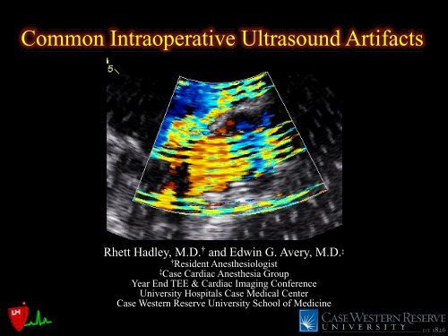
<strong>Common</strong> <strong>Intraoperative</strong> <strong>Ultrasound</strong> <strong>Artifacts</strong>Rhett Hadley, M.D. † and Edwin G. Avery, M.D. ‡†Resident Anesthesiologist‡Case Cardiac Anesthesia GroupYear End TEE & Cardiac Imaging ConferenceUniversity Hospitals Case Medical CenterCase Western Reserve University School of Medicine
Disclosures – Dr. Hadley
Disclosures – Dr. AveryConsultant, speaker’s bureau andfunded research with CovidienParticipated in research funded byMedtronic
OverviewIntroduce the appearance of<strong>com</strong>mon ultrasound artifactsencountered in the intraoperativesettingProvide a brief physics-basedexplanation of the artifacts presented
<strong>Artifacts</strong> - DefinedArtifact – any structure in an ultrasound imagethat does not have a corresponding anatomictissue structureGenerally there are 4 classes of ultrasoundartifacts:Missing structuresDegraded imagesFalsely perceived objectsStructures with a misregistered location
Acoustic Shadowing – Missing Structure
Acoustic Shadowing – Missing StructureAcoustic shadowing occurswhen the US signal encounters astrong reflector that results indecreased beam intensity distalto the strong reflectorAcoustic shadowing is alsoreferred to as an anechoic spaceProsthetic valve material andheavily calcified tissue createthis artifactAcoustic Shadow
Reverberation– Degraded Image
Reverberation– Degraded ImageReverberations are secondaryreflections that occur along thepath of the US beam as a resultof the signal bouncing between astructure and a reflective surface(i.e. the US probe itself)Reverberation may create asecond, third, fourth and so onobject at increasing distancesfrom the probe that do not reallyexist-they may appear to be onestructure referred to as a <strong>com</strong>ettailReverberationSignalBouncing
Enhancement– Degraded Image
Enhancement– Degraded ImageEnhancement is reciprocal ofacoustic shadowing in that itoccurs when the US beamencounters an interface with aweak reflector and the beam isthus increased in intensity distalto this pointIt is <strong>com</strong>monly seen when theUS signal interfaces with thethin pericardial tissue and can bemitigated by decreasing the timegain <strong>com</strong>pensationEnhancement
Noise– Degraded Image
Noise– Degraded ImageNoise may result from multipleetiologies such as excessive gainor most <strong>com</strong>monly in the ORfrom electrocauteryNoise appears as multiple smallechos on the scan (like in theMatrix)Noise
Refraction– Falsely Perceived Objects
Refraction– Falsely Perceived ObjectsRefraction results from the US beambeing deflected from its original path as aresult of passing through a medium witha different acoustic impedanceThe transducer assumes that the reflectedimage originated from the initial scan lineand the image is displayed as such,possibly in a location laterally orotherwise displaced from its true locationThese may be referred to as mirror imageartifacts and may be mitigated bychanging the angle of the US beamMirror Image
Side Lobe Artifact– Misregistered Locations
Side Lobe Artifact– Misregistered LocationsSide lobe artifacts resultfrom multiple US beamsbeing emitted at severaldifferent angles from thetransducer and may result instructures being placed inthe wrong location on thedisplayed imageRapid Oscillation of theecho beam in the transducermay also give rise to theside lobe artifacts
Side Lobe Artifact– Misregistered LocationsSide lobe artifacts are morelikely to be seen if these sidelobe/beams interface with astrong specular reflector asthe reflected energy will bestrong and will be added tothe reflected energy of themain beamThe crista terminalis is actingas a strong specular reflectorin this example
Anatomic “Artifact”–In<strong>com</strong>plete Knowledge of Cardiac AnatomyMid to Upper Esophageal 2C view with ~70-90° omniplaneCoumadin Ridge
The End – Thank YouPlease visit www.casecag.<strong>com</strong> for a copy of this presentationClickHere


