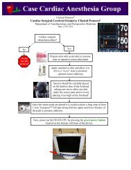
Persistent Left Superior Vena Cava: What is the ... - Casecag.com
Persistent Left Superior Vena Cava: What is the ... - Casecag.com
Persistent Left Superior Vena Cava: What is the ... - Casecag.com

You also want an ePaper? Increase the reach of your titles
YUMPU automatically turns print PDFs into web optimized ePapers that Google loves.
<strong>Pers<strong>is</strong>tent</strong> <strong>Left</strong> <strong>Superior</strong> <strong>Vena</strong> <strong>Cava</strong>:<strong>What</strong> <strong>is</strong> <strong>the</strong> Significance?Mark W. Lucia, M.D.Anes<strong>the</strong>siology House Officer – PGY32011 Year End Cardiac Anes<strong>the</strong>sia TEE Conference
D<strong>is</strong>closuresUnfortunately…”No”
OverviewD<strong>is</strong>cuss <strong>the</strong> prevalence, variants, diagnos<strong>is</strong> andpractical significance of <strong>the</strong> incidental finding of apers<strong>is</strong>tent left superior vena cavaPresent a clinical case of an incidental pers<strong>is</strong>tentLSVC in a patient undergoing double valve repair
LSVC Defined
LSVC DefinedThe LSVC <strong>is</strong> <strong>the</strong> anatomical variant that results when <strong>the</strong> leftanterior cardinal vein fails to involute in embryologicdevelopmentEstimated prevalence 0.1% - 0.3% in general population<strong>Left</strong>AnteriorCardinalVeinInvolutesNormalAnn Thorac Surg 2001;71:1389-1395
LSVC DefinedThe LSVC most <strong>com</strong>monly (90%) empties into <strong>the</strong> coronary sinusA LSVC <strong>is</strong> associated with <strong>the</strong> presence of o<strong>the</strong>r congential cardiacanomalies (e.g., ASD, VSD, cor triatriatum, mitral atresia)+/- Innominate veinPosterior View90%Anterior ViewAnn Thorac Surg 2001;71:1389-1395
LSVC VariantsThe LSVC may also drain into <strong>the</strong> left atrium or left upperpulmonary veinThe innominate vein <strong>is</strong> variably present+/- Innominate veinAnn Thorac Surg 2001;71:1389-1395
LSVC Diagnos<strong>is</strong>
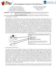
LSVC Diagnos<strong>is</strong>High fidelity imaging modalities (e.g., MRA) with contrast willclearly define <strong>the</strong> presence and variant of a pers<strong>is</strong>tent LSVCIndian Pacing Electrophysiol. J 2010;10(1):58-61
LSVC Diagnos<strong>is</strong>Angiography can also be useful to define LSVC anatomyRANephrol. Dial. Transplant. (2003) 18 (7): 1410-1411.
LSVC Diagnos<strong>is</strong>Both TEE and TTE can allow for <strong>the</strong> diagnos<strong>is</strong>Normally, <strong>the</strong> coronary sinus diameter <strong>is</strong> approximately 8.5 mm inadult normal subjects † , values > 10 mm should be suspectThe coronary sinus <strong>is</strong> most effectively imaged with TEE in ei<strong>the</strong>r <strong>the</strong>ME 4-C view (w/retroflexion) or <strong>the</strong> 90º bicaval view (w/right rotation &slight withdrawal)†Circulation 1995;92:436-441
LSVC TEE Diagnos<strong>is</strong>ME 4-C view (with retroflexion)
LSVC TEE Diagnos<strong>is</strong>ME bicaval view modified (with right rotation and withdrawal)
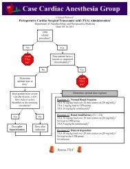
LSVC TEE Diagnos<strong>is</strong>Agitated saline contrast injection into a left upper extremity IV <strong>is</strong>re<strong>com</strong>mended to help define <strong>the</strong> presence and drainage pattern of aLSVC(1 mL air + 1 mL blood + 8 mLs of saline)
LSVC TEE Diagnos<strong>is</strong>Choose <strong>the</strong> TEE view that permits <strong>the</strong> best vantage point tosimultaneously assess <strong>the</strong> SVC and coronary sinusInject your 10 mLs of contrast medium and flush it in while yourintraoperative sonographer waits at <strong>the</strong> ready to capture <strong>the</strong> imageIf contrast exits <strong>the</strong> SVC first <strong>the</strong>n a LSVC <strong>is</strong> less likely (i.e. itcould still be present but emptying into <strong>the</strong> left atrium or <strong>the</strong> leftupper pulmonary vein)If contrast exits <strong>the</strong> dilated coronary sinus first <strong>the</strong>n a LSVC <strong>is</strong>likelyIf contrast ex<strong>is</strong>ts both <strong>the</strong> SVC and <strong>the</strong> coronary sinussimultaneously <strong>the</strong>n a LSVC <strong>is</strong> likely in <strong>the</strong> setting of an intactinnominate vein
Clinical Case Presentation
Clinical Case Presentation68 YOF with NYHA Class IV CHF presented for double valverepair (MV repair + TV repair)
Clinical Case PresentationUpon assessment of <strong>the</strong> TR a severely dilated coronary sinuswas noted. <strong>What</strong> <strong>is</strong> <strong>the</strong> significance?
Clinical Case PresentationBecause <strong>the</strong> degree of coronary sinus dilation was > 3x <strong>the</strong>expected diameter a LSVC should have been suspected27 mm(Normal Mean CS diameter = 8.5 mm)
Clinical Case PresentationThe remarkable dimension of <strong>the</strong> coronary sinus was<strong>com</strong>municated to <strong>the</strong> surgical team, but nei<strong>the</strong>r staff membersuggested provocative testing to rule out a LSVCUltimately CPB was initiated when shortly <strong>the</strong>reafter <strong>the</strong>surgeon d<strong>is</strong>covered <strong>the</strong> LSVC draining into <strong>the</strong> coronary sinusThe LSVC was temporarily ligated to permit safe delivery ofretrograde cardioplegia throughout <strong>the</strong> case.
Clinical Case PresentationFailure to ligate <strong>the</strong> LSVC will permit pressure and potentiallyair emboli to challenge <strong>the</strong> venous cerebral circulation because<strong>the</strong> LSVC <strong>is</strong> in continuity with <strong>the</strong> left internal jugular vein.Pressurizing <strong>the</strong> LIJ could result in a watershed stroke of <strong>the</strong> leftbrain by impeding cerebral venous drainage
Clinical Case PresentationDiagnos<strong>is</strong>: <strong>Pers<strong>is</strong>tent</strong> LSVC draininginto <strong>the</strong> coronary sinus with an intactinnominate vein.Innominate Vein
The End – Thank YouPlease v<strong>is</strong>it www.casecag.<strong>com</strong> for a copy of th<strong>is</strong> presentation andto view it in video formatClickHere