

Download - World Journal of Gastroenterology
Download - World Journal of Gastroenterology
Download - World Journal of Gastroenterology

You also want an ePaper? Increase the reach of your titles
YUMPU automatically turns print PDFs into web optimized ePapers that Google loves.
A<br />
B<br />
C<br />
D<br />
Figure 2 Henoch-Schönlein purpura in a 38-year-old man with hematochezia.<br />
A: Palpable purpura <strong>of</strong> the right foot; B: Contrast-enhanced computed tomography<br />
scan <strong>of</strong> the abdomen showed diffuse thickening <strong>of</strong> the ileum (target sign)<br />
with mesenteric hypervascularity in a palisading pattern (comb sign), suggesting<br />
ischemic ileitis; C, D: Single balloon enteroscopy showed edematous petechiae<br />
with linear ulcers in the affected ileum. All figures and legends are reproduced<br />
from Hokama et al [15] with permission from BMJ Publishing Group Ltd.<br />
wall thickening with the target sign, bowel dilatation, ascites,<br />
and engorgement <strong>of</strong> mesenteric vessels with comb<br />
WJGE|www.wjgnet.com<br />
Hokama A et al . Endoscopic and radiographic features <strong>of</strong> vasculitis<br />
A<br />
R<br />
B<br />
C<br />
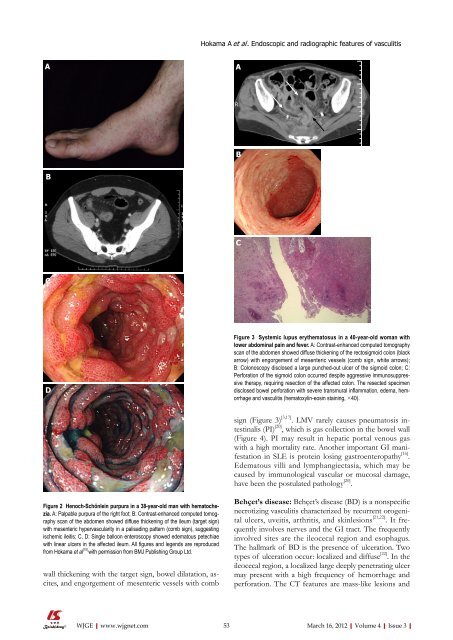
Figure 3 Systemic lupus erythematosus in a 40-year-old woman with<br />
lower abdominal pain and fever. A: Contrast-enhanced computed tomography<br />
scan <strong>of</strong> the abdomen showed diffuse thickening <strong>of</strong> the rectosigmoid colon (black<br />
arrow) with engorgement <strong>of</strong> mesenteric vessels (comb sign, white arrows);<br />
B: Colonoscopy disclosed a large punched-out ulcer <strong>of</strong> the sigmoid colon; C:<br />
Perforation <strong>of</strong> the sigmoid colon occurred despite aggressive immunosuppressive<br />
therapy, requiring resection <strong>of</strong> the affected colon. The resected specimen<br />
disclosed bowel perforation with severe transmural inflammation, edema, hemorrhage<br />
and vasculitis (hematoxylin-eosin staining, ×40).<br />
sign (Figure 3) [3,17] . LMV rarely causes pneumatosis intestinalis<br />
(PI) [20] , which is gas collection in the bowel wall<br />
(Figure 4). PI may result in hepatic portal venous gas<br />
with a high mortality rate. Another important GI manifestation<br />
in SLE is protein losing gastroenteropathy [16] .<br />
Edematous villi and lymphangiectasia, which may be<br />
caused by immunological vascular or mucosal damage,<br />
have been the postulated pathology [20] .<br />
Behçet’s disease: Behçet’s disease (BD) is a nonspecific<br />
necrotizing vasculitis characterized by recurrent orogenital<br />
ulcers, uveitis, arthritis, and skinlesions [21,22] . It frequently<br />
involves nerves and the GI tract. The frequently<br />
involved sites are the ileocecal region and esophagus.<br />
The hallmark <strong>of</strong> BD is the presence <strong>of</strong> ulceration. Two<br />
types <strong>of</strong> ulceration occur: localized and diffuse [22] . In the<br />
ileocecal region, a localized large deeply penetrating ulcer<br />
may present with a high frequency <strong>of</strong> hemorrhage and<br />
perforation. The CT features are mass-like lesions and<br />
53 March 16, 2012|Volume 4|Issue 3|


