

Download - World Journal of Gastroenterology
Download - World Journal of Gastroenterology
Download - World Journal of Gastroenterology

Create successful ePaper yourself
Turn your PDF publications into a flip-book with our unique Google optimized e-Paper software.
A B<br />
C<br />
was initially large because the endoscope is equipped<br />
with a zoom lens at the tip, but recent technical progress<br />
has reduced the diameter close to that <strong>of</strong> a conventional<br />
scope. Combining other techniques with magnifying<br />
endoscopy enables theidentification <strong>of</strong> the boundary <strong>of</strong><br />
lesions with an unclear margin <strong>of</strong> advancement undetectable<br />
using a single method.<br />
BIOPSY OF THE SURROUNDING REGION<br />
Although determination <strong>of</strong> the boundary by biopsy alone<br />
should be avoided, the boundary is still unclear even after<br />
sufficient observation in some lesions, and biopsy <strong>of</strong> the<br />
surrounding region may be useful in such cases.<br />
PREPARATION OF A COMPOSITE OF<br />
MAGNIFIED IMAGES BY TILING<br />
It is important to confirm whether the preoperative diagnosis<br />
<strong>of</strong> the range is correct by analyzing endoscopically<br />
resected specimens. When the diagnosis is incorrect, the<br />
correct range can be identified by investigating the reason<br />
for the failure. In addition, clarification <strong>of</strong> the conditions<br />
leading to an incorrect diagnosis <strong>of</strong> the range facilitates<br />
careful investigation in combination with other methods.<br />
We developed a method to prepare a composite <strong>of</strong><br />
magnified images by tiling in which the mucosal surface<br />
<strong>of</strong> the excised specimen can be closely observed. In this<br />
unique method, a magnifying endoscope was fixed, the<br />
WJGE|www.wjgnet.com<br />
D<br />
Ochiai Y et al . Boundary diagnosis <strong>of</strong> gastric cancer<br />
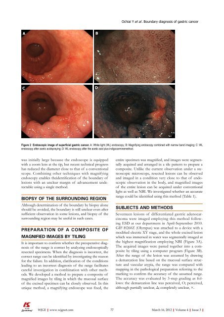
Figure 2 Endoscopic image <strong>of</strong> superficial gastric cancer. A: White light (WL) endoscopy; B: Magnifying endoscopy combined with narrow band imaging; C: WL<br />
endoscopy after acetic acidspraying; D: WL endoscopy after the acetic acid plus indigocarminemethod.<br />
entire specimen was magnified, and images were segmentally<br />
acquired and arranged in a tile pattern to prepare a<br />
composite. Unlike the current observation under a stereoscopic<br />
microscope, resected lesions can be observed<br />
and imaged in a condition very close to that <strong>of</strong> endoscopic<br />
observation in the body, and magnified images<br />
<strong>of</strong> the entire lesion can be acquired under conventional<br />
light as well as NBI. We investigated whether an accurate<br />
range could be identified using this method (Table 1).<br />
SUBJECTS AND METHODS<br />
Seventeen lesions <strong>of</strong> differentiated gastric adenocarcinoma<br />
were imaged employing this method following<br />
ESD at our department in April-September 2010.<br />
GIF-H260Z (Olympus) was attached to a device with a<br />
modified electric XY stage, and the whole excised lesion<br />
which was immersed in water was segmentally imaged at<br />
the highest magnification employing NBI (Figure 3A).<br />
The acquired images were pasted together into a composite<br />
by tiling using a computer program (Figure 3B).<br />
After the range <strong>of</strong> the lesion was assumed by drawing<br />
a demarcation line based on the mucosal surface structure<br />
and vascular atypia, the range was compared with<br />
mapping in the pathological preparation referring to the<br />
marking to confirm the accuracy <strong>of</strong> the assumed range.<br />
The accuracy was evaluated by 3-step grading as follows:<br />
the demarcation line was perceived, O; perceived,<br />
although partially unclear, Δ; completely unclear, ×.<br />
77 March 16, 2012|Volume 4|Issue 3|


