

Download - World Journal of Gastroenterology
Download - World Journal of Gastroenterology
Download - World Journal of Gastroenterology

You also want an ePaper? Increase the reach of your titles
YUMPU automatically turns print PDFs into web optimized ePapers that Google loves.
12 cm<br />
Figure 4 Systemic lupus erythematosus in a 23-year-old woman with<br />
abdominal pain and fever. Plain computed tomography scan <strong>of</strong> the abdomen<br />
showed intramural gas <strong>of</strong> the ascending colon, suggesting pneumatosis intestinalis<br />
(arrow). Hyperbaric oxygen therapy was effective for improvement <strong>of</strong> the<br />
pneumatosis.<br />
A C<br />
B<br />
Hokama A et al . Endoscopic and radiographic features <strong>of</strong> vasculitis<br />
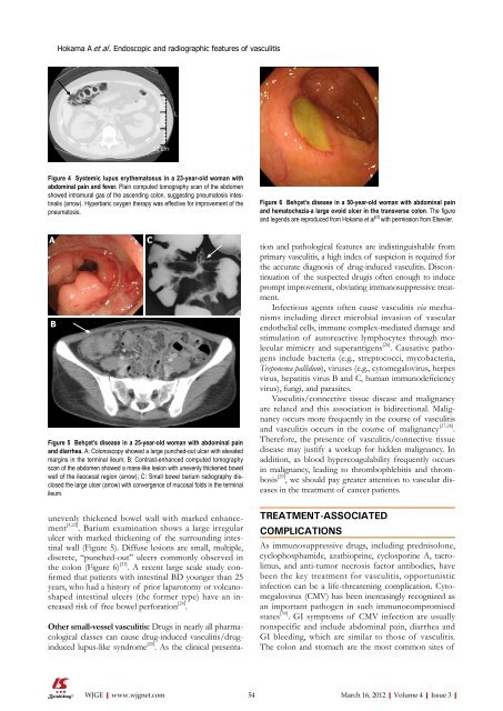
Figure 5 Behçet’s disease in a 25-year-old woman with abdominal pain<br />
and diarrhea. A: Colonoscopy showed a large punched-out ulcer with elevated<br />
margins in the terminal ileum; B: Contrast-enhanced computed tomography<br />
scan <strong>of</strong> the abdomen showed a mass-like lesion with unevenly thickened bowel<br />
wall <strong>of</strong> the ileocecal region (arrow); C: Small bowel barium radiography disclosed<br />
the large ulcer (arrow) with convergence <strong>of</strong> mucosal folds in the terminal<br />
ileum.<br />
unevenly thickened bowel wall with marked enhancement<br />
[3,22] . Barium examination shows a large irregular<br />
ulcer with marked thickening <strong>of</strong> the surrounding intestinal<br />
wall (Figure 5). Diffuse lesions are small, multiple,<br />
discrete, “punched-out” ulcers commonly observed in<br />
the colon (Figure 6) [23] . A recent large scale study confirmed<br />
that patients with intestinal BD younger than 25<br />
years, who had a history <strong>of</strong> prior laparotomy or volcanoshaped<br />
intestinal ulcers (the former type) have an increased<br />
risk <strong>of</strong> free bowel perforation [24] .<br />
Other small-vessel vasculitis: Drugs in nearly all pharmacological<br />
classes can cause drug-induced vasculitis/druginduced<br />
lupus-like syndrome [25] . As the clinical presenta-<br />
WJGE|www.wjgnet.com<br />
L<br />
Figure 6 Behçet’s disease in a 50-year-old woman with abdominal pain<br />
and hematochezia-a large ovoid ulcer in the transverse colon. The figure<br />
and legends are reproduced from Hokama et al [23] with permission from Elsevier.<br />
tion and pathological features are indistinguishable from<br />
primary vasculitis, a high index <strong>of</strong> suspicion is required for<br />
the accurate diagnosis <strong>of</strong> drug-induced vasculitis. Discontinuation<br />
<strong>of</strong> the suspected drugis <strong>of</strong>ten enough to induce<br />
prompt improvement, obviating immunosuppressive treatment.<br />
Infectious agents <strong>of</strong>ten cause vasculitis via mechanisms<br />
including direct microbial invasion <strong>of</strong> vascular<br />
endothelial cells, immune complex-mediated damage and<br />
stimulation <strong>of</strong> autoreactive lymphocytes through molecular<br />
mimicry and superantigens [26] . Causative pathogens<br />
include bacteria (e.g., streptococci, mycobacteria,<br />
Treponema pallidum), viruses (e.g., cytomegalovirus, herpes<br />
virus, hepatitis virus B and C, human immunodeficiency<br />
virus), fungi, and parasites.<br />
Vasculitis/connective tissue disease and malignancy<br />
are related and this association is bidirectional. Malignancy<br />
occurs more frequently in the course <strong>of</strong> vasculitis<br />
and vasculitis occurs in the course <strong>of</strong> malignancy [27,28] .<br />
Therefore, the presence <strong>of</strong> vasculitis/connective tissue<br />
disease may justify a workup for hidden malignancy. In<br />
addition, as blood hypercoagulability frequently occurs<br />
in malignancy, leading to thrombophlebitis and thrombosis<br />
[29] , we should pay greater attention to vascular diseases<br />
in the treatment <strong>of</strong> cancer patients.<br />
TREATMENT-ASSOCIATED<br />
COMPLICATIONS<br />
As immunosuppressive drugs, including prednisolone,<br />
cyclophosphamide, azathioprine, cyclosporine A, tacrolimus,<br />
and anti-tumor necrosis factor antibodies, have<br />
been the key treatment for vasculitis, opportunistic<br />
infection can be a life-threatening complication. Cytomegalovirus<br />
(CMV) has been increasingly recognized as<br />
an important pathogen in such immunocompromised<br />
states [30] . GI symptoms <strong>of</strong> CMV infection are usually<br />
nonspecific and include abdominal pain, diarrhea and<br />
GI bleeding, which are similar to those <strong>of</strong> vasculitis.<br />
The colon and stomach are the most common sites <strong>of</strong><br />
54 March 16, 2012|Volume 4|Issue 3|


