

Download - World Journal of Gastroenterology
Download - World Journal of Gastroenterology
Download - World Journal of Gastroenterology

You also want an ePaper? Increase the reach of your titles
YUMPU automatically turns print PDFs into web optimized ePapers that Google loves.
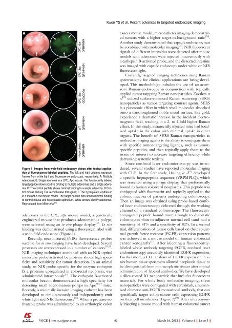
A<br />
B<br />
C<br />
D<br />
E<br />
adenomas in the CPC; Apc mouse model, a genetically<br />
engineered mouse that produces adenomatous polyps,<br />
were selected using an in vivo phage display [30] . In vivo<br />
binding was demonstrated using a fluorescein label with<br />
a wide-field endoscope (Figure 1).<br />
Recently, near-infrared (NIR) fluorescence probes<br />
suitable for in vivo imaging have been developed. Several<br />
proteases are overexpressed in a number <strong>of</strong> cancers [31,32] .<br />
NIR imaging techniques combined with an NIR optical<br />
molecular probe activated by protease shows high specificity<br />
and sensitivity for tumor detection. In an animal<br />
study, an NIR probe specific for the enzyme cathepsin<br />
B, a protease upregulated in colorectal neoplasia, was<br />
administered intravenously [11] . The cathepsin B-activated<br />
molecular beacon demonstrated a high specificity for<br />
detecting small adenomatous polyps in Apc Min/+ mice.<br />
Recently, a minimally invasive imaging catheter has been<br />
developed to simultaneously and independently emit<br />
white light and NIR fluorescence [33] . When a protease-activatable<br />
probe was administered to an orthotopic colon<br />
WJGE|www.wjgnet.com<br />
CPC; Apc<br />
QPIHPNNM<br />
CPC; Apc<br />
QPIHPNNM<br />
CPC; Apc<br />
GGGAGGGA<br />
WT<br />
QPIHPNNM<br />
Hyperplastic<br />
QPIHPNNM<br />
Figure 1 Images from wide-field endoscopy videos after topical application<br />
<strong>of</strong> fluorescence-labeled peptides. The left and right columns represent<br />
frames from white light and fluorescence endoscopy, respectively. A: Multiple<br />
adenomas; B: Single adenoma in a CPC; Apc mouse. The fluorescently labeled<br />
target peptide shows positive binding to multiple adenomas and a single adenoma;<br />
C: The control peptide shows minimal binding to a single adenoma; D:Control<br />
mouse lacking Cre recombinase transgene; E:The hyperplastic epithelium<br />
in a mutant K-ras mouse model. The target peptide also shows minimal binding<br />
to control mouse and hyperplastic epithelium. White arrows identify adenomas.<br />
Reproduced from Miller et al [30] .<br />
Kwon YS et al . Recent advances in targeted endoscopic imaging<br />
cancer mouse model, microcatheter imaging demonstrated<br />
tumors with a higher target-to-background ratio [34] .<br />
Another study demonstrated that capsule endoscopy can<br />
be combined with molecular imaging [35] . NIR fluorescent<br />
signals <strong>of</strong> different intensities were detected after mouse<br />
models with adenomas were injected intravenously with<br />
a cathepsin B-activated probe, and the dissected intestine<br />
was imaged with capsule endoscopy under white or NIR<br />
fluorescent light.<br />
Currently, targeted imaging techniques using Raman<br />
spectroscopy for clinical applications are being developed.<br />
This methodology includes the use <strong>of</strong> an accessory<br />
Raman endoscope in conjunction with topically<br />
applied tumor targeting Raman nanoparticles. Zavaleta et<br />
al [36] utilized surface-enhanced Raman scattering (SERS)<br />
nanoparticles as tumor targeting contrast agents. SERS<br />
is a plasmonic effect in which small molecules absorbed<br />
onto a nanoroughened noble metal surface, like gold,<br />
experience a dramatic increase in the incident electromagnetic<br />
field, resulting in a 2- to 4-fold higher Raman<br />
effect. In this study, intrarectally injected mice had localized<br />
uptake in the colon with minimal uptake in other<br />
organs. The benefit <strong>of</strong> SERS Raman nanoparticles as<br />
molecular imaging agents is the ability to conjugate them<br />
with specific tumor-targeting ligands, such as tumorspecific<br />
peptides, and then topically apply them to the<br />
tissue <strong>of</strong> interest to increase targeting efficiency while<br />
decreasing systemic toxicity.<br />
Since confocal laser endomicroscopy was introduced,<br />
several studies have reported molecular imaging<br />
with CLE. In the first study, Hsiung et al [15] developed<br />
a specific heptapeptide sequence (VRPMPLQ), which<br />
was screened using a phage display, that preferentially<br />
bound to human colorectal neoplasms. This peptide was<br />
conjugated with fluorescein and topically applied to the<br />
colonic mucosa <strong>of</strong> patients undergoing colonoscopy.<br />
Then an image was obtained using probe-based confocal<br />
laser endomicroscopy delivered through the working<br />
channel <strong>of</strong> a standard colonoscope. The fluoresceinconjugated<br />
peptide bound more strongly to dysplastic<br />
colonocytes than to adjacent normal cell sand had a<br />
sensitivity <strong>of</strong> 81% and a specificity <strong>of</strong> 82%. In a second<br />
trial, differentiation <strong>of</strong> tumor cells based on their epidermal<br />
growth factor receptor (EGFR)-expression patterns<br />
was achieved in a mouse model <strong>of</strong> human colorectal<br />
cancer xenografts [37] . After injecting a fluorescentlylabeled<br />
whole antibody targeting EGFR, confocal laser<br />
endomicroscopy accurately identified EGFR expression.<br />
Further more, a CLE analysis <strong>of</strong> EGFR expression in ex<br />
vivo human tissue specimens allowed neoplastic tissue to<br />
be distinguished from non-neoplastic tissues after topical<br />
administration <strong>of</strong> labeled antibodies. We have developed<br />
a silica-coated IO nanoparticle that includes fluorescent<br />
materials. For whole-body molecular imaging, these<br />
nanoparticles were conjugated with cetuximab, a humanized<br />
chimeric anti-EGFR monoclonal antibody, that can<br />
specifically target colon cancer cells expressing EGFR<br />
on their cell membranes (Figure 2) [38] . After intravenously<br />
injecting a mouse model with human colorectal cancer<br />
61 March 16, 2012|Volume 4|Issue 3|


