stimuli
Visual Image Filtering at the Level of Cortical Input
Visual Image Filtering at the Level of Cortical Input
- No tags were found...

Create successful ePaper yourself
Turn your PDF publications into a flip-book with our unique Google optimized e-Paper software.
INFORMATICA, 2004, Vol. 15, No. 4, 443–454 443<br />
© 2004 Institute of Mathematics and Informatics, Vilnius<br />
Visual Image Filtering at the Level of Cortical<br />
Input<br />
Aleksandr BULATOV, Algis BERTULIS<br />
Department of Biology, Kaunas University of Medicine<br />
Mickevičiaus 9, LT-3000, Kaunas, Lithuania<br />
e-mail: bulatov@vision.kmu.lt<br />
Received: October 2004<br />
Abstract. A computational model of the spatial-frequency filtering processes at the level of 4Cβ<br />
layer of the visual striate cortex is being proposed. The model does not interfere with the filtering<br />
performed by the cortical receptive fields itself, and the focus of attention is restricted to the<br />
cortical input. The model is based on the literature data concerning the conformal mapping of the<br />
visual field representation at the primary visual cortex and uniformity of short-range horizontal<br />
connections of cortical neurons. To test the model, the illusory figures were used as input <strong>stimuli</strong>,<br />
responses to which were computed and the output patterns constructed. The psychophysical<br />
experiments employing the same illusory figures were performed. A rather good correspondence<br />
between the model predictions and the experimental measurements of perceived size distortions<br />
was observed. In practice, the neurophysiological model provides a simplified and relatively fast<br />
algorithm of evaluation of distortions caused by filtering.<br />
Key words: visual recognition, image filtering, primary visual cortex, perceived size distortions.<br />
1. Introduction<br />
The functional efficiency of the visual system is related to its ability to remove redundant<br />
information from the messages sent by the retina to the brain similar to such of computer<br />
compression software used to transmit large images along networks. Among the others,<br />
one remove is due to the filtering procedure performed in the visual pathways in relation<br />
to various parameters of an image. A parameter of that kind is the spatial frequency<br />
spectrum of the image. On their own turn, the filtering parameters depend considerably<br />
on the visual field representation in the primary visual cortex (V 1 ) as well as on the<br />
lateral intracortical connections, also on density and distribution of the receptive fields of<br />
the cortical neurons.<br />
Pioneering electrophysiological recordings of the cortex activity in primates (Daniel<br />
and Whitteridge, 1961; Talbot and Marshall, 1941) have established a topographic map<br />
of the visual field in V 1 . The cortical representation of the central region of the visual<br />
field is relatively bigger than that of the peripheral part. Subsequent studies (Albus, 1975;<br />
Dow et al., 1981; Guld and Bertulis, 1976; Tootell et al., 1982; Tusa et al., 1978) of<br />
various biological subjects with application of metabolic markers, electro-sensitive stain-
444 A. Bulatov, A. Bertulis<br />
ing, positron-emission tomography, magnetic resonance imaging, various psychophysical<br />
procedures have set in order principles and details of topology of the primary visual<br />
cortex. Simultaneously, the experiments with microelectrode recordings of single units<br />
(Glezer et al., 1980; Hubel and Wiesel, 1968; 1974; 1977) have triggered an understanding<br />
of functional organization of cortical modules and various types of receptive fields.<br />
The obtained experimental data on organization of the lower pathways of the visual<br />
system have stimulated the testing and modelling of striking visual experience and phenomena:<br />
geometrical illusions and hallucinations (Bressloff et al., 2001; Bulatov et al.,<br />
1997; Earle and Maskell, 1995; Ginsburg, 1979; Grüsser, 1995; Morgan and Casco, 1990;<br />
Schwartz, 1980). The numerous geometrical illusions, well-known and well-documented<br />
distortions of perception (Gregory, 1970), illustrate the seeing conditions in which discrepancies<br />
between visual perception and real images are permanent and reproducible.<br />
The geometric hallucinations are recognized as a dynamic and less static process, “...<br />
the instability of which reflects an instability in its conditions in origin” (Klüver, 1966).<br />
Together, the two phenomena serve both as indications of the seeing limits and as the<br />
appropriate tools for studies of the functional structure of the visual system. For instance,<br />
the experimental investigations on illusions and modelling approaches have provided evidence<br />
that spatial filtering processes unavoidably produce distortions in size and shape<br />
relations in various parts of an image in accordance with the uncertainty principle which<br />
defines a certain interrelation between the extent of a signal and its spectral characteristics.<br />
The magnitude of the perceptual distortions depends directly on the parameters of the<br />
filtering system (Ginsburg, 1979; Eijkman, 1981; Morgan, Casco, 1990; Earle, Maskell,<br />
1995; Bulatov et al., 1997; 2001; Bulatov and Bertulis, 1999). The studies of geometric<br />
hallucinations like migraine phosphenes have helped in reckoning of quantitative characteristics<br />
of the topographic map of the visual field in V 1 (Grüsser, 1995; Schwartz, 1980).<br />
The investigations of illusions and hallucinations have been recognized as truly valid and<br />
convincing approaches (Klüver, 1966; Bressloff et al., 2001).<br />
Accordingly, we have made an attempt to examine whether the lateral connections and<br />
the visual field representation in the layer 4Cβ of V 1 are directly related to the perceptual<br />
distortions of images. We have constructed a simplified model of functional structure<br />
of the V 1 input at the 4Cβ morphological layer. The model employed the neurophysiological<br />
data on the properties of the retino-cortical pathways, the principles of spatial<br />
organization of concentric cortical receptive fields, and the scheme of the lateral connections<br />
of the neurons in the cortex. To test our model, we used illusory figures as the input<br />
<strong>stimuli</strong>, in response to which the output signal patterns were computed. Psychophysical<br />
experiments employing the same illusions were performed as well. The model predictions<br />
and the experimental results were found in a rather good correspondence to one another,<br />
thereby illustrating the parameters of the spatial-frequency filtering of the images at the<br />
cortical input level.<br />
2. Neurophysiologhical Basis<br />
According to the literature data, the anatomy of the striate cortex may be specified as<br />
being homogeneous. Density of the cortical neurons, in contrast with that of the retinal
Visual Image Filtering at the Level of Cortical Input 445<br />
ganglion cells, is practically the same overall the surface of the primary cortex (Hubel<br />
and Wiesel, 1974).<br />
The weighting function of a simple receptive field may be approximated by the twodimensional<br />
Gabor function, which provides minimum uncertainty of mutual spatial- frequency<br />
and orientation selectivity. Due to this property of the simple receptive fields, the<br />
filtering optimum may be achieved in 4C. Here, the vast number of neurons possesses the<br />
concentric and relatively small simple receptive fields. The outer and inner layers of the<br />
striate cortex comprise mainly large complex receptive fields.<br />
Using microelectrodes to record single units, Hubel and Wiesel (1968) examined position<br />
of the receptive fields in the retina. They found the changes of the positions being<br />
negligible and accidental when small steps of the cortical displacement of the microelectrode<br />
were made in directions perpendicular or parallel to the cortical surface. Therefore,<br />
they concluded the presence of the Gaussian distribution of the centres of the real receptive<br />
fields in a given recording site and postulated the existence of the aggregate receptive<br />
field with mean size plus scatter. On the contrary, the cortical projection of the average<br />
receptive field plus scatter is equivalent to a cortical point image, the distribution of cortical<br />
neural activity resulting from a point of light on the retina (Dow et al., 1981). The<br />
point image size is about 10 mm in the foveal projection, but it varies with eccentricity.<br />
Irrespective of random distribution of local cortical interconnections, the primary visual<br />
cortex is organized in a systematic topographic map, i.e., adjacent retinal areas are projected<br />
on to adjacent cortical regions as it has been established by Talbot and Marshal<br />
(1941) and, subsequently, confirmed by Daniel and Whitteridge (1961).<br />
The nonhomogeneity of the topographic map was termed (Daniel and Whitteridge,<br />
1961) as the cortical magnification factor to describe the amount of cortical surface devoted<br />
to a given portion of visual space (Dow et al., 1981; Hubel and Wiesel, 1974).<br />
The ratio value decreases with eccentricity. In the foveal region of V 1 , the magnification<br />
factor achieves a maximum of about 6 mm/deg, and in the far periphery region (90 ◦ ) it<br />
reduces to 0.15 mm/deg. The magnification factor varies slightly from species to species<br />
in mammals, nevertheless, the hyperbolic function fits well with the experimental data<br />
average (Grüsser, 1995; Hubel and Wiesel, 1974; Schwartz, 1980).<br />
The receptive field size growth observed with its displacement toward periphery indicates<br />
the visual system to have properties of the spatial-frequency filters with, substantially,<br />
nonhomogeneous parameters. The sizes of the cortical receptive fields are smaller<br />
in the fovea than that in the retinal periphery. The complex neurons in the cortical projection<br />
of the very centre of the fovea possess the receptive fields of about 0.25 ◦ × 0.5 ◦<br />
size while the majority of the neurons in the projections of the retinal periphery take up<br />
the receptive fields as big as 2 ◦ × 4 ◦ .<br />
The local cortical interconnections are found as being relatively short. In both horizontal<br />
and oblique orientations, they do not exceed 2 mm length in most cases, and occasionally,<br />
achieve 4 to 5 mm size (Blasdel, 1992; Bosking et al., 1997; Budd and Kisvárday,<br />
2001; Eysel, 1992, 1999). The vertical interconnections between the neurons of various<br />
layers are limited by the natural cortex boundaries. According to Hubel’s and Wiesel’s<br />
assumptions (Hubel and Wiesel, 1968; 1977), a segment of the cortex of 2 × 2 mm size
446 A. Bulatov, A. Bertulis<br />
should carry a complete set of mechanisms of processing of information transmitted from<br />
a certain portion of the visual space. The hypothetical functional subunit of 2 mm size<br />
is a proper characteristic of the 3rd layer, though. In the 5th and 6th layers, the cortical<br />
subunits ought appear larger since the size and scatter of the receptive fields of the<br />
neurons are twice bigger. In the 4C layer, size and scatter of the receptive fields of the<br />
neurons are of much smaller extent than those, and the functional subunits might measure<br />
up to 0.2 mm. In accordance with the most recent findings (Bosking et al., 1997; Eysel,<br />
1992; 1999), the horizontal cortical connections which do not exceed 0.5 mm distance<br />
are to be considered isotropic and having a standard organization of excitatory centre and<br />
inhibitory surround. Consequently, an additional procedure of spatial filtering of images<br />
appears to be performed at the level of 4C layer of V 1 .<br />
3. Modelling and Discussion<br />
Models of filtering processes at the cortical level face numerous neurophysiological factors<br />
of influence, such as nonhomogeneity of filtering parameters, weighting function<br />
shape variations, changes in the receptive field size and overlap degree. Yet, uniformity<br />
and short-range horizontal connections of the 4Cβ neurons may reduce some modelling<br />
expenditure, and therefore two assumptions might come forward.<br />
The first assumption is concerned with the cortical magnification factor as a function<br />
of eccentricity. In the studies of the topographic map of the visual field in V 1 (Grüsser,<br />
1995; Hubel and Wiesel, 1974; Letelier and Varela, 1984; Rovamo and Virsu, 1984;<br />
Schwartz, 1980), a linear characteristic of the inverse magnification factor,<br />
M −1 (ρ) = ρ + a<br />
k , (1)<br />
is used, where ρ is eccentricity, k and a are constants. Since the magnification factor is a<br />
derivative,<br />
M(ρ) = dL<br />
dρ , (2)<br />
the distance on the cortex is determined by the following equation:<br />
L(ρ) =<br />
∫ ρ<br />
0<br />
M(r)dr =<br />
∫ ρ<br />
0<br />
k dr<br />
= k ln(ρ + a). (3)<br />
r + a<br />
Supposing that the local magnification factor is isotropic, the topography of retinal<br />
representation on the cortex might be shown in a conformal transformation appearance,<br />
w(z) =k · ln(z + a), (4)
Visual Image Filtering at the Level of Cortical Input 447<br />
where w(z) =u(z) +iv(z) is a vector in the cortex plane, and z(x, y) =x + iy is a<br />
vector in the visual field plane. By means of simple calculations, the equations for the<br />
components of the cortical vector may be obtained:<br />
(√ [ ( x<br />
) ] 2 ( y<br />
) ) 2<br />
u(x, y) =k · ln arctg + a +arctg ,<br />
D D<br />
( (<br />
arctg<br />
y<br />
) )<br />
D<br />
v(x, y) =k · arctg<br />
arctg ( )<br />
x<br />
, (5)<br />
D + a<br />
and the inverse relations for the components of the visual field vector as well:<br />
( (<br />
x(u, v) =Dtg e u v<br />
) )<br />
k · cos − a<br />
k<br />
( (<br />
and y(u, v) =Dtg e u v<br />
) )<br />
k · sin , (6)<br />
k<br />
where D is a distance to the plane of an image.<br />
The next assumption is related to the character of the horizontal links between the 4C<br />
neurons and the local centre-surround organizations, the linear dimensions of which do<br />
not exceed 0.1–0.2 mm (Aleksander et al., 1999; Bosking et al., 1997; Eysel, 1992, 1999).<br />
The functional properties of these organizations with an excitatory centre and inhibitory<br />
surround correspond to those of spatial-frequency filters, the weighting function of which<br />
is determined by the difference of the Gauss functions DoG:<br />
(<br />
DoG(u, v) =exp − (u2 + v 2 )<br />
) (<br />
2σ 2 − A exp − (u2 + v 2 )<br />
)<br />
2(nσ) 2 , (7)<br />
where σ determines the size of the summation area of excitation; A and n are coefficients.<br />
The condition,<br />
∫+∞<br />
∫<br />
+∞<br />
−∞ −∞<br />
DoG(u, v)du dv =0 indicates that A is equal to n −2 . (8)<br />
There were four main stages of image processing in the present modelling:<br />
1. Formation of the input matrix I. The matrix I corresponds to a grayscale BMP<br />
image with dimensions of 600×600 pixels. Simple images of polar and orthogonal<br />
grids have been selected to illustrate in an easy way the geometry changes caused<br />
by the conformal transformation.<br />
2. Representation of the matrix I onto the matrix C in correspondence to the excitatory<br />
pattern by means of (5) (Fig. 2).<br />
3. Convolution of the matrix C with the matrix F , the elements of which are determined<br />
by the weighting function (7), and the size of which is the same as C<br />
(Fig. 3).<br />
4. Reconstruction of the matrix I ′ of the image by means of the inverse relations (6)<br />
from the matrix obtained by convolution in the previous step (Fig. 4).
448 A. Bulatov, A. Bertulis<br />
Fig. 1. Matrices I of the input patterns.<br />
Fig. 2. Representation of matrices I obtained by the conformal transformation (5).<br />
Fig. 3. Convolution of the matrices shown in Fig. 2 and a matrix the elements of which are determined by the<br />
filter weighting function (7).<br />
Due to uniformity of short-range horizontal links in the 4Cβ of V 1 , the filtering parameters<br />
(7) remain invariable overall the extent of the visual field. Nevertheless, the logarithmic<br />
character of the transformation (5) inflicts geometry of the image and, therefore,<br />
the parts of the image exposed in the central area of the visual field expand, and those in<br />
the periphery shrink. In other words, the resulting filtering does not remain homogeneous
Visual Image Filtering at the Level of Cortical Input 449<br />
Fig. 4. The resulting patterns obtained from Fig. 3 matrices by the inverse transformations (6).<br />
but increases with eccentricity. Together with eye movements and displacement of the<br />
gaze fixation point, the stimulus slides in various directions in accordance with the image<br />
matrix centre and hits the areas with various filtering parameters causing corresponding<br />
distortions (Fig. 4).<br />
4. Experimental Testing<br />
To test the model, psychophysical experiments were performed. The experiments were<br />
controlled by computer programs of our own design that arranged the order of the <strong>stimuli</strong>,<br />
presented them on the monitor, implemented alterations according to the subject’s command,<br />
recorded the subject’s responses, and computed the results. The experiments were<br />
carried out in a dark room and the display frame could not be discerned. The subjects<br />
viewed the <strong>stimuli</strong> monocularly through an artificial 3 mm pupil. The viewing distance<br />
was 400 cm. A chin holder limited movements of the subject’s head. To measure the<br />
perceived length distortions, the Müller-Lyer figure was generated against a dark background<br />
on the EIZO T562 monitor with gamma correction. For generation of the patterns,<br />
a Cambridge Research Systems VSG 2/3 was used. The <strong>stimuli</strong> patterns were oriented<br />
horizontally with the reference and test intervals arranged on the left and the right sides<br />
(Fig. 5A). In the experiments, the tilt angle of the wings of the Müller-Lyer figure varied<br />
from 10 ◦ to 170 ◦ . The subjects estimated the perceived length of the test part of the figure<br />
and adjusted it to be equal in length with the reference one. No instructions concerning<br />
the gaze fixation point were given. In presentations, the subjects were asked to disregard<br />
the random changes in the reference value. The length of the test part of the figure was<br />
also randomized, and subjects did not know in advance whether the computer program<br />
would show it longer or shorter and how much different it might be from the length of<br />
the reference part. The errors of the subjects were considered as illusion strength values.<br />
A hundred and seventy-five presentations were included in a single experiment, i.e., 35<br />
values of each parameter were repeated five times. The experiment was repeated two or<br />
three times during a session.
450 A. Bulatov, A. Bertulis<br />
Fig. 5. The Müller-Lyer <strong>stimuli</strong> (A), and the output patterns of the model (B). The normalized transverse sections<br />
of the output patterns on the axis X − X | illustrates the size relations of two parts of the patterns (C). The zero<br />
values on the abscissa axes indicate the gaze fixation points.<br />
In Fig. 6, the measurements of the visual illusion function are shown together with the<br />
modelling curve. The modelling curve is obtained by estimating the size relations in the<br />
output patterns (Fig. 5B and C). The model parameters (a =1.083; k =15.57; σ =0.2)<br />
have been obtained from the averaged literature data, and the experimental results of two<br />
tested observers indicate well known individual variability of the pshychophysical measurements<br />
of the illusion. These individual differences can be explained by: (i) differences<br />
of filtering parameters of the visual systems of various subjects, (ii) eye movements with<br />
preference of gaze fixation position within a stimulus during the experimental procedure<br />
(Bulatov et al., 1997). Irrespective of the individual differences, the shape of the modelling<br />
curve resembles that of the experimental data, thereby illustrating the parameters<br />
of the spatial-frequency filtering of the images at the 4Cβ cortical level.<br />
The present modelling approach is far from a complete picture of filtering procedures<br />
because of simplifications applied. Generally, the model is not related to the filtering performed<br />
by the cortical receptive fields per se. The latter processes have been tested separately<br />
(Bulatov et al., 1997). A comprehensive construction of the spatial image filtering<br />
in the visual pathways would be the next step in following studies.
Visual Image Filtering at the Level of Cortical Input 451<br />
Fig. 6. The perceived length distortion as a function of the wing tilt angle for the subjects’ UL (triangles) and<br />
NB (squares). The thin dashed line shows the predictions of the model.<br />
5. Conclusions<br />
1. The reported spatial filtering model has been developed basing on the literature<br />
data concerning with the principles of cortical representation of the visual field and<br />
properties of horizontal interconnections in the primary visual cortex.<br />
2. The model quantitatively describes the spatial-frequency filtering parameters at the<br />
cortical input level.<br />
3. A rather good correspondence between the model predictions and the psychophysical<br />
measurements of distortions of the perceived size was determined.<br />
References<br />
Aleksander, D.M., P. Sheridan, P. Bourke, O. Konstandatos and J.J. Wright (1999). Emergent symmetry of<br />
local and global maps in the primary visual cortex: Self-organization of orientation preference. Complexity<br />
International (Australia), 6, ISSN 1320-0682.<br />
Albus, K. (1975). A quantitative study of the projection area of the central and paracentral visual field in area<br />
17 of the cat. Exp Brain Res, 24, 159–179.<br />
Blasdel, G.G. (1992). Orientation selectivity, preference, and continuity in monkey striate cortex. JNeuroscience,<br />
12, 3139–3161.<br />
Bosking, W.H., Y. Zhang, B. Schofield and D. Fitzpatrick (1997). Orientation selectivity and arrangement of<br />
horizontal connections in tree shrew striate cortex. JNeuroscience, 17, 2112–2127.<br />
Bressloff, P.G., J.D. Cowan, M. Golubitsky, P.J. Thomas and M.C. Wiener (2001). Geometric visual hallucinations,<br />
Euclidean symmetry and the functional architecture of striate cortex. Philos Trans R Soc Lond B Biol<br />
Sci, 356(1407), 299–330.
452 A. Bulatov, A. Bertulis<br />
Budd, J.M.L., and Z.F. Kisvárday (2001). Local lateral connectivity of inhibitory clutch cells in layer 4 of cat<br />
visual cortex (area 17). Exp Brain Res, 140, 245–250.<br />
Bulatov, A., and A. Bertulis (1999). Distortions of length perception. Biol Cybern, 80, 185–193.<br />
Bulatov, A., A. Bertulis and L. Mickiene (1997). Geometrical illusions: study and modeling. Biol Cybern, 77,<br />
305–407.<br />
Bulatov, A., A. Bertulis and V. Strogonov (2001). Distortions of length perception in a combination of illusory<br />
patterns. Human Physiol, 27, 274–283.<br />
Daniel, P.M., and D. Whitteridge (1961). The representation of the visual field in the cerebral cortex in monkeys.<br />
J Physiol (Lond), 159, 302–321.<br />
Dow, B.M., A.Z. Snyder, R.G. Vautin and R. Bauer (1981). Magnification factor and receptive field size in<br />
foveal striate cortex of the monkey. Exp Brain Res, 44, 213–228.<br />
Earle, D.C., and S.J. Maskell (1995). Spatial filtering and the Zöllner-Judd geometrical illusion: further studies.<br />
Perception, 24(12), 1397–1406.<br />
Eysel, U.T. (1992). Lateral inhibitory interactions in area 17 and 18 of the cat visual cortex. Prog Brain Res,<br />
90, 407–422.<br />
Eysel, U. (1999). Turning a corner in vision research. Nature, 399, 641–644.<br />
Ginsburg, A.P. (1979). Visual perception based on spatial filtering constrained by biological data. In Proceed.<br />
of the International Conference on Cybern. and Society (IEEE Cat. N. 79CH1424-1SMC).<br />
Glezer, V.D., T.A. Tsherbach, V.E. Gauzelman and V.M. Bondarko (1980). Linear and non-linear properties of<br />
simple and complex receptive fields in area 17 of the cat visual cortex. A model of the field. Biol Cybern,<br />
37(4), 195–208.<br />
Gregory, R.L. (1970). The Intelligent Eye. Wiedenfeld and Nicolson, London.<br />
Grüsser, O.J. (1995). Migraine phosphenes and the retino-cortical magnification factor. Vision Res, 35, 1125–<br />
1134.<br />
Guld, C., and A. Bertulis (1976). Representation of fovea in the striate cortex of vervet monkey, Cercopithecus<br />
aethiops pygerythrus. Vision Res, 16, 629–631.<br />
Hubel, D.H., and T.N. Wiesel (1968). Receptive fields and functional architecture of monkey striate cortex. J<br />
Physiol (Lond), 195, 215–243.<br />
Hubel, D.H. and T.N. Wiesel (1974). Uniformity of monkey striate cortex. A parallel relationship between field<br />
size, scatter, and magnification factor. JCompNeurol, 158, 295–306.<br />
Hubel, D.H., and T.N. Wiesel (1977). Functional architecture of macaque monkey visual cortex. Proc R Soc<br />
Lond [Biol], 198, 1–59.<br />
Klüver, H. (1966). Mescal and Mechanisms and Hallucinations. University of Chicago Press.<br />
Letelier, J.C., and F. Varela (1984). Why the cortical magnification factor in rhesus is isotropic? Vision Res, 24,<br />
1091–1095.<br />
Morgan, M.J., and C. Casco (1990). Spatial filtering and spatial primitives in early vision: An explanation of<br />
the Zöllner-Judd class of geometrical illusion. Proc R Soc London [Biol], 242(1303), 1–10.<br />
Rovamo, J., and V. Virsu (1984). Isotropy of cortical magnification and topography of striate cortex. Vision Res,<br />
24, 283–286.<br />
Schwartz, L. (1980). Computational anatomy and functional architecture of striate cortex: a spatial mapping<br />
approach to perceptual coding. Vision Res, 20, 645–669.<br />
Talbot, S.A., and W.H. Marshall (1941). Physiological studies on neural mechanisms of visual localization and<br />
discrimination. Am J Ophthalmol, 24, 1255–1263.<br />
Tootell, R.B.H., M.S. Silverman, E. Switkes and R.L. DeValois (1982). Desoxyglucose analysis of retinotopic<br />
organization in primate striate cortex. Science, 218, 902–904.<br />
Tusa, R.J., L.A. Palmer and A.C. Rosenquist (1978). The retinotopic organization of area 17 (striate cortex) in<br />
the cat. JCompNeurol, 177, 213–236.
Visual Image Filtering at the Level of Cortical Input 453<br />
A. Bulatov is a university lecturer, D. Sc. at Kaunas University of Medicine. His research<br />
interests include visual recognition, distortions of perception, modelling of the sensory<br />
information processing in the neural networks.<br />
A. Bertulis, professor of biology, MD. D. Sc. habil is a head of Department of Biology at<br />
Kaunas University of Medicine. His research interests include visual recognition, distortions<br />
of perceived image size and shape, colour vision, neurophysiology of the retina and<br />
primary visual cortex. In 1991, he became the organizer of the XIV European Conference<br />
on Visual Perception held in Lithuania. In 1994, became a winner of the National Award<br />
on Science.
454 A. Bulatov, A. Bertulis<br />
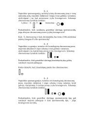
Vaizdo filtracija regos pirminės žievės lygyje<br />
Aleksandr BULATOV, Algis BERTULIS<br />
Pasiūlytas matematinis erdvinės-dažninės filtracijos, vykstančios regos pirminės žievės 4Cβ<br />
morfologinio sluoksnio lygyje, modelis. I ↩<br />
modelio struktūrane ↩<br />
itrauktos ↩<br />
neuronu ↩<br />
recepcijos lauku<br />
↩<br />
savybės, ir dėmesys koncentruojamas ties žievės neuronu ↩<br />
sluoksniu, i ↩ kur i↩ ateina signalai iš<br />
žemesniuj ↩<br />
u ↩<br />
regos centru. ↩<br />
Naudojami literatūriniai duomenys apie regos lauko projekcijos regos<br />
žievėje konformine ↩<br />
transformacijairžievės ↩<br />
neuronu ↩<br />
horizontaliutrump ↩<br />
uj ↩<br />
uryši ↩<br />
u ↩<br />
vienoduma. ↩<br />
Modeliui<br />
patikrinti panaudoti geometrines iliuzijas sukeliantys stimulai. Tie patys stimulai naudojami<br />
psichofizikiniuose eksperimentuose, ir gauti duomenys lyginami su modelio skaičiavimais. Žmogaus<br />
suvokimo deformaciju ↩<br />
ir modelio apskaičiuotu ↩<br />
pakitimu ↩<br />
charakteristikos yra artimai panašios.<br />
Modelis leidžia paprastai ir greitai apskaičiuoti filtracijos inešamas ↩<br />
deformacijas.